

Lupus & Autoimmune Disease: Complete Beginner’s Guide
Unexplained joint pain, profound exhaustion, and shifting symptoms often leave patients searching for answers long before receiving an official diagnosis. For many, the journey to understanding what is happening inside their own body spans months or even years of inconclusive tests and specialist referrals.
Navigating the medical system with vague, overlapping symptoms can be deeply isolating. Many patients fear the worst while struggling to articulate exactly what they are experiencing clearly enough for a specialist to act on it.
By the end of this guide, you will understand the fundamental biology of systemic autoimmune diseases so you can communicate effectively with a rheumatologist. We break down the exact mechanisms of lupus, outline symptom tracking methods, and review current medical management strategies.
This article is for educational purposes only and does not replace consultation with a qualified medical professional.
Key Takeaways
Systemic lupus erythematosus (SLE) is a chronic autoimmune condition where the body’s immune system attacks healthy tissue, affecting multiple organ systems simultaneously.
- Immune dysfunction: SLE involves the loss of B-cell tolerance and the production of autoantibodies that target the body’s own nuclear antigens.
- Symptom tracking: Using the Systemic Mapping Framework helps distinguish lupus flares from normal fatigue by categorizing symptoms into visible, systemic, and clinical buckets.
- Baseline treatment: The American College of Rheumatology recommends Hydroxychloroquine to reduce flares and prevent organ damage in all lupus patients.
- Diagnostic clarity: A rheumatologist uses ANA blood testing combined with physical symptoms and secondary immunological markers to confirm a diagnosis.
Clinical Integrity Note: The medical information in this guide synthesizes peer-reviewed data from the National Institutes of Health (NIH), the American College of Rheumatology (ACR), and the National Institute of Diabetes and Digestive and Kidney Diseases (NIDDK) to provide an objective, evidence-led baseline for patient education.
Lupus & Systemic Autoimmune Disease Fundamentals
Systemic lupus erythematosus (SLE) is a chronic autoimmune disease where the immune system mistakenly attacks healthy tissue across multiple organ systems. Unlike localized conditions, a systemic disease (one that circulates throughout the body via the bloodstream) can affect the skin, joints, kidneys, and brain simultaneously — often at the same time and with varying severity.
Understanding why this happens at the cellular level is the first step toward productive conversations with specialists. The biology is complex, but it can be explained clearly.
What Is an Autoimmune Disease?
Your immune system’s primary function is to detect and destroy foreign threats — bacteria, viruses, and damaged cells. It does this by producing proteins called antibodies, which act like identification tags that bind to and neutralize specific targets. Under normal conditions, this system is extraordinarily precise.
In an autoimmune disease, that precision fails. The immune system begins producing autoantibodies — proteins that mistakenly target the body’s own healthy tissues rather than external pathogens. Think of it like a home security system that has been misconfigured to flag the homeowners themselves as intruders, triggering alarms and locking doors against the very people it was designed to protect.
Why is SLE called a “systemic” disease? Because autoantibodies travel through the bloodstream, they can reach virtually any organ system. This is what separates systemic lupus erythematosus from a localized autoimmune condition like Hashimoto’s thyroiditis, which primarily targets the thyroid gland. In SLE, the attack is bodywide and unpredictable.
This widespread nature is exactly why a structured approach to tracking symptoms is essential. The Systemic Mapping Framework is a method for patients to categorize their vague, shifting symptoms into three distinct buckets before attending a specialist appointment:
- Visible signs: Skin rashes, hair loss, oral ulcers — anything observable externally
- Internal sensations: Fatigue, joint pain, chest tightness — what you feel but cannot see
- Clinical markers: Lab results, urine changes, blood pressure readings — measurable data only tests reveal
By mapping symptoms across these three categories, patients arrive at appointments with organized, clinically relevant information rather than a fragmented collection of complaints. This transforms a rushed 15-minute consultation into a focused diagnostic conversation.
Pathophysiology and B-cell tolerance in systemic lupus erythematosus confirms that SLE is characterized by the loss of B-cell tolerance and the production of autoantibodies targeting nuclear antigens, establishing the biological foundation for the widespread tissue damage patients experience (NIH, 2021).
While the biological mechanism explains the “why,” understanding how this cellular confusion translates into physical reality is crucial for patients.
The Pathophysiology of SLE
Systemic lupus erythematosus causes harm through a specific sequence of cellular failures that researchers have mapped in increasing detail over the past three decades. Understanding this chain of events demystifies why the disease behaves so unpredictably.
At the center of SLE pathology is a concept called B-cell tolerance — the immune system’s learned ability to distinguish “self” from “non-self.” B-cells are white blood cells that produce antibodies. In a healthy immune system, B-cells undergo a strict selection process that eliminates or silences any cell programmed to attack the body’s own tissue.
When B-cell tolerance breaks down, these self-reactive B-cells survive and proliferate. They begin producing autoantibodies that specifically target nuclear antigens (the proteins and DNA found inside the nucleus of every healthy cell). Imagine the nucleus of a cell as a locked vault containing your genetic blueprint. In SLE, the immune system begins treating that vault as a threat and constantly attempting to break it open.
This relentless attack generates immune complexes — clusters of autoantibodies bound to nuclear material — that deposit in organ tissues, particularly in the kidneys, skin, and blood vessel walls. These deposits trigger chronic, systemic inflammation (persistent, body-wide tissue irritation driven by immune activation), which is the direct cause of the symptoms patients experience: joint swelling, fatigue, rashes, and, in severe cases, organ dysfunction.
The key clinical insight: Because this inflammation is circulating and systemic rather than fixed in one location, symptoms in lupus shift and migrate in ways that localized conditions do not. A flare may present primarily as joint pain one month and a skin rash the next, making consistent symptom tracking — the foundation of the Systemic Mapping Framework — essential for building a clinical picture over time.

Caption: Autoantibodies in SLE travel through the bloodstream to deposit immune complexes in joints, skin, kidneys, and other organs — explaining the disease’s unpredictable, multi-system symptom pattern.
Because these autoantibodies can develop in anyone with a predisposing genetic background, researchers continually analyze demographic data to understand who is most vulnerable.
Who Is Most at Risk? (Demographics)
Lupus does not affect all populations equally. Clinical data reveals clear patterns in who develops the disease, and understanding these patterns helps patients contextualize their own risk profile.
Gender is the dominant risk factor. Approximately 90% of lupus cases occur in women, with the highest incidence during the childbearing years between ages 15 and 45. Researchers attribute this largely to the role of estrogen in modulating immune activity — estrogen appears to amplify B-cell responses, which may lower the threshold for autoimmune activation (NIH, 2021).
Ethnicity significantly affects both prevalence and severity. African American women develop lupus at roughly three times the rate of white women and tend to experience more severe kidney involvement and higher disease activity at diagnosis. Hispanic, Asian, and Native American populations also face elevated risk compared to non-Hispanic white populations. These disparities likely reflect a combination of genetic susceptibility, socioeconomic barriers to early diagnosis, and differential access to rheumatology care.
Genetic inheritance plays a supporting role, but is not determinative. Having a first-degree relative with lupus increases personal risk, but having the genetic predisposition does not guarantee disease onset. Environmental triggers — prolonged UV light exposure, certain viral infections (particularly Epstein-Barr virus), hormonal shifts, and specific medications — appear to be required to activate the disease in genetically susceptible individuals. This gene-environment interaction explains why identical twins do not always share a lupus diagnosis, even with identical DNA.
Knowing you fit the demographic profile is only the first step; the critical next phase is identifying the physical warning signs.
Recognizing Lupus Symptoms & Disease Manifestations
Lupus symptoms vary drastically between patients, earning the disease the nickname “the great imitator.” However, most patients experience a core triad of autoimmune disease symptoms: profound fatigue, unexplained joint pain, and photosensitive skin rashes. Tracking these indicators accurately is the first step toward a definitive clinical diagnosis.
No two patients will present identically. One patient may experience primarily dermatological symptoms for years before kidney involvement develops. Another may have severe fatigue as their dominant complaint with minimal skin findings. This variability is clinically intentional — SLE affects whatever organ system the circulating autoantibodies happen to target most heavily at any given time.
Visible Signs: The Malar Rash and Discoid Lupus
The most recognized physical hallmark of SLE is the malar rash, commonly known as a butterfly rash because of the shape it forms across the face. This rash appears on the bridge of the nose and extends symmetrically across both cheeks, resembling butterfly wings. It presents as a flat or slightly raised, reddish-to-purple discoloration that does not cross the nasolabial folds (the creases running from the nose to the corners of the mouth) — a detail that clinicians use to distinguish it from other facial rashes.
Characteristics of the malar rash that patients should document:
- Trigger: Most commonly provoked or worsened by sun or UV light exposure
- Duration: Can last from hours to weeks; may fade and recur without clear pattern
- Sensation: Often described as warm and slightly tender, occasionally itchy
- Appearance: Distinct from acne or rosacea in that it respects the nasolabial fold boundary
- Photography note: Because the rash may fade before a rheumatology appointment, patients should photograph it immediately upon appearance — this is a direct application of the Systemic Mapping Framework’s “visible signs” category
Beyond the malar rash, patients may develop photosensitive skin reactions in sun-exposed areas (arms, neck, décolletage) and oral ulcers on the roof of the mouth or inner cheeks, which are often painless and go unnoticed until pointed out by a clinician.
Cutaneous or discoid lupus is a distinct form of the disease isolated to the skin, without the internal organ involvement that defines SLE. Discoid lupus produces circular, coin-shaped lesions that can scar permanently if untreated. These lesions are raised, scaly, and tend to appear on the face, scalp, and ears. Critically, discoid lupus does not automatically indicate systemic disease — approximately 10–15% of patients with discoid lupus eventually develop SLE, but the majority do not (Office on Women’s Health, 2022). A rheumatologist or dermatologist uses skin biopsy and ANA testing to differentiate the two conditions.
While skin issues are the most visible sign of lupus, the invisible symptoms often cause the most daily disruption.
Systemic Fatigue vs. Normal Tiredness
Fatigue is reported by the vast majority of lupus patients and is frequently cited as the most debilitating symptom — yet it is also the most commonly dismissed. Understanding the biological distinction between lupus fatigue and ordinary tiredness is critical for both patients and the people supporting them.
Normal tiredness is proportional to activity and resolves with adequate rest. Sleep a full eight hours and you wake feeling restored. Lupus fatigue is physiologically different. It is driven by the ongoing metabolic cost of systemic inflammation — the body is constantly activating immune responses, producing inflammatory proteins called cytokines (cell-signaling molecules that drive inflammation), and attempting to repair tissue damage. This chronic immune activation consumes enormous energy resources, leaving patients feeling exhausted regardless of how much sleep they get.
Key distinguishing features of lupus fatigue:
- Unrestorative sleep: Rest does not fully relieve the exhaustion
- Concurrent physical symptoms: Fatigue almost always accompanies joint pain, stiffness, or cognitive fog (“brain fog”)
- Activity sensitivity: Modest physical exertion can trigger disproportionate exhaustion lasting days
- Unpredictability: Fatigue levels fluctuate significantly, correlating with disease activity
Musculoskeletal symptoms are nearly universal in SLE. Patients experience joint pain (arthralgia), swelling, and morning stiffness that typically affect the small joints of the hands, wrists, and knees symmetrically. Importantly, lupus arthritis is generally non-erosive — meaning it does not cause the severe bone and joint destruction seen in Rheumatoid Arthritis. This distinction matters clinically when determining the appropriate therapeutic approach.
Differentiating lupus fatigue from thyroid fatigue is a common diagnostic challenge. Differentiating profound fatigue in lupus from thyroid conditions from the Office on Women’s Health explains that extreme fatigue is common in both lupus and thyroid conditions like Hashimoto’s disease, but lupus is more often accompanied by joint pain and a distinctive malar rash across the cheeks and nose (2022). Thyroid fatigue typically appears alongside weight changes, temperature sensitivity, and hair thinning — without the joint involvement or photosensitivity that characterize lupus.
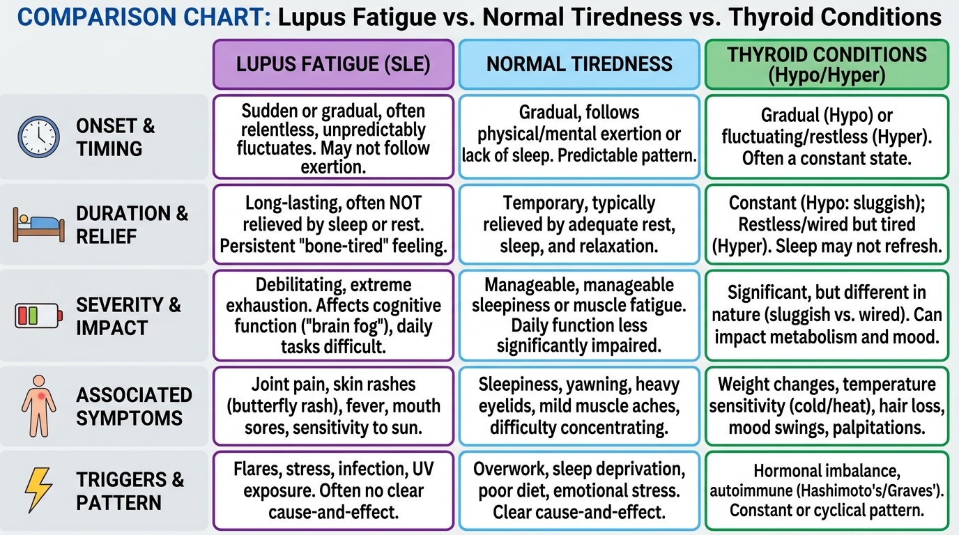
Caption: Distinguishing lupus fatigue from normal tiredness and thyroid-related exhaustion requires comparing concurrent symptoms — joint pain and photosensitivity point toward SLE.
Because these symptoms fluctuate daily, patients must learn to recognize when a bad day transitions into a clinical flare.
What Constitutes a Disease Flare?
A disease flare is medically defined as a measurable increase in lupus disease activity and systemic inflammation after a period of relative stability. Flares are not simply “bad days” — they represent a clinically significant shift in immune activity that often requires medical intervention to control before permanent organ damage occurs.
Common flare triggers identified in clinical literature include:
- UV light exposure: Even brief, unprotected sun exposure can trigger skin and systemic flares
- Psychological stress: Elevated cortisol disrupts immune regulation and can provoke inflammatory cascades
- Infections: Viral and bacterial illnesses activate the immune system broadly, which can cross-activate lupus pathways
- Physical exhaustion: Sustained overexertion depletes the body’s regulatory capacity
- Medication changes: Abrupt discontinuation of immunosuppressive drugs is a particularly dangerous trigger
How to identify a flare using the Systemic Mapping Framework: Patients should log symptoms across all three categories — visible, internal, and clinical — and note when symptoms are both new and rapidly worsening rather than stable or slowly evolving. A flare is characterized by acceleration, not merely the presence of symptoms.
Practically, patients should keep a symptom log that records the date, symptom type, severity on a 1–10 scale, any potential triggers from the preceding 24–48 hours, and whether the symptom is new or a known pattern. This documentation gives a rheumatologist the longitudinal data needed to adjust treatment proactively rather than reactively.
While recognizing flares is critical for daily comfort, understanding how lupus mimics or overlaps with other conditions is essential for an accurate initial diagnosis.
Understanding Connective Tissue & Overlap Autoimmune Disorders
Lupus belongs to a broader family of conditions known as connective tissue diseases — a group of autoimmune disorders that primarily affect the proteins and structures holding the body together, including collagen, tendons, cartilage, and blood vessels. Other members of this family include Sjogren’s disease, Systemic Sclerosis (scleroderma), and Polymyositis.
What makes autoimmune diagnostics particularly challenging is the existence of overlap syndromes — clinical situations where a patient simultaneously meets diagnostic criteria for more than one connective tissue disease. The most formally recognized of these is Mixed Connective Tissue Disease (MCTD), a condition defined by overlapping features of systemic lupus, systemic sclerosis, and polymyositis. Patients with MCTD often test positive for anti-U1-RNP antibodies (a specific immunological marker) while presenting with features of multiple separate diagnoses at once.
Clinical markers of mixed connective tissue disease and overlap syndromes confirms that mixed connective tissue disease is characterized by overlapping clinical features of systemic lupus erythematosus, systemic sclerosis, and polymyositis (MedlinePlus, 2020).
Lupus and Sjogren’s disease co-occur at a particularly high rate. Sjogren’s disease is an autoimmune condition that attacks the moisture-producing glands, causing chronic dry eyes and dry mouth. Studies estimate that 15–20% of lupus patients also carry a Sjogren’s diagnosis, and both conditions share the ANA-positive profile on blood tests, making differential diagnosis reliant on more specific immunological markers. A patient with profound fatigue, joint pain, and dry mucous membranes requires careful investigation for both conditions simultaneously.
For a comprehensive clinical exploration of overlap conditions, diagnostic criteria, and the nuances of differentiating these disorders, the dedicated cluster article on understanding connective tissue and overlap disorders provides the full clinical picture needed.
Severe Complications: Kidney Disease & Renal Failure
Among the most serious internal consequences of long-term SLE is Lupus Nephritis — a condition in which the autoantibodies characteristic of SLE specifically target the delicate filtration structures within the kidneys, known as glomeruli (tiny capillary clusters responsible for filtering waste from the blood). When immune complexes deposit in these structures, they trigger localized inflammation that progressively impairs filtration capacity.
The progression of kidney involvement in lupus follows a staged pathway. Early stages are often clinically silent — detectable only through routine urine and blood tests — which is why regular screening is not optional for lupus patients but a medical necessity. Key monitoring markers include creatinine levels (a waste product that accumulates when filtration declines) and proteinuria (the presence of excess protein in urine, detectable as foaming or frothing).
NIDDK data showing up to 30 percent of lupus nephritis patients develop end-stage renal disease reports that up to 30 percent of people who have lupus nephritis eventually develop end-stage renal disease (ESRD), requiring dialysis or a kidney transplant if disease activity is not adequately controlled (NIDDK, 2022). Early detection through routine screening is the single most effective intervention available.
Clinically actionable screening intervals: Most rheumatologists recommend urinalysis and creatinine testing at every routine appointment for SLE patients, regardless of whether kidney symptoms are present. Patients should report any sudden changes in urine appearance, unexpected fluid retention in the legs or ankles, or unexplained blood pressure elevations immediately.
For a detailed examination of lupus nephritis staging, creatinine monitoring protocols, and the long-term prognosis of renal disease in autoimmune patients, the cluster article on lupus kidney disease and renal failure covers this complex territory comprehensively.
Differentiating Thyroid & Liver Autoimmune Conditions
Profound, chronic fatigue is a symptom shared by multiple autoimmune conditions — and not all severe exhaustion indicates lupus. Two frequently confused categories are organ-specific autoimmune conditions, which attack a single gland or organ, and systemic autoimmune diseases like SLE, which circulate dysfunction throughout the body.
Thyroid autoimmunity encompasses two primary conditions: Hashimoto’s thyroiditis (where the immune system attacks and progressively destroys the thyroid, slowing metabolism and causing hypothyroidism) and Graves’ disease (where autoantibodies over-stimulate the thyroid, causing hyperthyroidism). Both produce profound fatigue — but the associated symptoms differ meaningfully from lupus. Hashimoto’s fatigue accompanies weight gain, cold intolerance, and hair thinning. Graves’ disease fatigue accompanies weight loss, heat intolerance, and heart palpitations. Neither routinely involves the joint pain or photosensitive rashes that characterize SLE.
Autoimmune hepatitis targets the liver specifically — autoantibodies attack hepatocytes (liver cells), causing progressive liver inflammation without the broader systemic involvement seen in lupus. Clinical distinctions between autoimmune hepatitis and lupus liver involvement confirms that while both conditions share systemic inflammatory markers, autoimmune hepatitis (AIH) primarily targets hepatocytes, whereas hepatic involvement in SLE typically manifests without severe liver failure (NIH, 2019). Elevated liver enzymes in a lupus patient are more often a medication side effect than a sign of autoimmune hepatitis.
If your symptoms include fatigue but do not clearly align with the joint pain, malar rash, and photosensitivity pattern of SLE, your rheumatologist will need a comprehensive thyroid panel and liver function tests before reaching a diagnosis. The cluster article on differentiating thyroid and liver autoimmune conditions provides the full differential diagnosis framework.
Treatment, Medication & Patient Lifestyle Management
Effectively managing systemic lupus erythematosus requires a dual approach: rigorous clinical diagnosis followed by a combination of pharmaceutical intervention and daily lifestyle modification. Because SLE is a chronic systemic disease, treatment focuses on suppressing hyperactive immune responses, minimizing organ damage, and improving long-term functional outcomes rather than achieving a cure.
No two lupus treatment plans are identical. A rheumatologist builds a personalized strategy based on which organ systems are involved, the current level of disease activity, the patient’s tolerance for specific medications, and their broader health profile. Understanding the tools available in that strategy — from diagnostic tests to drug classes — positions patients to participate actively in their own care.
The ANA Testing and Diagnostic Process
Antinuclear Antibody (ANA) testing is the primary blood screening for autoimmunity. The ANA test measures whether a patient’s blood contains antibodies that react against nuclear components of cells — a broad marker of autoimmune activity. Results are reported as a titer (a measurement of concentration, such as 1:80 or 1:320) and a pattern (how the antibodies are distributed within the nucleus under a microscope).
A critical point that prevents unnecessary alarm: a positive ANA does not automatically mean a patient has lupus. Approximately 15–20% of healthy individuals test ANA-positive without any autoimmune disease, and ANA positivity is also associated with other conditions including Sjogren’s disease, Rheumatoid Arthritis, and even certain medications (NIH, 2021). This is why clinical symptoms must always be evaluated alongside bloodwork — the laboratory result alone cannot carry a diagnosis.
When an ANA is positive and clinical symptoms are consistent with lupus, rheumatologists order more specific secondary tests:
- Anti-dsDNA (antibodies against double-stranded DNA): Highly specific for SLE and useful for tracking disease activity — elevated levels often correlate with flares
- Anti-Sm (antibodies against Sm nuclear proteins): Lower sensitivity but very high specificity for SLE, meaning a positive result strongly suggests the diagnosis
- Complement levels (C3, C4): These proteins are consumed during immune complex formation; low levels indicate active immune attack
In cases involving suspected organ damage, tissue biopsy becomes essential. A kidney biopsy graded on the WHO/ISN classification system (a standardized scoring framework for lupus nephritis) reveals the degree of glomerular inflammation and guides treatment intensity. Skin biopsies help differentiate discoid lupus from other dermatological conditions. Biopsy results provide information that no blood test alone can supply.
What to expect at your first rheumatology appointment: Bring a complete symptom log organized using the Systemic Mapping Framework — visible signs, internal sensations, and any clinical results you already have. Include records of previous blood tests, any medications taken, and a timeline of when symptoms began and evolved. Rheumatology appointments are often time-limited; arriving with organized, specific data significantly increases the clinical value of the consultation.
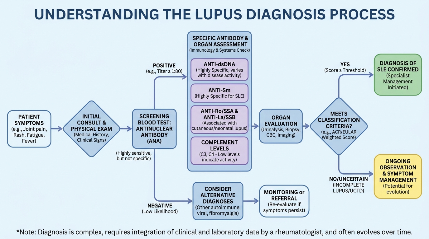
Caption: The lupus diagnosis pathway moves from ANA screening to specific immunological markers and, where needed, tissue biopsy — no single test provides a standalone diagnosis.
Once diagnostic bloodwork confirms autoimmune activity, rheumatologists typically deploy a tiered medication strategy.
Core Medications: From Hydroxychloroquine to Biologics
Lupus pharmacology operates on a tiered principle: begin with the safest, most broadly beneficial agents and escalate to more powerful immunosuppressive therapies only when clinical evidence requires it.
Hydroxychloroquine (Plaquenil) is the foundational anchor of lupus treatment. Originally developed as an antimalarial drug, it was discovered to have potent anti-inflammatory properties in autoimmune conditions. Hydroxychloroquine works by disrupting the activation of immune cells within acidic cellular compartments, reducing the inflammatory signaling that drives lupus activity. American College of Rheumatology treatment guidelines for lupus recommend Hydroxychloroquine for all patients with lupus, as it reduces flares, improves survival, and prevents organ damage (ACR, 2023). It is considered safe for long-term use, with the primary monitoring requirement being an annual eye examination — high cumulative doses over many years can rarely cause retinal changes.
Corticosteroids (most commonly Prednisone) are deployed for rapid control of acute inflammation during flares. They are highly effective at suppressing the immune response quickly but carry significant long-term side effects — including bone density loss, weight gain, elevated blood sugar, and increased infection risk — that make prolonged use medically undesirable. The clinical goal is always to use the lowest effective dose for the shortest necessary period, then taper the patient off as the flare resolves.
Immunosuppressants enter the treatment strategy when lupus causes significant organ involvement or when corticosteroid use cannot be adequately reduced:
- Methotrexate: Commonly used for skin and joint-dominant lupus; blocks folate-dependent pathways that fuel rapidly dividing immune cells
- Azathioprine (Imuran): Used for maintenance therapy and as a steroid-sparing agent; suppresses the production of new immune cells
- Mycophenolate mofetil (CellCept): Frequently used for lupus nephritis management; selectively inhibits lymphocyte (immune cell) proliferation
Biologics represent the most targeted tier of lupus therapy. Belimumab (Benlysta) — the first biologic specifically approved for lupus — works by blocking a protein called BLyS (B-lymphocyte stimulator) that promotes the survival of self-reactive B-cells. It is prescribed for patients with persistent, moderate-to-severe disease activity despite standard therapies.
Every medication plan must be individualized by a specialist. Consult a rheumatologist before starting, changing, or discontinuing any lupus treatment.
While medications suppress the internal biological attack, environmental triggers can still provoke the immune system, making lifestyle management non-negotiable.
The Systemic Mapping Framework for Daily Life
Lifestyle management in lupus is not supplementary advice — it is a clinical requirement that directly affects disease trajectory and medication effectiveness. The Systemic Mapping Framework applied to daily life means actively monitoring environmental inputs across the three symptom categories and making evidence-based adjustments.
Photosensitivity management is a medical priority. UV light triggers lupus flares through a specific mechanism: ultraviolet radiation induces a process called apoptosis (programmed cellular death) in skin cells. In lupus patients, the cellular debris released during this process exposes nuclear antigens that circulating autoantibodies then attack — directly triggering both cutaneous and systemic inflammation. This is not a cosmetic concern. Sun protection for lupus patients includes:
- Broad-spectrum SPF 50+ sunscreen applied daily, even on overcast days (UV penetrates cloud cover)
- Protective clothing: long sleeves, wide-brimmed hats, UV-blocking fabrics
- Avoiding peak UV hours (10 AM–4 PM) where practical
- Window film for car and home glass in high-UV environments
Pacing and energy management addresses the reality of living within an “energy envelope” — the concept that lupus patients have a finite pool of available energy that, if exceeded consistently, triggers immune dysregulation and flares. Applying the Systemic Mapping Framework means tracking daily energy expenditure in the “internal sensations” category, identifying activities that reliably precede exhaustion, and building scheduled rest periods into the day as non-negotiable rather than optional.
Diet and stress management function as supportive rather than curative interventions. An anti-inflammatory dietary pattern (emphasizing omega-3 fatty acids, vegetables, and minimizing processed food and refined sugars) reduces the baseline inflammatory load the immune system must contend with. Psychological stress elevates cortisol and disrupts regulatory immune pathways — patients benefit from evidence-based stress reduction techniques such as mindfulness-based cognitive therapy and structured relaxation. Neither diet nor stress management replaces pharmaceutical therapy but both reduce the frequency and severity of flares when practiced consistently.

Caption: A structured daily routine — built around the Systemic Mapping Framework’s three categories — helps lupus patients proactively manage triggers rather than react to flares.
Despite strict adherence to medications and lifestyle changes, living with a chronic illness involves inevitable challenges that require professional oversight.
Common Pitfalls & When to Seek Expert Help
Common Pitfalls in Autoimmune Management
Even well-informed, motivated patients encounter predictable failure points in long-term lupus management. Awareness of these patterns prevents the most damaging outcomes.
Pitfall 1: Self-discontinuing medications when feeling better. This is the most clinically dangerous error. Hydroxychloroquine and immunosuppressants suppress disease activity — they do not cure the underlying immune dysfunction. Patients who stop medication during periods of remission frequently experience severe rebound flares within weeks, sometimes involving organ systems that had previously been unaffected. The absence of symptoms is a sign that treatment is working, not a sign that it can be stopped. Any medication changes must be directed by a rheumatologist based on bloodwork, not on subjective wellbeing alone.
Pitfall 2: Pursuing unverified alternative treatments. Patients experiencing months or years of inadequate symptom control are understandably vulnerable to claims about “miracle cures,” unregulated stem-cell therapies, or supplement protocols. These alternatives carry real risks: financial exploitation, delayed access to evidence-based care during which organ damage may progress, and in some cases direct physical harm from unregulated substances. Patients should evaluate any non-pharmaceutical intervention by asking their rheumatologist directly whether evidence supports it.
Pitfall 3: Misattributing all new symptoms to lupus. Patients living with a chronic condition sometimes develop a cognitive pattern of assuming every new symptom is “just the lupus.” This is dangerous because lupus patients are immunosuppressed and therefore at elevated risk for serious infections, medication side effects, and secondary conditions. A new symptom that deviates from established personal patterns — particularly fever, localized pain, or neurological changes — requires medical evaluation rather than home management.
When to Seek Emergency Expert Help
Certain symptom presentations in lupus patients require immediate medical attention rather than routine appointment scheduling. Seek emergency care if you experience:
- Sudden, severe chest pain or shortness of breath: These symptoms may indicate lupus-related pericarditis (inflammation of the membrane surrounding the heart) or pleuritis (lung membrane inflammation), both of which require urgent evaluation. Do not wait to see if the symptom resolves.
- Signs of kidney distress: Foamy or frothy urine (indicating proteinuria), sudden extreme swelling in the legs or ankles, or unexplained and rapid increases in blood pressure signal potential renal involvement. Consult a doctor or visit an emergency room immediately.
- High fever unresponsive to standard management: Lupus patients on immunosuppressive therapy have reduced ability to fight infections. A fever that does not respond to standard measures — particularly one accompanied by chills, confusion, or localized pain — may signal a serious bacterial or opportunistic infection requiring urgent treatment. Do not delay seeking care.
Frequently Asked Questions
What Is Systemic Lupus Erythematosus (SLE)?
Systemic lupus erythematosus (SLE) is the most common and serious type of lupus, where the immune system attacks its own healthy tissues across multiple organ systems. This systemic attack circulates inflammation throughout the body via the bloodstream, distinguishing it from localized autoimmune conditions. Because of this widespread inflammation, it can damage major organs including the kidneys, brain, and heart. Approximately 70% of all lupus cases are classified as systemic. Patients require lifelong monitoring by a rheumatologist to manage disease activity and prevent permanent organ damage.
How Does Lupus Affect the Immune System?
Lupus causes the immune system to lose its ability to distinguish between foreign pathogens and healthy cellular tissue. Instead of producing protective antibodies, the body generates destructive autoantibodies that target the nuclei of healthy cells — a process driven by the loss of B-cell tolerance. This biological friendly fire triggers chronic inflammation and tissue damage across various organs throughout the body. Clinical blood tests, particularly ANA and anti-dsDNA screenings, detect these specific autoantibodies to confirm immune dysfunction. Treatment relies on medications designed to suppress this hyperactive immune response while minimizing long-term side effects.
How Is Lupus Diagnosed by a Rheumatologist?
A rheumatologist diagnoses lupus by combining detailed symptom history with specific immunological blood tests. The process typically begins with an Antinuclear Antibody (ANA) screening to detect generalized autoimmunity — but a positive ANA alone does not confirm lupus. If the ANA is positive and clinical symptoms are consistent, doctors order highly specific markers like anti-dsDNA and anti-Sm tests to confirm SLE. In cases involving suspected organ damage, doctors may require a tissue biopsy to assess cellular destruction directly. There is no single definitive test for lupus, making the combined judgment of an experienced rheumatologist essential to an accurate diagnosis.
Conclusion
For patients navigating unexplained chronic symptoms, systemic lupus erythematosus (SLE) represents a complex biological misfire where autoantibodies attack healthy tissue (NIH, 2021). The most effective approach to management combines rigorous diagnostic bloodwork — beginning with ANA testing and extending to organ-specific markers — adherence to baseline medications like Hydroxychloroquine, and strict environmental protections against UV exposure and known flare triggers.
Implementing the Systemic Mapping Framework allows patients to categorize their visible and internal symptoms accurately into observable, felt, and measurable categories. This structured tracking replaces health anxiety with objective data, ensuring that consultations with specialists are focused on clinical evidence rather than fragmented accounts of a “bad few months.”
Do not attempt to diagnose systemic autoimmune diseases at home. If you are experiencing chronic fatigue, persistent joint pain, or unexplained rashes, compile your medical records and organize your symptoms using the three-category framework described in this guide. Then schedule a formal evaluation with a board-certified rheumatologist to establish an evidence-based care plan. Early diagnosis, combined with consistent monitoring, remains the most powerful tool available for preventing irreversible organ damage.