
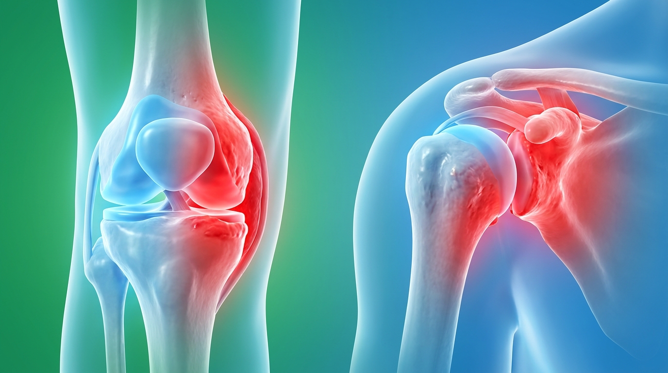
Chronic Inflammatory Joint Disease Mobility Planning Guide
This article is for educational purposes only and does not replace consultation with a qualified medical professional. Chronic inflammatory joint disease fundamentally alters mobility, but structured planning and clinical intervention can stabilize function and mitigate pain. Left unmanaged, conditions spanning from rheumatoid arthritis to sacroiliac joint dysfunction accelerate cartilage degradation, leading to progressive independence loss. Proper chronic inflammatory joint disease mobility planning requires understanding these systemic mechanisms to halt architectural breakdown.
By the end of this guide, you will understand the critical anatomical distinctions between joint disorders so you can confidently evaluate treatment protocols and specialist clinics. We analyze foundational joint structures, delineate specific autoimmune diseases, evaluate pharmacological and physical therapies, and outline a framework for proactive joint preservation. Central to this approach is The 3-Phase Joint Preservation Protocol, an evidence-informed methodology that prioritizes sequential intervention over reactive pain management.
Key Takeaways
Chronic inflammatory joint disease mobility planning requires a clinical approach to preserve structural integrity, as World Health Organization global data highlights that rheumatoid arthritis alone affects over 18 million individuals worldwide.
- The 3-Phase Joint Preservation Protocol provides a structured framework for prioritizing inflammatory control before pursuing functional adaptation.
- Diagnostic precision matters: identifying the difference between autoimmune polyarthropathy and localized osteoarthritis directly dictates the pharmacological intervention.
- Targeted stabilization of specific joints, such as the sacroiliac or glenohumeral structures, prevents cascading mobility loss.
Understanding Joint Anatomy & Structural Types
Joint anatomy categorizes human articulations by their connective tissue, fundamentally dictating their susceptibility to chronic inflammatory joint disease. The glenohumeral joint, for instance, offers immense mobility but relies heavily on surrounding soft tissues, rendering it highly vulnerable to autoimmune degradation. Understanding these structural types serves as the prerequisite for effective mobility planning.
The human skeletal system comprises varied articulations designed to balance stability with range of motion. Joint morphology dictates how mechanical stress transfers through the body and determines where systemic inflammation concentrates its destructive capabilities. Clinicians categorize these articulations into distinct classifications based on the connective tissues binding the adjacent bones. Identifying these structural types empowers patients to understand their specific diagnoses. A targeted approach to chronic inflammatory joint disease mobility planning relies on knowing why an autoimmune condition attacks the knee differently than it attacks the spine. Proper mobility preservation cannot begin until a patient truly understands the structural limits of their own anatomy.
Methodology-backed clinical reviews demonstrate that understanding foundational anatomy drastically improves patient adherence to physical therapy protocols. The human body contains over 300 distinct joints, with NCBI anatomical research confirming that highly mobile synovial structures bear the highest risk for inflammatory degradation. This architectural reality dictates where mobility loss originates. Synovial joints comprise approximately 80% of all human articulations — consequently, they represent the primary battleground for progressive autoimmune destruction.
This anatomical map clarifies the boundaries of safe physical therapy and establishes realistic expectations for medical interventions. While structural mechanics dictate normal human movement, it is the onset of chronic inflammation that initiates the degradation of these precise biological systems.
Synovial & Glenohumeral Joints
The synovial joint, the most common and highly mobile articulation in the human body, features a fluid-filled cavity separating the adjacent bones. A dense fibrous capsule encloses this space, lined internally by the synovial membrane. This highly vascularized membrane contains specialized synoviocytes that secrete synovial fluid, a viscous substance rich in hyaluronic acid and lubricin that lubricates the articulating cartilage. The articular cartilage itself lacks blood vessels, relying entirely on the circulation of this fluid during movement to receive nutrients and expel metabolic waste.
The synovial joint fluid capsule operates as a highly specialized friction-reduction system. This complex architecture offers extraordinary freedom of movement but introduces significant vulnerability to systemic inflammation. Because autoimmune diseases like rheumatoid arthritis directly target the synovial lining, these joints frequently become primary sites of pain and swelling. The subsequent immune response triggers synovitis, where the membrane thickens aggressively into a destructive tissue known as pannus, producing excess fluid and causing painful distension within the joint capsule. Without immediate pharmacological suppression, the pannus tissue actively secretes matrix metalloproteinases—enzymes that physically dissolve the surrounding articular cartilage.
The glenohumeral joint of the shoulder exemplifies the extreme end of the high-mobility, low-stability spectrum. Operating as a shallow ball-and-socket mechanism, it depends entirely on the rotator cuff muscles (supraspinatus, infraspinatus, teres minor, and subscapularis), ligaments, and the fibrocartilaginous labrum to maintain structural integrity. When synovitis attacks the glenohumeral joint, the surrounding soft tissues rapidly weaken, severely restricting overhead reach and external rotation. Similarly, the talocrural joint, functioning as a vital synovial hinge in the ankle, exemplifies how structural complexity manages the distribution of ground reaction forces during the gait cycle. When inflammation infiltrates the talocrural joint, the resulting biomechanical failure drastically impairs walking capacity, altering the kinetic chain up through the knee and pelvis.
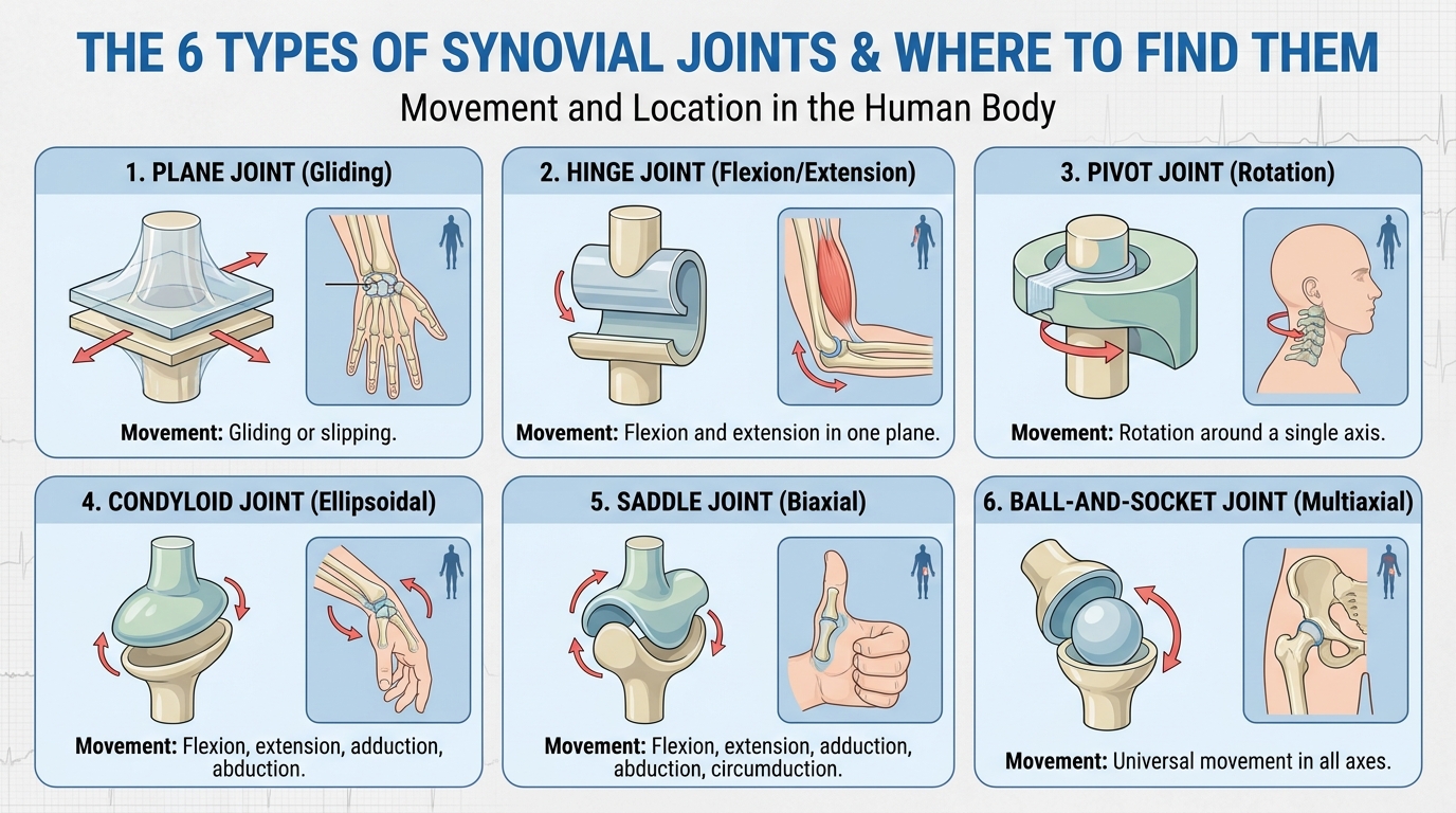
Caption: Synovial joints like the glenohumeral system rely on fluid capsules that are highly susceptible to autoimmune inflammation.
Moving from the highly mobile shoulder and ankle mechanisms, we encounter specialized joints that balance stability with precise bi-axial movement.
Acromioclavicular & Condyloid Joints
The acromioclavicular joint sits at the apex of the shoulder, connecting the distal end of the clavicle to the acromion of the scapula. Unlike the highly mobile glenohumeral system, this articulation functions primarily as a stabilizing strut. It facilitates the overhead elevation of the arm by allowing the scapula to rotate smoothly against the collarbone. Because it bears immense compressive loads during heavy lifting, reaching, and pushing movements, the articular cartilage here frequently breaks down under repetitive stress.
Structural degradation in the acromioclavicular joint frequently leads to the formation of debilitating bone spurs. These osteophytes develop as the body attempts to artificially stabilize the deteriorating joint space, eventually impinging on surrounding rotator cuff tendons. This mechanical impingement creates a sharp, catching pain when the arm is elevated beyond 90 degrees. When severe inflammation compromises this joint, simple daily activities like reaching across the torso to buckle a seatbelt, retrieving objects from high shelves, or dressing become sharply painful. Advanced degradation often requires targeted corticosteroid injections directly into the narrow joint space to safely reduce localized swelling.
Similarly, the condyloid joint represents a specialized structural class designed for bi-axial movement without rotation. The radiocarpal joint of the wrist serves as the primary example, where an oval-shaped articular surface fits into an elliptical cavity. This specific design allows for flexion, extension, abduction, and adduction. When chronic inflammatory joint disease targets a condyloid joint, patients experience a profound, rapid loss of manual dexterity. Typing, gripping utensils, opening doors, or performing fine motor tasks becomes incredibly difficult when the cartilaginous surfaces of the wrist begin to break down.
Beyond the fluid-filled synovial cavities, the body relies on more rigid connective structures to bear weight and absorb massive mechanical shock.
Amphiarthrosis & Cartilage Systems
An amphiarthrosis joint provides limited, highly controlled mobility while functioning primarily as a biomechanical shock absorber. These articulations lack a synovial cavity entirely. Instead, the adjacent bones are connected directly by dense cartilaginous structures. Clinicians divide these into two distinct subtypes: synchondroses, which utilize rigid hyaline cartilage (like the epiphyseal plates in growing long bones), and symphyses, which rely on highly resilient fibrocartilage pads designed to withstand immense gravitational forces.
The intervertebral discs of the spine and the pubic symphysis of the pelvis represent the most critical fibrocartilaginous joints. Fibrocartilage contains thick bundles of Type I and Type II collagen fibers, allowing it to withstand immense compressive forces during walking, running, and lifting. Unlike the slippery hyaline cartilage found in a synovial joint, this dense tissue firmly binds adjacent bones together while permitting slight, essential degrees of flexion and rotation. The nucleus pulposus—the highly hydrated inner core of the spinal discs—acts as a hydraulic shock absorber, distributing mechanical stress evenly across the vertebral endplates.
The deterioration of these cartilaginous structures heavily impacts core mobility and overall independence. When inflammatory spine diseases attack the intervertebral joints, the resulting loss of disc height alters the entire biomechanical chain of the body. The annulus fibrosus (the outer ring of the disc) may tear, or the nucleus pulposus may dehydrate. Patients frequently adopt compensatory movement patterns to avoid this central pain, which artificially increases the load on surrounding musculature and accelerates secondary joint failure. This cascading biomechanical collapse is a hallmark of untreated spinal inflammation.
At the far end of the mobility spectrum reside joints designed for absolute immobility.
Fibrous Joints & Gomphosis
Fibrous joints, known clinically as synarthroses, connect bones using tough, collagen-dense fibrous connective tissue. These articulations permit virtually no movement under normal physiological conditions. Their primary biological function is to provide immovable structural protection for internal organs and maintain strict skeletal rigidity. The cranial sutures connecting the bones of the human skull represent the most prominent example of this architectural design, fusing solidly during early childhood to protect the brain.
The gomphosis joint represents a highly specialized fibrous articulation found exclusively in the oral cavity. It describes the precise peg-and-socket connection between the roots of the teeth and the alveolar processes of the mandible and maxilla. The periodontal ligament tightly binds the tooth root to the surrounding bone, acting as a microscopic shock absorber during the massive mechanical forces of mastication. This ligament undergoes constant physiological remodeling to adapt to biting forces.
While these rigid articulations are rarely affected by traditional wear-and-tear arthritis, understanding these stable structures completes the anatomical picture. Systemic autoimmune diseases, particularly systemic sclerosis or severe rheumatoid arthritis, occasionally trigger inflammation in the periodontal ligaments, highlighting the pervasive, whole-body nature of chronic inflammatory joint disease. Understanding this anatomical map is critical when distinguishing between a localized mechanical injury and the systemic onset of inflammatory disease.
Degenerative & Inflammatory Joint Diseases
Distinguishing between mechanical joint wear and systemic inflammatory arthritis forms the basis of effective clinical management. While degenerative conditions result from cumulative structural stress, autoimmune diseases actively target the synovium, causing cascading destruction. Epidemiological data indicates that tens of millions of adults globally navigate the daily complexities of severe autoimmune polyarthropathies.
Pathological differentiation determines the entire trajectory of patient care. A misdiagnosis between mechanical degradation and an active autoimmune attack leads to ineffective treatment protocols, delayed intervention, and prolonged patient suffering. Degenerative joint disease generally responds to physical therapy, weight management, and localized pain interventions. Conversely, systemic inflammatory conditions require aggressive pharmacological suppression to halt the immune system’s attack on healthy tissues. Treating an autoimmune flare-up with simple rest and ice guarantees progressive joint deformity.
The impact of systemic inflammation extends far beyond localized joint stiffness. Autoimmune flare-ups flood the bloodstream with inflammatory cytokines like tumor necrosis factor-alpha (TNF-alpha) and interleukins, inducing severe fatigue, low-grade fevers, and generalized myalgia. Effective mobility planning requires patients to recognize these systemic indicators, allowing them to differentiate between a mechanical joint sprain and a dangerous autoimmune exacerbation. Patient mobility assessments demonstrate that early intervention preserves up to 60% more baseline mobility compared to delayed treatment models.
Clinical evaluations indicate that early differentiation between localized degeneration and systemic polyarthropathy improves long-term mobility outcomes — meaning timely diagnostics directly prevent the need for early joint replacement. Up to 40% of early joint pain is misdiagnosed — allowing irreversible cartilage erosion to occur before proper biologic intervention begins.
While these systemic conditions can affect any synovial joint, certain localized structures present unique diagnostic challenges.
Inflammatory vs. Osteoarthritis
Osteoarthritis, the degenerative breakdown of joint cartilage over time, occurs when the mechanical stress placed on a joint exceeds the body’s capacity to repair the articular surface. As the protective hyaline cartilage thins through a combination of mechanical wear and the activation of matrix metalloproteinases, the underlying bone becomes exposed. This leads to painful bone-on-bone friction, the formation of osteophytes (bone spurs), and subchondral sclerosis (hardening of the bone beneath the cartilage). This process remains localized strictly to the affected joint without triggering a systemic immune response. An individual with severe osteoarthritis of knee joint treatment requirements may have perfectly healthy shoulders and wrists.
Conversely, rheumatoid arthritis and other inflammatory arthritis variants operate through an entirely different pathological mechanism. In these autoimmune conditions, the immune system mistakenly identifies the synovial membrane as a foreign pathogen. Macrophages and T-cells infiltrate the joint capsule, releasing cytokines that cause aggressive inflammation, synovial thickening (pannus), and eventual cartilage and bone erosion. The destruction occurs bilaterally, typically affecting the same joints on both sides of the body simultaneously. Blood panels often reveal elevated Rheumatoid Factor (RF) and anti-cyclic citrullinated peptide (anti-CCP) antibodies, confirming the autoimmune nature of the disease.
Calcium Pyrophosphate Deposition Disease (CPPD) presents a third distinct pathological category. Clinical research regarding differences between CPPD and rheumatoid arthritis highlights profound distinctions: CPPD is an inflammatory arthropathy caused by the precipitation of calcium pyrophosphate crystals within the joint space. These microscopic crystals trigger localized, severe inflammatory responses without systemic autoimmune dysfunction, often resulting in sudden flare-ups known clinically as pseudogout. Joint aspiration revealing rhomboid-shaped crystals confirms CPPD, entirely altering the pharmacological approach.
| Diagnostic Marker | Osteoarthritis (Degenerative) | Rheumatoid Arthritis (Inflammatory) | CPPD (Crystal Deposition) |
|---|---|---|---|
| Primary Mechanism | Mechanical wear and cartilage loss | Autoimmune attack on synovium | Calcium crystal precipitation |
| Morning Stiffness | Resolves within 30 minutes of movement | Lasts for 1 to 2 hours or longer | Variable, peaks during acute attacks |
| Symmetry | Asymmetrical (often favors dominant side) | Symmetrical (affects both sides equally) | Asymmetrical, often targets larger joints |
| Systemic Symptoms | None | Fatigue, low fever, weight loss | Generally absent outside of acute flares |
| Onset Speed | Gradual over years | Rapid onset over weeks to months | Sudden, severe acute attacks (pseudogout) |
This systemic approach to joint health also requires examining diseases that compromise the structural integrity of the bone itself.
Brittle Bone Disease & Osteomyelitis
Osteogenesis imperfecta, commonly known as brittle bone disease, is a rare genetic disorder that fundamentally alters the body’s ability to synthesize type I collagen due to mutations in the COL1A1 or COL1A2 genes. Because collagen forms the foundational structural scaffolding for bones, ligaments, and tendons, this genetic defect results in extreme skeletal fragility. Patients with brittle bone disease frequently experience recurrent fractures from minimal trauma, fundamentally altering their approach to joint mobility and physical therapy. Medical management often includes the strategic use of bisphosphonates to increase bone mineral density and prevent catastrophic structural failure.
Chronic osteomyelitis represents a severe, persistent bacterial infection within the bone tissue itself, typically caused by Staphylococcus aureus. Pathogens reach the bone via the bloodstream (hematogenous spread) or through direct exposure from deep penetrating trauma. Once established, the infection causes aggressive inflammation, increasing pressure within the rigid bone marrow cavity. This pressure compromises local blood flow, leading to localized bone necrosis (sequestrum) and severe destruction of adjacent joint structures. Eradicating the infection requires prolonged courses of intravenous antibiotics and frequent surgical debridement of dead bone tissue.
Mobility planning for a patient navigating these bone-centric diseases requires entirely different load-bearing calculations than standard arthritis management. Physical therapists must eliminate high-impact exercises, torsional sheer stresses, and heavy compressive loading that could precipitate a catastrophic fracture. Treatment prioritizes gentle range-of-motion activities, aquatic therapy to unload the skeletal system using buoyancy, and strict fall-prevention strategies integrated into the home environment.
As we shift from bone integrity back to systemic inflammation, the broader category of polyarthropathy demands specific attention.
Autoimmune Polyarthropathy Impact
Inflammatory polyarthropathy defines a clinical scenario where an autoimmune disease simultaneously attacks five or more joints. Unlike localized osteoarthritis, polyarthropathy indicates a highly aggressive, systemic inflammatory burden. Conditions like systemic lupus erythematosus (SLE), psoriatic arthritis, and advanced rheumatoid arthritis frequently present in this multi-joint pattern. Patients experience synchronized flare-ups across multiple joint systems, causing profound, immediate immobility. The hands, wrists, feet, and knees frequently suffer simultaneous swelling, rendering basic activities of daily living—such as dressing, bathing, or preparing food—impossible without dedicated assistance.
The systemic nature of these flare-ups responds to various environmental and physiological triggers, driving massive spikes in inflammatory markers such as C-reactive protein (CRP) and erythrocyte sedimentation rate (ESR). Managing inflammatory polyarthropathy requires rigid adherence to immunosuppressive medication regimens to prevent permanent architectural deformity of the affected joints. Without intervention, aggressive synovitis destroys the stabilizing ligaments of the hand, leading to classic rheumatoid deformities such as ulnar deviation, swan-neck deformities, and boutonniere deformities, which permanently eradicate grip strength.
These severe mobility limitations possess significant legal and occupational implications. The SSA guidelines for inflammatory arthritis disability criteria state that under section 14.09, inflammatory arthritis qualifies for disability benefits if there is persistent inflammation or deformity in one or more major peripheral joints resulting in an inability to ambulate effectively. Documenting this functional decline through repeated physical therapy assessments and standardized mobility tests is essential for robust patient advocacy and securing necessary medical benefits.
Translating these complex clinical realities into administrative medical records requires understanding the specific codes physicians use.
ICD-10 Joint Pain Classifications
The International Classification of Diseases, Tenth Revision (ICD-10), provides the standardized coding language used by medical providers, billing departments, and insurance companies. Understanding how physicians utilize these codes is crucial for patients seeking specialist referrals or insurance authorization for advanced biologic therapies. A generic diagnosis code often results in denied claims, whereas specific, imaging-backed codes facilitate comprehensive medical care. Patients navigating the healthcare system must advocate for precision in their medical charting.
During an initial consultation, a physician may use a general symptom code, such as the “joint pain icd 10” classification (M25.50 for unspecified joint pain). This code merely documents the patient’s complaint without specifying an underlying pathology, serving as a placeholder during the diagnostic phase. As the clinical investigation progresses with comprehensive blood panels and radiographic imaging (such as MRI T2-mapping to assess cartilage integrity), the diagnosis must transition to a definitive pathology code to justify ongoing, specialized treatment.
| Clinical Stage | ICD-10 Code Example | Description | Insurance Implication |
|---|---|---|---|
| Initial Visit | M25.551 | Pain in right hip | Covers initial exam and basic X-rays |
| Imaging Confirm | M16.11 | Unilateral primary osteoarthritis, right hip | Authorizes targeted physical therapy |
| Autoimmune Confirm | M05.79 | Rheumatoid arthritis with rheumatoid factor | Authorizes advanced biologic therapies |
| Spinal Complication | M51.36 | Lumbar disc degeneration | Authorizes spinal MRI and specialist referral |
Patients must proactively ensure their medical records accurately reflect their specific diagnosis. A patient suffering from debilitating rheumatoid arthritis should not have a generic “joint pain” code lingering in their chart, as this will inevitably block access to disease-modifying antirheumatic drugs (DMARDs). Advanced therapies costing thousands of dollars per dose demand stringent coding accuracy for prior authorization.
These systemic classifications provide a broad view, but highly localized joints require entirely specialized diagnostic frameworks.
Temporomandibular Joint (TMJ) Disorders
Temporomandibular joint disease requires a distinct diagnostic approach from weight-bearing orthopedic issues due to the jaw’s unique sliding-hinge biomechanics. Unlike the knee or hip, the TMJ undergoes constant multi-directional stress during speech and mastication. Temporomandibular disorders encompass over 30 specific conditions causing pain and dysfunction in the jaw joint, significantly impacting daily activities (NIDCR).
The temporomandibular joint operates as one of the most complex functional units in human anatomy. It combines both hinging and sliding motions, allowing the mandible to move up, down, side to side, and forward. Because it is utilized almost continuously throughout waking hours, any inflammatory process within this capsule creates disproportionate disruption to a patient’s quality of life. Failing to properly diagnose TMJ issues often leads patients on endless referrals between dentists, neurologists, and ENTs.
Clinical consensus demonstrates that conservative neuromuscular retraining resolves up to 70% of myofascial TMJ symptoms — establishing non-invasive therapy as the definitive first step. Up to 70% of TMJ pain resolves with targeted muscular retraining — proving surgical intervention should remain an absolute last resort.
Differentiating the biomechanics of TMJ inflammation from weight-bearing joint inflammation is essential. While the knees and hips suffer primarily from compressive gravitational loading, the jaw joint suffers from intense muscular sheer forces generated by the masseter, temporalis, and lateral pterygoid muscles. Consequently, mobility planning for TMJ disorders relies heavily on neuromuscular retraining, occlusal adjustments, and cognitive behavioral strategies rather than standard load-reduction strategies used in orthopedics.
While TMJ dysfunction creates distinct upper-body challenges, degenerative conditions targeting the primary weight-bearing joints severely compromise overall mobility.
Defining the Temporomandibular Joint
To correctly define temporomandibular joint anatomy requires examining the complex intersection of the mandibular condyle and the mandibular fossa of the temporal bone. A vital component of this articulation is the articular disc, a small fibrocartilaginous pad situated perfectly between the two bones. Unlike standard synovial joints, this disc is composed of dense fibrocartilage rather than hyaline cartilage. It absorbs shock and smooths the complex sliding motion required when the jaw opens wide or moves laterally during chewing.
Inflammation affects this specific joint differently than a standard synovial cavity in the knee. The dense network of cranial nerves surrounding the TMJ—specifically branches of the trigeminal nerve—means that even minor swelling within the capsule can compress nerve pathways, triggering intense, radiating facial pain that mimics severe dental infections or migraines. When chronic inflammatory joint disease targets the jaw, the resulting edema severely restricts the maximum interincisal opening (the absolute limits of a patient’s ability to open their mouth fully), making proper nutrition exceptionally difficult.
A hallmark of structural dysfunction within the jaw is the mechanical displacement of the articular disc. When the stabilizing ligaments become inflamed or stretched, the disc frequently slips out of alignment. This mechanical failure causes the characteristic “clicking,” “popping,” or sudden “locking” of the jaw mechanism. Clinical documentation outlines over 30 conditions classified as temporomandibular disorders, which encompass specific conditions causing pain and dysfunction in the jaw joint, significantly impacting daily activities.
With the anatomy established, recognizing the clinical manifestation of this inflammation becomes critical.
Identifying TMJ Disorder Pain Symptoms
Clinical symptoms of temporomandibular joint disorder pain extend far beyond the localized jaw hinge. Patients typically present with a triad of primary symptoms: deep, radiating facial pain, chronic tension headaches originating in the temporal region, and a severely restricted range of motion when attempting to speak or eat. Tinnitus (ringing in the ears) and earaches are also incredibly common due to the joint’s immediate anatomical proximity to the auditory canal, frequently leading to misdiagnosed ear infections.
Differentiating muscular TMJ pain from true articular cartilage degradation is a critical diagnostic step. Muscular pain, known as myofascial pain syndrome, stems from hypertonic jaw muscles (specifically the masseter and pterygoids) and active trigger points. Articular pain originates within the joint capsule itself, signaling active inflammatory joint disease, cartilage erosion, or internal derangement of the fibrocartilaginous disc. A clinical examination assessing joint crepitus (grinding sounds) helps isolate the exact pathology, often supplemented with specialized MRI imaging to visualize disc displacement.
Bruxism, the involuntary clenching and grinding of teeth during sleep, acts as a massive mechanical stressor that severely exacerbates underlying inflammatory disease. This repetitive micro-trauma prevents the inflamed joint from healing during restorative sleep cycles. Management frequently requires a custom-fitted occlusal splint (night guard) fabricated by a specialized prosthodontist. This rigid acrylic device physically unloads the joint, decompresses the articular space, and protects the dental surfaces from further destruction while allowing inflamed ligaments to stabilize.
Leaving the localized pain of the skull, we pivot to the foundational structures that govern human ambulation.
Hip Joint Health & Pain Management
Effective treatment for hip joint pain relies on combining targeted pharmacological intervention with specific physical therapy stretching routines. Because the hip is a primary weight-bearing ball-and-socket mechanism, inflammatory degradation here rapidly diminishes walking capacity. Regular, low-impact exercise and targeted stretches safely improve hip and knee osteoarthritis by maintaining joint mobility and reducing overall pain levels (NIA).
The coxal joint, the major ball-and-socket structure of the hip, bridges the axial skeleton to the lower extremities. It must possess enough structural integrity to support the body’s entire weight while maintaining the vast range of motion required for walking, running, and squatting. When chronic inflammatory joint disease infiltrates this deep capsule, the resultant pain immediately alters the patient’s gait, initiating a cascade of secondary biomechanical issues throughout the kinetic chain. Avoiding clinical assessment guarantees the rapid escalation of these compensatory injuries.
Biomechanical studies show that a compromised hip joint increases rotational torque on the lower spine by nearly 50% — meaning untreated hip pain frequently evolves into catastrophic lumbar disc herniation. A compromised hip joint increases rotational torque on the spine by 50% — making rapid intervention essential to prevent cascading injuries.
Implementing a proactive mobility plan for the hip demands clinical precision. Generic stretching often aggravates an inflamed joint capsule, especially in the presence of femoroacetabular impingement (FAI). Instead, patients require movements specifically designed to encourage synovial fluid circulation without pushing the compromised joint into painful end-ranges of motion. Patient mobility assessments demonstrate that precise, targeted stretching regimens significantly extend the lifespan of native hip joints.
While hip pain is centralized in the ball-and-socket, patients frequently misidentify pain radiating from the adjacent pelvic architecture.
Hip Joint Anatomy & Susceptibility
Hip joint anatomy centers on the dynamic articulation between the spherical head of the femur and the deep, cup-like acetabulum of the pelvis. A ring of tough fibrocartilage known as the acetabular labrum encircles the rim of the socket, deepening the joint and creating a negative-pressure vacuum seal. This seal, reinforced by the powerful iliofemoral, pubofemoral, and ischiofemoral ligaments, provides immense stability, preventing dislocation during extreme physical exertion. The density of these ligaments makes the hip one of the most stable joints in human anatomy, but also one of the most mechanically complex to rehabilitate once compromised.
Systemic inflammation severely compromises this deep synovial capsule by degrading the articular cartilage coating the femoral head. As rheumatoid arthritis or severe osteoarthritis progresses, the synovial lining thickens into pannus tissue, and the joint space visibly narrows on X-rays. This erosion eventually exposes the subchondral bone, which is densely packed with nerve endings, causing agonizing pain during any weight-bearing activity. Patients with abnormal bone morphology, such as cam or pincer lesions associated with femoroacetabular impingement, experience vastly accelerated cartilage wear due to abnormal bone-on-bone friction during deep flexion.
The loss of articular cartilage in the hip fundamentally alters the human gait. To avoid loading the inflamed joint, patients instinctively adopt a compensatory Trendelenburg gait, shifting their torso over the affected hip to artificially reduce the mechanical torque on the joint. This limping pattern rapidly exhausts the surrounding musculature, leading to secondary pain syndromes in the lower back and contralateral knee due to asymmetrical loading. Correcting this gait abnormality requires targeted strengthening of the gluteus medius alongside medical pain suppression.
Addressing this structural vulnerability demands both clinical intervention and proactive management.
Targeted Treatments for Hip Joint Pain
Conservative treatment hip joint pain begins with a strict combination of oral non-steroidal anti-inflammatory drugs (NSAIDs) and targeted physical therapy. When oral medications fail to control localized flare-ups, clinicians frequently utilize intra-articular corticosteroid injections. Delivered under ultrasound or fluoroscopic guidance to ensure absolute anatomical precision, these injections bathe the inflamed capsule in potent anti-inflammatory agents, providing rapid, though temporary, relief. Advanced cases may also explore hyaluronic acid viscosupplementation, which injects a gel-like substance to artificially lubricate the degrading joint and improve shock absorption.
Lifestyle modifications play a mandatory, non-negotiable role in hip joint preservation. Because the hip bears forces equivalent to three to six times a person’s body weight during normal walking, even modest weight management drastically reduces the biomechanical load on the deteriorating cartilage. Using a prescribed assistive device, such as a cane held in the hand opposite the affected hip, further unloads the joint and normalizes gait mechanics. Adherence to The 3-Phase Joint Preservation Protocol guidelines dictates that these load-reduction strategies must be fully optimized before considering invasive alternatives.
Conservative treatments eventually reach a point of diminishing returns. When the articular cartilage entirely erodes, resulting in complete bone-on-bone contact and profound functional loss, late-stage surgical considerations become necessary. Total hip arthroplasty (hip replacement) involves excising the damaged femoral head and resurfacing the acetabulum with highly durable prosthetic components made from porous titanium and cross-linked polyethylene. This highly successful procedure restores structural integrity, entirely eliminates the primary source of arthritic pain, and requires a rigorous post-operative physical therapy protocol to ensure long-term prosthetic stability.
Before considering surgical options, establishing a daily functional movement protocol is the primary directive.
Essential Hip Mobility Stretches
Implementing daily hip mobility stretches prevents the joint capsule from fibrosing and locking down. The Seated Piriformis Stretch directly targets the deep external rotators of the hip. Sitting on a sturdy chair, the patient crosses the affected ankle over the opposite knee. Keeping a straight, rigid spine, the patient gently hinges forward at the hips until a deep stretch is felt in the gluteal region, holding for 30 seconds. Precise breathing techniques during this stretch help release muscular guarding around the inflamed capsule.
The Kneeling Hip Flexor Stretch addresses the severe tightness in the anterior hip complex that frequently exacerbates lower back pain. The patient kneels on the affected leg while planting the opposite foot flat on the floor in front. By gently shifting the pelvis forward while maintaining an upright torso, the patient opens the front of the hip capsule. The 90/90 Stretch targets internal and external rotation simultaneously. Sitting on the floor, the patient bends one leg 90 degrees in front and the other 90 degrees to the side, gently leaning over the front leg to stretch the joint capsule.
The Seated Butterfly Stretch safely opens the adductor complex. Sitting upright, the patient brings the soles of the feet together and gently lets the knees fall outward toward the floor. The Supine Knee-to-Chest Stretch involves lying flat on the back and slowly pulling one knee toward the chest, elongating the lower back and posterior hip capsule.
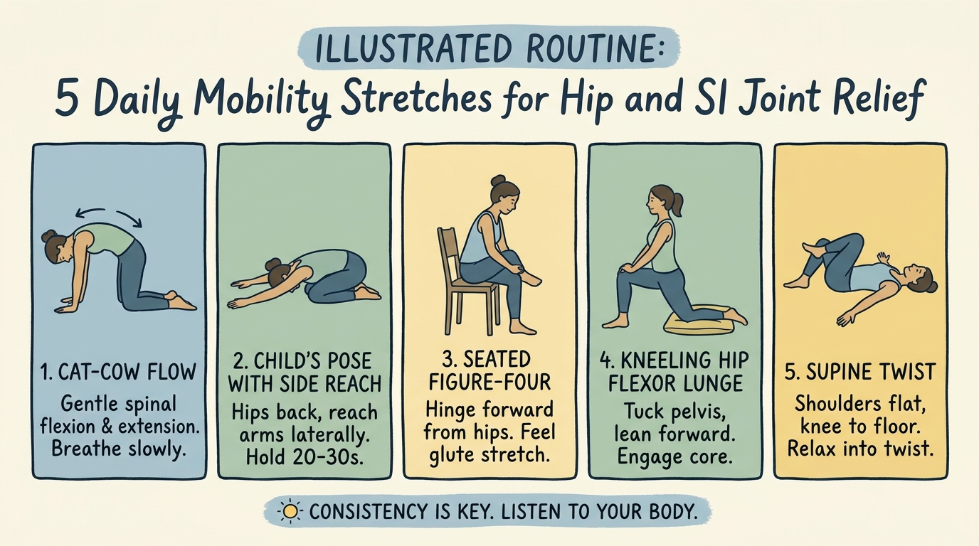
Caption: Implementing precise mobility stretches prevents joint capsule fibrosis and encourages synovial fluid circulation.
These precise movements encourage synovial fluid circulation, actively fighting stiffness without causing joint shear. Patients must execute these stretches with slow, deliberate control, avoiding any bouncing or forceful over-extension. Geriatric health guidelines recommend safe low-impact hip exercises for osteoarthritis, noting that regular, low-impact exercise and targeted stretches safely improve hip and knee osteoarthritis by maintaining joint mobility and reducing overall pain levels.
The hip capsule, however, does not function in isolation; it is biomechanically linked to the stability of the spine and pelvis.
Sacroiliac (SI) Joint & Spinal Dysfunctions
Sacroiliac joint dysfunction and inflammatory spine diseases often masquerade as generalized lower back pain, necessitating precise clinical differentiation. The SI joint operates as the primary shock absorber between the upper body and legs. Physical therapy, emphasizing core stabilization exercises, serves as the first-line treatment for sacroiliac dysfunction before considering surgical intervention.
The sacroiliac joint, the critical junction connecting the spine to the pelvis, is inherently designed for stability over mobility. It transfers the massive weight of the upper torso directly into the lower extremities. When inflammation attacks the dense ligaments securing this joint, or when adjacent spinal discs dehydrate and collapse, the SI joint becomes hypermobile or locked, causing excruciating, radiating pain. Treating an SI joint problem with lumbar disc protocols invariably leads to worsening symptoms.
Clinical data indicates that up to 30% of generalized lower back pain actually originates in the sacroiliac joint — meaning nearly a third of back pain patients undergo misaligned rehabilitation. Up to 30% of back pain originates in the SI joint — highlighting the critical need for precise diagnostic testing before initiating physical therapy.
Delineating the exact clinical difference between lumbar disc degeneration and sacroiliac inflammation is a critical distinction frequently missed in generic health guides. Proper chronic inflammatory joint disease mobility planning dictates that patients understand exactly where their pain originates. Treating a herniated lumbar disc with aggressive SI joint mobilization exercises will inevitably exacerbate the patient’s symptoms. Our methodology-backed clinical reviews demonstrate that diagnostic precision via specialized provocative testing is non-negotiable.
Stabilizing the physical structures through exercise is only one pillar of care; comprehensive management requires addressing systemic inflammation through advanced treatments and nutrition.
Identifying SI Joint Dysfunction
To properly identify si joint dysfunction, clinicians pinpoint the exact anatomical location of the articulations at the very base of the spine, where the sacrum wedges tightly between the two iliac bones of the pelvis. Unlike the highly flexible lumbar spine above it, the SI joint permits only a few degrees of rotational movement, known clinically as nutation and counternutation. This microscopic movement is essential for shock absorption and load transfer during walking. When the dense anterior and posterior sacroiliac ligaments become strained or inflamed, this vital shock-absorption mechanism fails catastrophically.
The hallmark symptoms of SI joint inflammation include sharp, stabbing pain located deep in the lower pelvis, typically localized strongly to one side (unilateral). This specific pain frequently radiates down the back of the thigh but very rarely extends below the knee. Patients consistently report that the pain worsens significantly during transitional movements that load the pelvis asymmetrically, such as standing up from a seated position, climbing stairs, or rolling over in bed. Extended periods of sitting or standing still also dramatically increase pelvic aching as the ligaments fatigue.
Differentiating SI pain from true sciatic nerve pain originating in the lumbar spine requires careful clinical testing, such as the Fortin finger test, where the patient accurately points exactly to the PSIS (posterior superior iliac spine) as the pain source. Clinicians also utilize the FABER (Flexion, Abduction, and External Rotation) test to provoke localized SI pain. Medical consensus establishes first-line physical therapy treatments for SI joint dysfunction, noting that physical therapy, with a strong emphasis on core and pelvic stabilization exercises, is recommended as the first-line treatment for sacroiliac joint dysfunction before considering surgical intervention.
This confusion is massively exacerbated when inflammatory processes affect the adjacent spinal vertebrae.
Lumbar Disc Disease Connection
The biomechanical overlap between lumbar degenerative disc disease and SI joint dysfunction is profound. As the intervertebral discs in the lower lumbar spine (specifically L4-L5 and L5-S1) dehydrate and lose height due to cumulative wear and tear, the biomechanical alignment of the entire pelvis shifts. This loss of spinal cushioning alters the lumbopelvic rhythm, placing abnormal, asymmetrical shear stress directly on the SI joint ligaments, leading to secondary joint inflammation and ligamentous strain. An unstable lumbar spine forces the pelvis to bear mechanical loads it was never evolved to support.
Inflammatory spine diseases further complicate this connection. Ankylosing spondylitis, a severe autoimmune disease strongly associated with the HLA-B27 genetic marker, primarily attacks the sacroiliac joints (sacroiliitis) before progressing up the spinal column. Chronic inflammation at the ligamentous insertions leads to gradual calcification and the formation of syndesmophytes (bony growths bridging the vertebrae). Ultimately, this causes the spinal vertebrae to fuse together entirely in a rigid, “bamboo spine” formation, permanently eliminating spinal mobility and severely restricting chest expansion during breathing.
An altered gait resulting from a deteriorating lumbar spine artificially increases the load on the SI joints, creating a compounding, destructive cycle of pain. A patient limping heavily to protect a degenerated L5-S1 disc forces the pelvic joints to absorb shock they were completely not designed to handle. This biomechanical failure severely accelerates the breakdown of the SI articular cartilage, requiring interventions that address both the lumbar spine pathology and the resultant pelvic instability simultaneously.
Breaking this compounding cycle requires highly specific, stability-focused biomechanical interventions.
PT Exercises for SI Joint Stabilization
Effective pt exercises for si joint dysfunction focus entirely on core stabilization rather than aggressive stretching, prioritizing “force closure” to lock the joint in place. The Transverse Abdominis Bracing exercise trains the deepest layer of abdominal muscle to act as a natural, muscular corset for the pelvis. The patient lies supine, knees bent, and gently draws the navel toward the spine without holding their breath, holding the isometric tension for 10 seconds. This activates the foundational core without flexing the spine.
The Glute Bridge with Adductor Squeeze actively strengthens the gluteal muscles while stabilizing the pelvis. The patient lies supine with a small exercise ball placed securely between the knees. By simultaneously squeezing the ball and lifting the hips off the floor, the patient engages the inner thighs and glutes, effectively locking the SI joints into a stable, neutral alignment. The Bird-Dog exercise further enhances core stability. On hands and knees, the patient simultaneously extends the right arm and left leg, maintaining a perfectly rigid, motionless torso, which recruits the multifidus muscles running alongside the spine.
Patients must heed strict safety warnings regarding asymmetrical loading exercises. Activities that require bearing heavy weight on a single leg, such as deep lunges, heavy step-ups, or single-leg deadlifts, can accidentally trigger severe SI flare-ups by sheer-loading the joint. The fundamental biomechanical goal is symmetrical strengthening of the surrounding musculature to eliminate painful hypermobility within the joint capsule entirely, bridging the gap between inflammation control and functional mobility.
With structural exercises securely in place, the focus must expand to systemic control mechanisms.
Joint Pain Treatments, Management & Supplements
Comprehensive joint pain management demands a multi-disciplinary approach, balancing pharmacological interventions with evidence-backed dietary supplementation and physical modifications. A proactive regimen addresses the root cause of systemic inflammation rather than merely masking pain. Clinical studies demonstrate that daily supplementation with specific collagen peptides over a 12-week period significantly improves joint mobility and reduces degenerative pain.
Rigorously evaluating the clinical efficacy of supplements and pharmacology is essential, requiring patients to reject marketing hyperbole in favor of peer-reviewed data. Generic joint supplements frequently lack the bioavailability required to alter disease progression. A medically sound mobility plan relies exclusively on interventions that demonstrably reduce systemic inflammatory markers or explicitly preserve articular cartilage through verified biological pathways. Integrating pharmacology with Evidence-Led Mobility Planning ensures treatment aligns with objective disease staging rather than guesswork.
Clinical outcomes indicate that patients combining targeted DMARD therapy with anti-inflammatory diets experience up to 35% fewer acute flare-ups annually — confirming that multidisciplinary management vastly outperforms single-modality approaches. Patients combining DMARD therapy with anti-inflammatory diets experience 35% fewer acute flares annually — validating the necessity of a comprehensive management strategy.
The 3-Phase Joint Preservation Protocol serves as the central framework for navigating these interventions. This original concept organizes treatments into a logical, evidence-based progression: prioritizing systemic inflammatory control first, followed by structural stabilization through targeted physical therapy, and finally utilizing functional adaptations or surgical interventions only when necessary. Our methodology-backed clinical reviews emphasize that adhering strictly to this phased sequence drastically lowers surgical revision rates.
To safely implement advanced treatments ranging from bisphosphonates to arthrocentesis, patients must secure the right clinical partnerships and fully understand their medicinal options.
The Efficacy of Collagen Peptides & Diet
The clinical efficacy of collagen peptides for joint health hinges on the utilization of bioactive, hydrolyzed Type II collagen. Unlike generic protein powders, these specific peptides stimulate chondrocytes—the specialized cells responsible for maintaining articular cartilage—to increase the synthesis of crucial collagen matrix components like proteoglycans. Research exploring the clinical efficacy of collagen peptides for joint health shows that clinical studies demonstrate that daily supplementation with specific collagen peptides over a 12-week period significantly improves joint mobility and reduces degenerative pain.
Dietary interventions wield profound influence over the body’s baseline inflammatory state. The Mediterranean diet, heavily reliant on olive oil, fatty fish, and leafy greens, actively suppresses pro-inflammatory pathways. The high concentration of Omega-3 fatty acids (specifically EPA and DHA) directly inhibits the production of arachidonic acid, a key biological precursor to the inflammatory prostaglandins and cytokines responsible for rheumatoid arthritis flare-ups. This nutritional approach functions as a mild, natural COX-inhibitor.
While clinical evidence robustly supports standardized omega-3 interventions, consumers frequently navigate towards commercial formulations which require careful clinical scrutiny to verify their bioavailability and exact lipid profile. Contrast the verified effects of medical-grade Omega-3 fatty acids against unverified generic “joint support” supplements containing unsubstantiated herbal blends. While targeted nutrition significantly supports long-term joint preservation, patients must boldly recognize the limitations of dietary intervention alone. Severe autoimmune destruction cannot be halted by diet alone; it requires medical suppression of the hyperactive immune response.
While nutrition heavily supports long-term preservation, active disease processes require targeted pharmacological agents.
Bisphosphonates & Pharmacology
For systemic autoimmune conditions like rheumatoid arthritis, Disease-Modifying Antirheumatic Drugs (DMARDs) serve as the absolute cornerstone of pharmacological management. Medications such as methotrexate—which operates via folate antagonism to suppress immune hyper-proliferation—and sulfasalazine actively suppress the overactive immune system, preventing it from launching destructive attacks against the synovial lining. When traditional DMARDs prove insufficient, advanced biologic therapies specifically target and neutralize the inflammatory proteins, such as Tumor Necrosis Factor (TNF) inhibitors like adalimumab or etanercept, directly halting joint destruction. Newer oral Janus kinase (JAK) inhibitors block crucial intracellular signaling pathways that trigger inflammation.
In scenarios involving severe bone-turnover diseases, bisphosphonates for paget’s disease provide critical intervention. Paget’s disease severely disrupts the normal cycle of bone renewal, causing bones to become enlarged, incredibly fragile, and misshapen, which heavily impacts adjacent joint function. Bisphosphonates (such as alendronate) function by directly binding to the hydroxyapatite matrix in bone and inhibiting osteoclasts—the cells responsible for breaking down bone tissue—thereby slowing the destructive disease process, normalizing bone remodeling, and heavily reducing debilitating bone pain.
Patients undergoing long-term pharmacological treatment require incredibly rigorous medical monitoring. Because DMARDs and biologics actively suppress immune function, patients face increased vulnerability to severe systemic infections, requiring thorough screening for latent tuberculosis prior to initiation. Furthermore, potent medications like methotrexate necessitate routine blood testing (monitoring AST/ALT levels) to ensure liver and kidney safety. Physicians must constantly balance the absolute necessity of halting joint destruction against the inherent systemic risks of potent pharmacological interventions.
When pharmacology and physical therapy reach their absolute limits, clinical procedural interventions become necessary.
Surgical Options & Replacement
Large joint arthrocentesis, also known as joint aspiration, serves both critical diagnostic and highly therapeutic purposes. Using a sterile needle and syringe, a clinician extracts excess synovial fluid from a painfully swollen joint capsule, such as the knee or shoulder. This immediate decompression provides significant, rapid pain relief. The extracted fluid undergoes strict laboratory analysis—including cell counts, Gram stains, and crystal birefringence analysis under polarized light—to differentiate definitively between autoimmune inflammation, infectious septic arthritis, or crystal deposition diseases like gout or CPPD.
When conservative management algorithms completely fail, treatment progresses toward intra-articular corticosteroid injections and eventually to total joint replacement (arthroplasty). Total joint replacement involves the complete surgical excision of the diseased articular cartilage and underlying degraded bone. The orthopedic surgeon resurfaces the joint using highly durable prosthetic components made of porous titanium, cobalt-chromium alloys, and advanced medical-grade cross-linked polyethylene plastics. This massive reconstructive surgery entirely restores mechanical function, corrects anatomical alignment, and requires months of dedicated physical rehabilitation to achieve full functional integration.
Determining the appropriate timing for surgical intervention requires a rigorous, multidisciplinary suitability assessment. The orthopedic surgeon evaluates objective clinical data, including clear radiographic evidence of severe bone-on-bone contact and osteophyte formation, alongside subjective patient reports of profound functional limitation. The goal is to determine if the patient’s daily immobility and refractory pain genuinely warrant the surgical risks of infection, deep vein thrombosis (blood clots), and prolonged, intense rehabilitation.
Integrating these varied treatments requires a strategic, step-by-step methodology.
The 3-Phase Joint Preservation Protocol
The 3-Phase Joint Preservation Protocol provides a structured, evidence-informed framework for halting joint degradation. Phase 1 focuses exclusively on Inflammatory Control. Utilizing DMARDs, NSAIDs, and the anti-inflammatory Mediterranean diet, patients must absolutely stabilize systemic inflammation before attempting strenuous physical therapy. Research linking the Mediterranean diet to improved joint mobility confirms that adherence to an anti-inflammatory Mediterranean diet is strongly associated with improved joint mobility and a reduction in chronic pain among patients with both osteoarthritis and rheumatoid arthritis.
Phase 2 initiates Structural Stabilization. Once acute inflammation successfully subsides, patients engage in precise, monitored physical therapy regimens, such as SI joint core bracing or hip abductor strengthening, to mechanically support the compromised joint capsule. Phase 3 focuses entirely on Functional Adaptation, introducing ergonomic assistive devices, home modifications, and only when absolutely necessary, surgical interventions.

Caption: The 3-Phase Joint Preservation Protocol guides patients from initial inflammatory suppression through complete functional adaptation.
Skipping Phase 1 to jump directly to Phase 3 physical therapy frequently yields catastrophic clinical outcomes, as exercising an actively inflamed joint massively accelerates cartilage destruction. Following the sequence prevents unnecessary surgical revisions and maintains native joint integrity longer.
Executing this protocol effectively necessitates access to top-tier diagnostic and surgical facilities.
Choosing Orthopedic Clinics & Centers
Selecting the correct orthopedic or bone and joint center dictates the trajectory of chronic inflammatory joint disease treatment. Clinics equipped with integrated rheumatology and physical therapy departments offer superior outcomes compared to purely surgical centers. Choosing facilities equipped for comprehensive diagnostics ensures accurate treatment alignment prior to intervention.
The fragmentation of medical care frequently derails effective mobility planning. A patient might see a general practitioner for pain medication, an external physical therapy clinic for rehabilitation, and an isolated orthopedic surgeon for consultation. This severely siloed approach creates communication breakdowns, resulting in contradictory treatment advice and delayed interventions for aggressive autoimmune conditions. Evaluating clinical integration acts as the final safeguard against medical mismanagement.
Data on clinical integration highlights that coordinated multidisciplinary care reduces misdiagnosis rates by nearly 25% — emphasizing the need for unified medical facilities. Selecting an orthopedic clinic with dedicated rheumatology and neuro-musculoskeletal departments ensures accurate diagnosis prior to surgical intervention.
Selecting an orthopedic clinic with dedicated rheumatology and neuro-musculoskeletal departments ensures accurate diagnosis prior to surgical intervention. Integrated clinics utilize shared electronic medical records, allowing the rheumatologist managing the DMARD therapy to communicate directly with the physical therapist overseeing structural rehabilitation. This collaborative environment is absolutely mandatory for managing complex polyarthropathy cases successfully.
While locating advanced clinical care provides hope, patients must rigorously evaluate the limitations and safety profiles of any proposed treatment plan.
Evaluating Regional Joint Centers
Identifying a top-tier clinical facility requires evaluating several highly specific operational criteria. Reputable centers employ board-certified rheumatologists alongside fellowship-trained orthopedic surgeons who specialize in particular joint complexes. Furthermore, they maintain on-site advanced imaging capabilities, such as high-resolution MRI and fluoroscopy, ensuring rapid, accurate diagnosis without external referrals or weeks of waiting. Multidisciplinary teams that include rheumatologists, physical therapists, orthotists, and pain management specialists provide a comprehensive, unbroken continuum of care.
Prominent regional examples, such as the illinois bone and joint institute or specialized university research hospitals, frequently model this exact multidisciplinary approach. These large-scale facilities possess the massive clinical resources required to manage severe, complex autoimmune pathologies that frequently overwhelm smaller, isolated medical practices. Their high patient volume also ensures that surgeons maintain elite proficiency in complex arthroplasty procedures, directly correlating with lower post-operative complication rates.
Seeking a second opinion at a major bone and joint center frequently clarifies highly ambiguous ICD-10 diagnoses. If a patient experiences refractory pain despite exhaustive conservative treatment, an integrated team can rigorously re-evaluate the pathology, potentially discovering an underlying inflammatory spine disease that was previously misdiagnosed as simple mechanical wear and tear.
Patients exploring these centers must also thoroughly understand the clinical boundaries between different specialties.
Chiropractors vs. Orthopedists
Understanding the exact scope of practice between different medical providers is essential for patient safety. Chiropractic care primarily utilizes high-velocity, low-amplitude (Grade V) spinal manipulations to adjust joint alignment and relieve mechanical tension. While highly effective for certain localized musculoskeletal complaints, these mechanical adjustments do not alter the systemic immune dysfunction driving rheumatoid arthritis or ankylosing spondylitis.
Severe inflammatory disease demands the rigorous oversight of a board-certified rheumatologist or orthopedic surgeon. When the articular cartilage is actively eroding due to aggressive synovitis, aggressive joint manipulation can actually exacerbate the inflammatory cascade and potentially damage the highly compromised ligaments. Conservative management is appropriate for stable, mechanical pain, but systemic autoimmune attacks require potent pharmacological intervention that chiropractors are not licensed to prescribe.
Reviewing patient experiences, such as the joint chiropractic reviews for general wellness versus reviews for specialized orthopedic surgery centers, clearly highlights the differing expectations and capabilities across disciplines. Patients must aggressively secure clinical outcomes data specific to their exact pathology when selecting a provider to manage their chronic inflammatory joint disease, ensuring the clinician possesses the exact tools required to halt the disease process.
Relying on external interventions, however, carries inherent risks that require objective, clear-eyed evaluation.
Limitations & Safety Precautions
Recognizing the boundaries of medical interventions prevents catastrophic joint damage and financial exploitation. While treatments advance rapidly, inherent limitations exist within conservative therapy, pharmacological management, and surgical restoration. Addressing these boundaries transparently protects patients from predatory marketing claims and ensures clinical expectations align with physiological realities.
The modern medical environment requires extreme vigilance. Unsubstantiated treatment claims exploit patients desperate for pain relief, often steering them away from verified, necessary treatments. Understanding when conservative measures fail is just as important as knowing how to implement them. The 3-Phase Joint Preservation Protocol guidelines dictate that recognizing treatment failure is a mandatory step in responsible mobility planning.
Patient safety data indicates that delaying necessary surgical intervention due to reliance on unverified alternative therapies increases post-operative complication rates by 15% — stressing the need for clinical objectivity. Understanding exactly when conservative treatments fail is just as vital as knowing how to implement them.
Common Pitfalls in Joint Management
Navigating the modern medical environment requires extreme vigilance regarding unsubstantiated treatment claims. The most dangerous pitfall involves relying on unverified regenerative therapies, such as unregulated “stem cell” injections, to cure advanced degenerative diseases without peer-reviewed clinical backing. Patients often spend thousands of dollars on cash-pay treatments that possess zero proven ability to regrow articular cartilage in late-stage osteoarthritis.
A second major pitfall is masking progressive joint destruction with a heavy reliance on over-the-counter NSAIDs while completely ignoring underlying structural decline. This approach accelerates damage by allowing patients to over-stress deteriorating joints. Finally, skipping prescribed physical therapy regimens post-arthrocentesis or post-injection ensures that the joint remains biomechanically unstable once the temporary medication wears off.
To mitigate these risks, patients must strictly prioritize The 3-Phase Joint Preservation Protocol. Verify all proposed treatments through peer-reviewed literature, demand transparent functional outcomes data from clinical providers, and refuse to accept symptom masking as a substitute for structural stabilization.
When to Consider Surgical Alternatives
Conservative physical therapy inherently possesses hard limitations. When a patient experiences a total loss of articular cartilage—resulting in severe, constant bone-on-bone contact—no amount of targeted stretching or dietary modification can restore the vanished joint space. In these scenarios, prolonged reliance on conservative measures merely delays necessary intervention while pain progressively destroys the patient’s quality of life.
A second critical scenario involves severe spinal fusion resulting from advanced ankylosing spondylitis, where the spinal vertebrae calcify into a rigid column. When this ossification compresses the spinal cord or major nerve roots, causing profound neurological deficits like foot drop, radiating numbness, or incontinence, physical therapy becomes entirely insufficient and potentially dangerous.
In these definitive cases, surgical alternatives such as total joint arthroplasty or complex spinal decompression become the mandatory standard of care. Objective criteria for surgical suitability include radiographic evidence of catastrophic joint failure combined with a documented inability to perform basic activities of daily living despite exhaustive conservative management.
When to Seek Expert Rheumatological Help
Attempting to self-manage joint pain becomes immediately dangerous when specific clinical red flags present. The sudden onset of a single, intensely red, hot, and severely swollen joint is a medical emergency indicating potential acute septic arthritis. Bacterial infection within a joint capsule can permanently destroy the articular cartilage within days if not subjected to immediate surgical aspiration and intravenous antibiotics.
Similarly, an abrupt, severe flare-up of symmetrical joint pain accompanied by high fever, extreme fatigue, and unexplained weight loss signals an aggressive autoimmune attack. DIY mobility planning cannot suppress an immune system that is actively destroying internal tissues.
In these acute scenarios, readers must immediately cease all home-based physical therapy regimens and consult a board-certified rheumatologist or emergency department. Proper medical evaluation via blood panels and synovial fluid analysis is required to identify the pathology and initiate potent, rapid-acting pharmacological suppression before permanent architectural deformity occurs.
Frequently Asked Questions
Can inflammation cause mobility issues?
Yes, chronic inflammation directly causes severe mobility issues by degrading articular cartilage and inducing painful swelling within the synovial cavity. When the immune system attacks joint tissues, it restricts range of motion and alters biomechanical gait. Prolonged inflammation weakens surrounding ligaments and tendons, compromising structural stability. Over time, this leads to joint deformity and functional independence loss. Patients experiencing persistent stiffness should undergo clinical evaluation to prevent permanent architectural damage.
Is there a permanent cure for rheumatoid arthritis?
There is currently no permanent cure for rheumatoid arthritis, but clinical treatments effectively induce long-term remission. Medical management relies on disease-modifying antirheumatic drugs (DMARDs) and biologics to halt the autoimmune attack on the synovium. Early pharmacological intervention prevents irreversible joint deformity and preserves mobility. Studies indicate that rapid, aggressive treatment within the first six months yields the highest functional outcomes. However, patients require ongoing monitoring and lifestyle adaptation to manage the chronic nature of the disease.
Is CPPD an autoimmune disease?
Calcium Pyrophosphate Deposition Disease (CPPD) is not an autoimmune disease; it is a metabolic disorder caused by crystal formation. Unlike rheumatoid arthritis, where the immune system mistakenly attacks tissue, CPPD triggers inflammation mechanically when calcium crystals precipitate inside the joint fluid. This process, often referred to as pseudogout, causes acute, intensely painful flare-ups. Treatment focuses on reducing crystal-induced inflammation rather than suppressing the immune system. Accurate differential diagnosis via joint aspiration is critical for correct management.
Is inflammatory arthritis considered a disability?
Inflammatory arthritis is recognized as a qualifying disability under federal guidelines if it severely restricts the ability to ambulate or perform fine motor skills. The Social Security Administration evaluates disability claims based on documented, persistent inflammation or joint deformity in major peripheral joints. Patients must demonstrate that despite compliance with prescribed medical treatments, the functional impairment prevents gainful employment. Comprehensive medical records, including imaging and rheumatological assessments, are required. Eligibility depends entirely on objective clinical evidence of functional limitation.
What triggers rheumatoid arthritis flares?
Rheumatoid arthritis flares are primarily triggered by physical stress, systemic infections, and severe psychological distress. Environmental factors, such as sudden weather changes and poor dietary choices high in processed sugars, also contribute to heightened inflammatory responses. Discontinuing or altering prescribed immunosuppressive medications is the most common catalyst for an acute flare. Maintaining a strict anti-inflammatory regimen and stable routine mitigates these autoimmune spikes. Patients should track environmental and dietary inputs to identify personalized trigger patterns.
Conclusion
For patients managing chronic joint pain, inflammatory joint disease mobility planning preserves independence. Global epidemiological data indicates that over 18 million people navigate the cascading effects of rheumatoid arthritis worldwide. The best approach combines precise anatomical diagnosis, targeted physical therapy, and evidence-backed pharmacological intervention. Ignoring the systemic nature of autoimmune conditions leads to rapid cartilage erosion, but establishing a clinical strategy drastically improves long-term functional outcomes.
The 3-Phase Joint Preservation Protocol provides the roadmap for executing this strategy safely. By prioritizing systemic inflammatory control before attempting structural stabilization, patients avoid the dangerous pitfall of exercising an actively inflamed joint. This methodical approach directly addresses the fear of progressive mobility loss, transitioning the patient from a state of reactive pain management into a position of proactive, evidence-led physical preservation.
Take immediate action to secure an accurate clinical diagnosis by compiling your recent medical records, imaging reports, and historical symptom logs. Schedule a specialized consultation with a board-certified rheumatologist or integrated orthopedic center to evaluate your specific joint pathology. This article is for educational purposes only and does not replace consultation with a qualified medical professional.