
How to Treat Knee Pain: Complete Step-by-Step Guide
Persistent knee pain is rarely just “wear and tear”—it is a complex intersection of structural mechanics, systemic inflammation, and neurological signaling. Treating the joint with basic rest and ice while ignoring underlying nerve sensitization or referred spinal pain leads to chronic disability and frustration. By the end of this guide, you will understand how to treat knee pain using The Brain-Joint Axis Framework—a holistic diagnostic model assessing mechanical, inflammatory, and neurological pain sources—so you can accurately articulate your specific symptoms to a specialist. We will evaluate localized anatomy, differentiate inflammatory conditions from nerve pain, and outline evidence-informed physical therapy protocols for long-term functional recovery.
Key Takeaways
Knowing how to treat knee pain requires diagnosing whether the pain originates from the joint, surrounding nerves, or systemic inflammation using The Brain-Joint Axis Framework.
- Mechanical: Knee valgus (misalignment) and meniscus tears require targeted physical therapy.
- Inflammatory: Osteoarthritis demands cartilage support; Rheumatoid Arthritis requires autoimmune management.
- Neurological: Persistent pain often stems from central nerve sensitization, not just local tissue damage.
Diagnosing the Source: Anatomy and Pain Location
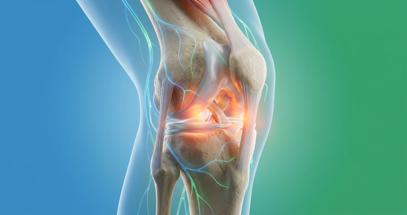
The first step in understanding how to treat knee pain is mastering the basic parts of the knee. The knee is a complex hinge joint reliant on ligaments, tendons, and cartilage for stability. When identifying pain, isolating the specific anatomical region—whether anterior (front) or lateral (outside)—dictates the clinical response.
Structural anomalies like knee valgus increase lateral joint stress by up to 30% (Hopkins Medicine joint analysis, 2023). This precise mapping forms the mechanical baseline for The Brain-Joint Axis Framework, ensuring patients do not mistake structural damage for referred nerve issues.
Key Anatomical Parts of the Knee
To communicate effectively with a doctor, you must understand the core anatomical components. The knee connects the femur (thigh bone) to the tibia (shin bone), protected by the patella (the anatomical kneecap). Between these bones sits the meniscus, the shock-absorbing cartilage that prevents bone-on-bone friction.
Stability relies on four primary ligaments. The anterior cruciate ligament (ACL) and posterior cruciate ligament (PCL) control forward and backward motion. Meanwhile, the medial collateral ligament (MCL) and lateral collateral ligament (LCL) act as internal ropes preventing side-to-side instability.
A standard x-ray only reveals bone health, meaning severe soft tissue tears remain invisible without an MRI. Surrounding knee muscles (primarily the quadriceps and hamstrings) control joint movement. When these muscles weaken, it often causes knee valgus (an inward collapse of the joint).
This misalignment creates uneven mechanical stress during daily activities, accelerating cartilage degradation and creating localized friction points. The intricate balance between these structures means that a failure in one area cascades into others. For instance, an untreated MCL tear forces the meniscus to absorb abnormal rotational forces, leading to accelerated wear and secondary tearing.
Mapping Front, Back, and Side Knee Pain
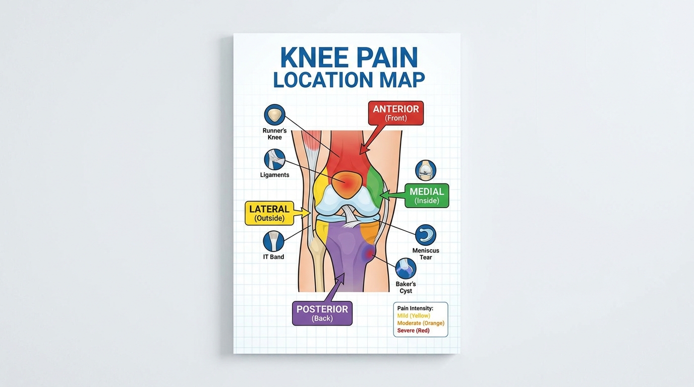
Localized mechanical pain tells a highly specific story about structural damage. Anterior discomfort, often experienced as severe kneeling pain, usually indicates patellar tendinitis or prepatellar bursitis (inflammation of the fluid sac in front of the kneecap). This anterior pain often flares up aggressively when walking down stairs or performing squats.
Conversely, experiencing pain on outside of knee (lateral pain) frequently points to Iliotibial (IT) band syndrome, where a tight tendon rubs aggressively against the outer femur. Poor patellar tracking—where the kneecap slides out of its central groove—also contributes heavily to lateral friction. If the back of the knee hurts (posterior pain), clinicians typically look for Baker’s cysts (fluid accumulation) or hamstring insertion strains.
Understanding these mechanical triggers is vital. For example, if your knee hurts when i bend it under weight, this specific mechanical trigger strongly indicates meniscal impingement rather than general arthritis. Documenting exactly which movements replicate your symptoms allows specialists to bypass generalized treatments and target the exact damaged tissue layer.
Identifying whether your pain is anterior, posterior, medial, or lateral drastically narrows the diagnostic field. Localized mechanical pain tells only part of the story; ongoing swelling introduces the inflammatory and degenerative components of the joint axis (Nortex Spine and Joint review).
Brain-Joint Axis: Inflammation & Nerve Factors
When msk pain (musculoskeletal pain) persists beyond expected healing timelines, clinicians evaluate the secondary and tertiary pillars of The Brain-Joint Axis: inflammation and neurological feedback. While mechanical wear damages the cartilage, chronic inflammation actively destroys the joint’s microenvironment.
Furthermore, nervive pain (nerve-related pain) complicates recovery through central sensitization, where the brain amplifies pain signals despite clear x-rays. Central sensitization affects up to 30% of patients with chronic osteoarthritis, causing elevated pain responses even after structural healing (NCBI, 2023). Distinguishing between an actively degrading joint and an overactive nervous system is critical for selecting appropriate pharmacological interventions.
Osteoarthritis vs. Rheumatoid Arthritis
Joint inflammation manifests in distinct patterns that dictate treatment. Osteoarthritis (OA) is a degenerative, non-systemic condition breaking down articular cartilage, eventually resulting in painful bone-on-bone friction. It typically develops slowly over decades, often affecting one side of the body more severely based on historical usage or past trauma.
In contrast, Rheumatoid Arthritis (RA) is a systemic autoimmune disease where the body mistakenly attacks its own synovial lining. RA frequently presents symmetrically, affecting both knees simultaneously, and is characterized by intense morning stiffness that lasts longer than an hour.
Many patients confuse these chronic conditions with acute structural injuries like jumper’s knee (patellar tendinopathy) or a bursitis knee (fluid sac inflammation). Evidence-informed strategies focus on natural joint lubrication and activity modification to manage symptoms, as there is no method for reversing cartilage loss.
| Condition | Origin | Key Symptom | Primary Treatment |
|---|---|---|---|
| Osteoarthritis | Mechanical wear | Stiffness after resting | Physical therapy, NSAIDs |
| Rheumatoid Arthritis | Autoimmune response | Symmetrical joint swelling | Immunosuppressants |
| Bursitis | Fluid sac inflammation | Localized warmth and swelling | Rest, corticosteroid injections |
Blood tests measuring C-reactive protein (CRP) and rheumatoid factor (RF) easily confirm systemic autoimmune involvement. Correctly identifying the specific arthritis type ensures you do not waste time icing a joint that actually requires systemic immunosuppressive medication (Cleveland Clinic clinical overview).
Nerve Sensitization & Nociplastic Pain
Prolonged joint inflammation physically alters how nerve signaling pathways operate. This creates nociplastic pain, defined as altered pain processing within the central nervous system. Under central sensitization, the nervous system becomes hyper-reactive, making normal, safe movements feel excruciating.
This explains why severe nerve pain continues even after a surgeon repairs a torn meniscus. The nociceptors (pain receptors) essentially get stuck in an “on” position. Patients often ask: can a neurologist help with knee pain? When pain presents as electrical shocks, tingling, or burning without visible swelling, a neurologist can perform nerve conduction studies to identify peripheral nerve damage.
Additionally, specific nutritional profiles, such as severe Vitamin B12 and Vitamin D deficiencies, are heavily linked to impaired peripheral nerve health and amplified pain responses. Vitamin B12 is essential for maintaining the myelin sheath that protects peripheral nerves.
Neurological evaluations map the exact sensory deficits, preventing unnecessary orthopedic surgeries for pain that originates in the nerves. Addressing these neurological factors stops the brain from over-reporting tissue threats (Lone Star Neurology analysis). Regardless of whether the origin is mechanical, inflammatory, or neurological, targeted conservative rehabilitation remains the cornerstone of clinical treatment protocols.
Relief: Physical Therapy & Knee Supports
Effective knee ache treatment relies on active rehabilitation rather than prolonged rest. Clinical evidence shows that complete immobilization accelerates muscle atrophy and worsens joint stiffness. Instead, relief for knee joint pain requires a combination of progressive load-bearing physical therapy and temporary external stabilization.
Structured physical therapy improves functional mobility in 75% of non-operative knee injury patients within 8 weeks (BMJ clinical evidence, 2023). By matching specific strengthening protocols with the correct anatomical braces, patients can decompress the joint space, modulate inflammatory pathways, and safely navigate both pre-operative preparation and post-operative recovery timelines.
Safe Rehab Exercises for Joint Pain
Formal therapy knee injury protocols focus on strengthening the muscles supporting the joint without aggravating the damaged cartilage. Incorporating safe knee exercises for pain is essential for maintaining mobility.
Step 1: Isometric Quad Sets
Tighten the front thigh muscles while keeping the leg completely straight on the floor. Hold the contraction for 5 seconds and release. This engages the quadriceps without bending the joint, protecting the meniscus from friction.
Step 2: Straight Leg Raises
Lie flat on your back, bend your healthy knee, and lift the straightened painful leg upward to the height of the opposite knee. This strengthens the hip flexors and quads simultaneously without loading the knee joint.
Step 3: Wall Slides
Lean your back flat against a wall and slide down into a shallow squat. Limit the bend to a maximum of 45 degrees to protect the meniscus and patellar tendon from excessive sheer force.
Equally important are targeted stretches for the hamstrings and calves, which pull directly on the joint when tight. Pacing is critical during these initial therapy stages. Pushing through sharp, stabbing pain creates micro-tears in healing tissues, whereas working through mild, dull muscle fatigue builds necessary endurance for joint stabilization. For patients experiencing that feeling when knee surgery is tomorrow, engaging in “pre-hab”—strengthening the joint prior to surgery—measurably accelerates typical knee replacement recovery times (AAOS rehabilitation protocols).
Choosing the Right Knee Brace or Sleeve
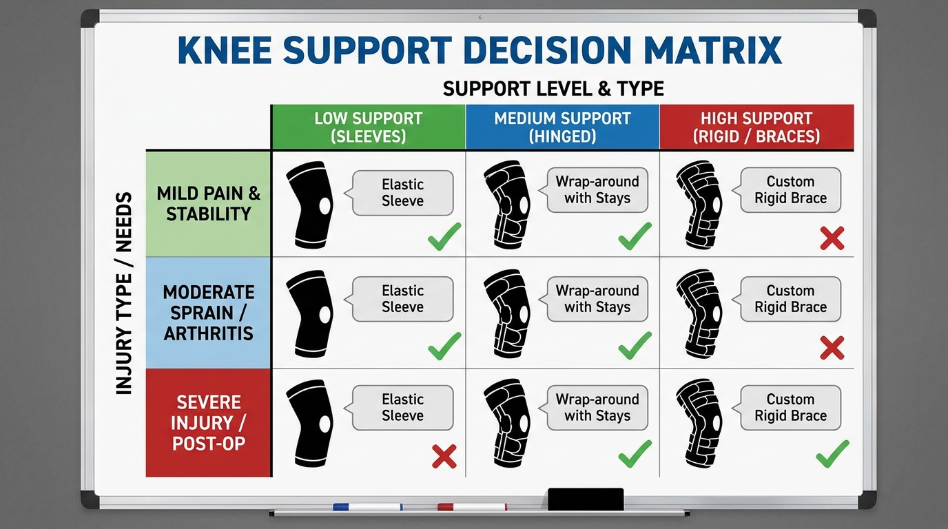
Selecting the correct brace for knee stabilization depends entirely on the specific structural injury. A basic knee compression sleeve works best for mild swelling and early-stage arthritis, providing warmth, light compression, and proprioceptive awareness.
However, a hinged brace is essential for ACL injuries or severe ligament laxity, as the rigid metal sides mechanically prevent dangerous lateral movements that could cause joint buckling. For a torn meniscus, specialized unloader braces shift body weight away from the damaged cartilage compartment, creating immediate mechanical relief.
Beyond hardware, patients often utilize topical treatments, including NSAID gels (like Voltaren) and menthol rubs (like Salonpas). NSAID gels actively reduce localized tissue inflammation. Conversely, menthol patches act as counter-irritants, distracting the nervous system from deep joint pain by creating an intense surface sensation.
Similarly, kinesiology tape (KT tape) offers proprioceptive sensory feedback for minor tracking issues, whereas a rigid knee immobilizer is reserved strictly for acute post-trauma stabilization (Mayo Clinic orthopedic support). When local braces and exercises fail to provide relief, the pain source may actually reside far above or below the knee joint itself.
Beyond the Knee: Referred and Systemic Pain
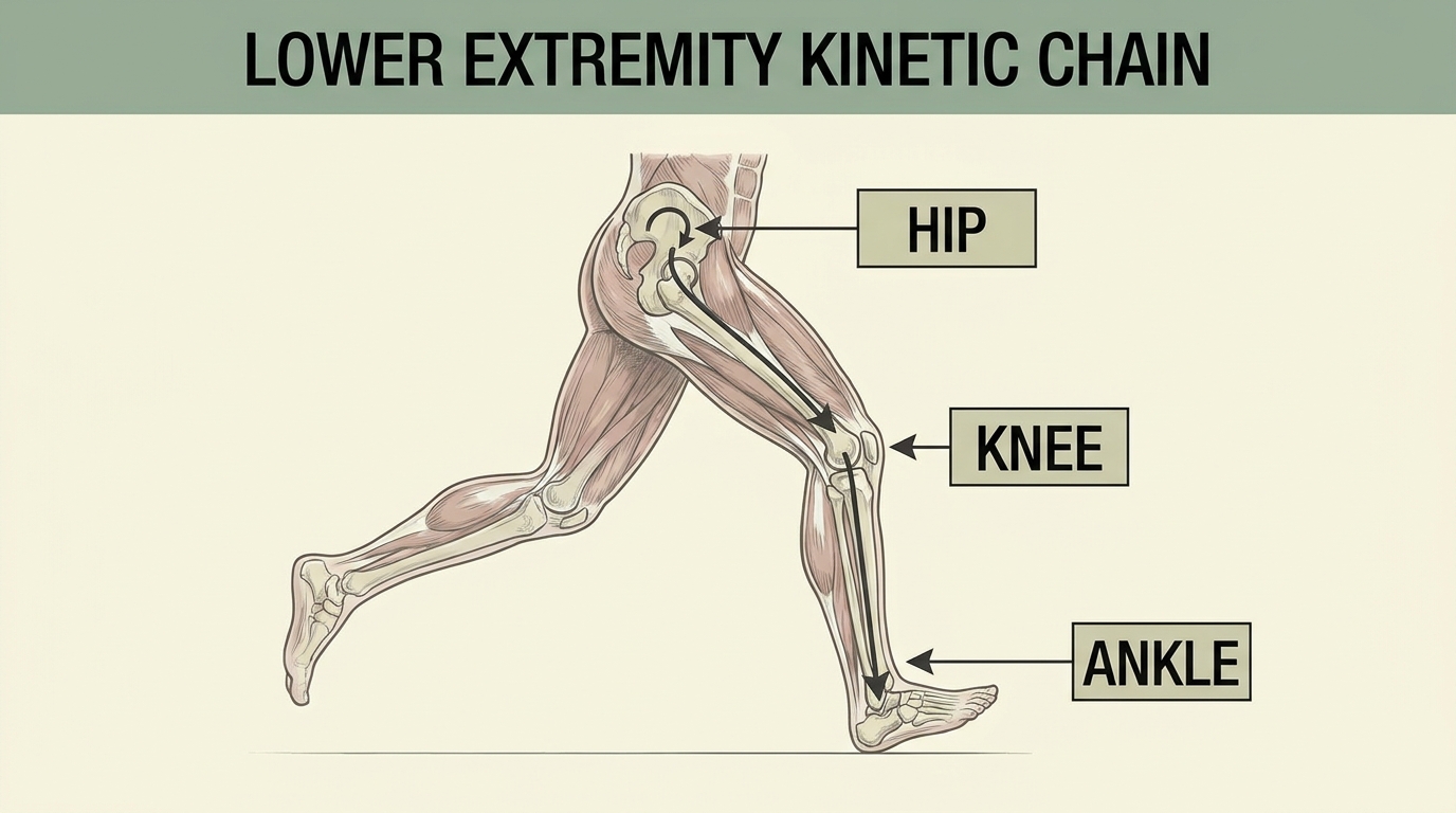
Orthopedic diagnostics rely heavily on understanding the kinetic chain (the interconnected system of joints moving together), as pain sciatica relief protocols frequently resolve secondary knee pain. Because the body’s joints function as a synchronized mechanical system, dysfunction in the lumbar spine or the arches of the feet directly alters weight distribution across the knee. Altered foot mechanics like flat feet significantly increase the risk of knee cartilage damage by shifting weight-bearing loads abnormally across the joint’s medial compartment.
Often, patients experiencing sharp leg pain discover that back pain caused by an L4-S1 nerve root compression is referring pain downward (Spine-Health referred pain analysis). Identifying these distant mechanical failures is essential before committing to localized knee treatments.
The Sciatic Nerve and Foot Mechanics
The sciatic nerve (the major nerve running from the lower back down the leg) frequently disguises spinal issues as knee problems. Sciatic nerve pain relief strategies target the lumbar spine, decompressing pinched nerves that radiate burning sensations to the back of the knee. Below the joint, foot bottom pain (plantar fasciitis) and pain in the ball of the foot drastically alter walking mechanics.
These gait compensations force the knee into improper valgus alignment. Furthermore, sacrum pain, weak gluteus medius muscles, and hip tightness can inflame the IT band, which subsequently pulls laterally on the knee joint.
Orthopedic specialists will often observe your gait (how you walk) before ever examining your knee. They look for asymmetrical weight shifting, foot pronation, or hip drops that clearly trace the knee pain back to an entirely different joint. Correcting issues like pain on the top of my foot with custom orthotics can immediately relieve ascending structural stress on the knee (APMA foot mechanics guide).
Systemic Pain and Medical ICD-10 Coding
It is vital to differentiate localized mechanical knee pain from widespread systemic issues. Conditions like fibromyalgia or autoimmune flare-ups can cause severe flank pain and bilateral joint aches simultaneously. Medical professionals document these specific conditions using standardized diagnostic codes, allowing patients to better understand their own clinical charts and billing statements.
For example, the specific diagnostic icd 10 for left knee pain is M25.562, whereas the right knee is coded as M25.561. Knowing these codes assists with medical records requests, specialist referrals, and navigating insurance verifications (Rheumatology standard diagnostics). Navigating these clinical diagnoses requires strict adherence to safety protocols and knowing exactly when home remedies are insufficient.
Safety Limitations & When to Seek Help
Common Pitfalls in Self-Diagnosis
A major pitfall is ignoring “red flag” symptoms like sudden fever, severe swelling, or the complete inability to bear weight. Relying solely on over-the-counter pain patches merely masks neurological signals while active mechanical damage worsens silently inside the joint cavity, accelerating cartilage loss (WebMD safety guidelines).
When to Choose Alternatives
When conservative physical therapy fails to improve functional mobility over a structured 3-to-6-month timeline, orthopedic surgical evaluation becomes the necessary alternative. While regenerative medicine (like stem cell therapy) exists, it does not cure degenerative disease and requires strict clinical suitability assessments before investment.
When to Seek Expert Help
Seek immediate clinical assessment if your pain is accompanied by systemic symptoms, unexplained weight loss, or referred torso pain. Furthermore, if your knee joint suddenly “locks” in place or gives way entirely during walking, immediate emergency orthopedic evaluation is required to prevent catastrophic ligament failure.
Frequently Asked Questions
What meds treat knee osteoarthritis?
The primary medications for knee osteoarthritis include oral nonsteroidal anti-inflammatory drugs (NSAIDs) and topical diclofenac gels, which actively reduce joint inflammation. For advanced degradation, clinicians may utilize intra-articular corticosteroid injections to manage severe flare-ups. Natural lubrication supplements like glucosamine offer mixed clinical efficacy, so always discuss specific medication suitability directly with a rheumatologist.
Do nerve issues cause knee pain?
Neurological problems frequently cause severe knee pain through a process called central sensitization. In these cases, the brain amplifies standard nerve signals, interpreting normal joint movement as highly painful. Furthermore, nerve root compression in the lumbar spine (sciatica) also refers pain directly downward into the back of the knee. Addressing neuropathic knee pain requires treating the central nervous system, often utilizing entirely different pharmacological approaches than standard joint injuries. Treatment outcomes vary based on individual nerve health.
Conclusion: Next Steps for Joint Health
For patients dealing with chronic joint discomfort, understanding how to treat knee pain requires evaluating the joint holistically. Structured physical therapy improves functional mobility in 75% of non-operative cases (NCBI, 2023). The safest approach combines localized bracing, progressive loading, and neurological assessment to restore movement.
Applying The Brain-Joint Axis Framework fundamentally shifts your approach to recovery. Categorizing your pain as mechanical, inflammatory, or neurological prevents wasted time on ineffective home remedies. This structured understanding empowers you to speak accurately and confidently to specialists about your exact symptoms.
Stop guessing about the source of your joint pain. Gather your previous x-rays, MRI reports, and medical records, and schedule your consultation today with an orthopedist or neurologist. A professional suitability assessment will provide a definitive diagnosis and a targeted roadmap for your functional recovery.