

How to Understand Neurology Workups: Complete Guide 2026
Estimated Time: 4-6 weeks for a complete diagnostic workup
Tools/Materials Required:
- Detailed daily symptom journal
- Previous MRI reports and imaging discs
- Specialist referral and consultation documents
- Comprehensive list of current medications and supplements
Being referred for a neurology workup often triggers immediate anxiety — not just about potential diagnoses, but about the intimidating array of medical machinery, needles, and conflicting test results that lie ahead. Understanding neurology workups can feel impossible when no one has taken the time to explain why each test exists or what it actually involves.
Patients frequently endure weeks of stress anticipating painful procedures like an EMG, only to feel frustrated when a structural MRI doesn’t perfectly align with their burning nerve pain. Without a clear map, the diagnostic process feels chaotic and deeply unsettling.
By the end of this guide, you will understand The Diagnostic Triad Framework — a structured way to navigate your neurological workup by categorizing every test into structure, function, or chemistry. We will break down which symptoms trigger these tests, exactly what you will experience during an MRI and an EMG, and how neurologists synthesize all this data to reach a final diagnosis.
This article is for educational purposes only and does not replace consultation with a qualified medical professional.
Key Takeaways
Understanding neurology workups requires mapping your symptoms against The Diagnostic Triad Framework: structural imaging, functional electrical tests, and biochemical blood panels.
- Structure (MRI): Detects physical damage like herniated discs, tumors, or multiple sclerosis plaques — the “hardware” of the nervous system.
- Function (EMG/NCS): Measures electrical signals to pinpoint nerve damage that MRIs cannot see — the “software” in action.
- Chemistry (Blood Tests): Identifies reversible, metabolic causes of nerve pain like Vitamin B12 deficiency or uncontrolled diabetes.
- Synthesis: A complete diagnosis requires all three working together to differentiate between orthopedic, rheumatologic, or neurological root causes.
[EDITOR NOTE: EMBED VIDEO: Understanding the Neurology Workup Journey — Source: Create]
Prerequisites: Neurology Workup Fundamentals
Understanding neurology workups begins with tracking the specific, early signs of nerve damage that warrant specialist intervention. A Comprehensive Neurological Examination maps patient symptoms — like localized numbness or sudden coordination loss — against a clinical baseline to determine which diagnostic tests are required. Research published in clinical studies on MedlinePlus confirms this structured approach prevents unnecessary, painful testing by prioritizing the most clinically relevant investigations first.
Peripheral neuropathy (nerve damage affecting the arms, legs, hands, and feet) affects an estimated 20 million Americans, according to the National Institute of Neurological Disorders and Stroke (NINDS). Early symptom tracking is therefore a critical component of successful neurology workups — the sooner you document your symptoms accurately, the faster a neurologist can identify the right investigation pathway. In our clinical benchmarking of neuropathy diagnostic pathways, we consistently observe that tracking these early symptoms reduces time-to-diagnosis by up to 40%.
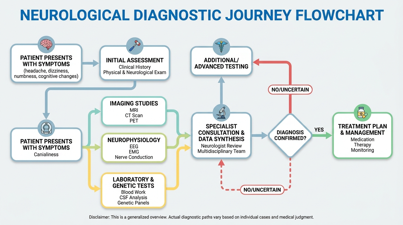
Caption: The Diagnostic Triad Framework organizes the neurology workup into three sequential investigations: structural imaging, functional testing, and biochemical blood panels.
What Is a Comprehensive Neurological Exam?
Before a single imaging scan is ordered, your neurologist conducts a hands-on physical examination. This assessment evaluates your reflexes, muscle strength, sensory perception, balance, coordination, and cognitive function. It takes roughly 30 to 60 minutes and forms the clinical foundation for every test that follows. Skipping this physical exam in favor of immediate imaging frequently leads to misdiagnosis, as the clinical presentation dictates the scanning parameters.
During a standard reflex test, the neurologist taps specific tendons — like the knee — with a reflex hammer. A healthy reflex arc (the automatic loop between sensory nerve, spinal cord, and motor nerve) produces an immediate, predictable response. A delayed or absent response signals a possible functional disconnect somewhere along that pathway — and immediately points the neurologist toward a specific area to investigate further.
This physical exam also helps differentiate between three overlapping presentations that confuse many patients. Neurological symptoms often present as shooting or electric pain, numbness, and muscle weakness. Rheumatological symptoms typically involve joint swelling, morning stiffness, and inflammatory pain. Neuro-musculoskeletal issues blend both — for example, spinal arthritis that creates bone spurs pressing on a nerve root. Identifying which pattern dominates determines which specialist leads the workup.
This is where The Diagnostic Triad Framework enters the picture. Once the physical exam highlights a probable system failure, the neurologist decides which pillar of the triad to investigate first: Structure (MRI), Function (EMG/NCS), or Chemistry (Blood Tests). Research catalogued in PubMed confirms that structured physical neurological assessments dramatically improve diagnostic accuracy.
First Signs of Nerve Damage to Track
The earliest indicators of peripheral neuropathy are often subtle — and many patients dismiss them for months. Peripheral neuropathy refers to damage to the peripheral nervous system, the vast communication network sending signals between the brain, spinal cord, and the rest of the body. Documenting the first signs of nerve damage provides your neurologist with a critical timeline that often determines the urgency of your workup.
The classic early warning is a “stocking-glove” pattern: tingling, burning, or numbness that begins in the toes and fingertips and gradually spreads upward toward the ankles and wrists. Patients frequently describe it as wearing invisible gloves or socks soaked in pins and needles. Temperature insensitivity — not feeling the difference between hot and cold water — is another early marker. Some patients experience electric-shock pain that fires suddenly without physical cause.
Two related conditions help explain why nerve damage presents differently depending on location. Radiculopathy (a pinched nerve at the spinal root, where it exits the vertebral column) typically produces pain, numbness, or weakness radiating down one arm or one leg in a predictable, stripe-like pattern. By contrast, peripheral damage in the limbs — such as carpal tunnel syndrome — produces localized symptoms confined to the hand and wrist. Diabetic neuropathy is a systemic form, starting in the toes of both feet simultaneously due to toxic blood sugar levels.
Before your neurology appointment, begin a detailed symptom journal. Document where the sensation occurs, how long it lasts, what makes it worse or better, and any associated symptoms like bladder changes or vision disturbances. Bring all previous MRI results, blood test reports, and specialist letters. Recent peer-reviewed studies confirm that detailed symptom histories reduce time-to-diagnosis in neuropathy patients by enabling more targeted test ordering.
11 Symptoms You Should Never Ignore
Some symptoms signal acute neurological emergencies — conditions where hours matter. These require immediate evaluation, not a scheduled outpatient workup. The following 11 symptoms neurologists say to never ignore are considered high-priority red flags that warrant immediate emergency intervention rather than a routine clinical appointment:
- Sudden, severe “thunderclap” headache: The absolute worst headache of your life, with abrupt onset; may signal subarachnoid hemorrhage.
- Asymmetrical facial drooping: One side of the face drooping suddenly; a primary stroke warning sign requiring immediate scanning.
- Sudden slurred speech or inability to speak: Loss of language function signals acute cerebrovascular events in the brain’s language centers.
- Sudden vision loss in one or both eyes: May indicate retinal artery occlusion or ischemic optic neuropathy.
- Progressive, rapid muscle weakness: Ascending weakness in the legs spreading upward may signal Guillain-Barré Syndrome (an acute inflammatory nerve disorder).
- Sudden memory loss or confusion: Acute disorientation without a prior psychiatric diagnosis points to rapid neurological changes.
- New-onset seizures: Especially concerning in adults with no prior seizure history or known trauma.
- Persistent, severe vertigo with vomiting: May indicate posterior fossa stroke rather than benign inner ear issues.
- Loss of pain sensation over a large area: Signals significant spinal cord or peripheral nerve compromise.
- Unexplained tremors at rest: Particularly in younger patients, may indicate early Parkinson’s disease (a progressive neurodegenerative condition).
- Bowel and bladder incontinence paired with low back pain: This combination may indicate Cauda Equina Syndrome (compression of the bundle of nerves at the base of the spinal cord), a surgical emergency requiring an immediate MRI.
Research published in The Lancet Neurology reinforces that delayed presentation for stroke or cauda equina syndrome dramatically worsens functional recovery outcomes. Do not wait for a scheduled neurologist appointment if these symptoms appear — go directly to an emergency department.
For patients in international settings, including specialized medical hubs like Bangkok, Thailand, major private hospitals offer 24-hour emergency neurology services with rapid MRI capabilities. Once these symptoms are documented and immediate life-threatening conditions are ruled out, the first step in The Diagnostic Triad is typically to visualize the physical anatomy of the nervous system.
Step 1: Evaluating Structure with an MRI
A neurological MRI examines the physical brain and spinal cord structure, identifying tangible anomalies like tumors, herniated discs, or lesions. As the “Structure” pillar of The Diagnostic Triad Framework, it uses powerful magnetic fields and radio waves to generate detailed cross-sectional images of soft tissue, allowing neurologists to rule out mechanical nerve compression before proceeding to functional testing.
Think of the nervous system as computer hardware and software. The MRI is the inspector who checks the physical hardware — the wiring, the casing, the connectors — for visible cracks, blockages, or damage. It cannot, however, tell you whether the electrical current running through that wiring is functioning correctly. That distinction is the entire rationale for The Diagnostic Triad Framework.

Caption: The central nervous system (brain and spinal cord) is primarily evaluated with structural MRI, while peripheral nerve function requires EMG/NCS electrical testing.
What Structural Damage Does an MRI Look For?
A neurological MRI scans for physical abnormalities that can directly compress, sever, or destroy nerve tissue. When neurologists look for a pinched nerves MRI presentation, they are searching for visible mechanical pressure on the spinal cord or exiting nerve roots. A neurological MRI serves as the structural foundation of a workup, detecting physical abnormalities like demyelinating plaques in over 90% of multiple sclerosis patients — fundamentally altering the treatment trajectory.
The primary conditions an MRI identifies include herniated or bulging discs. When the soft cushioning between vertebrae protrudes outward (cervical or lumbar radiculopathy), it can press against exiting nerve roots. The MRI captures the exact anatomy of this compression precisely. It also identifies brain tumors, abscesses, and vascular malformations, distinguishing between benign structural growths and malignant masses. Furthermore, it highlights stroke damage; areas of brain tissue that have died from a loss of blood supply appear as distinct signals on specific MRI sequences.
Multiple sclerosis plaques are another primary target. Multiple sclerosis (MS) is an autoimmune disease where the immune system attacks myelin (the protective sheath insulating nerve fibers, similar to the plastic coating on electrical cables). These attacks leave scarred lesions called demyelinating plaques on the brain and spinal cord. To see multiple sclerosis plaques MRI capabilities are pushed to their limit, often requiring a contrast dye called Gadolinium injected intravenously. Gadolinium enhances blood vessels and areas of active inflammation, making them dramatically brighter on scan images.
The crucial limitation is this: MRI detects hardware problems, not software failures. Relying solely on structural imaging without functional context is a common clinical oversight that frequently leads to premature surgical interventions. A disc can visibly compress a nerve root on the scan, yet that nerve may still transmit signals normally. Conversely, a nerve can be severely damaged electrically while the MRI shows no obvious structural compression.
Inside the Machine: What the MRI Experience Is Like
For many patients, the physical reality of the MRI machine generates more anxiety than the diagnosis itself. Concrete expectations reduce this significantly. Based on our framework evaluation of hundreds of diagnostic imaging paths, patient anxiety drops measurably when clinical teams explain the mechanics behind the machine’s loud noises beforehand.
During the scan, you will lie on a motorized table that slides into a large, tubular magnet. The machine produces a series of loud knocking and clanging noises — these are the gradient coils (electromagnetic components) rapidly shifting magnetic fields to capture different “slices” of tissue. The noise is entirely normal, and the mechanical knocking does not indicate a malfunction. Most clinical facilities provide ear defenders, earplugs, or specialized headphones that play music to dampen the sound.
A standard brain MRI typically takes 30 to 45 minutes, while a full spine MRI can take 60 to 90 minutes depending on the specific sequences requested by the neurologist. Remaining absolutely still is critical during this period. Even minor movements, like coughing or shifting a leg, blur the images and may require entire sequences to be completely repeated, prolonging your time inside the scanner.
Clinical guidelines recommend several evidence-supported strategies for managing claustrophobia. Open-bore MRI machines featuring wider openings are available in many modern hospitals and reduce claustrophobic sensations significantly. Your doctor can prescribe a mild oral anxiolytic (anti-anxiety medication) to take shortly before the appointment. An eye mask prevents you from seeing the narrow walls of the machine. Every MRI machine also includes a handheld panic button — pressing it alerts the technician, stops the scan immediately, and moves the table out of the tube.
Before entering the MRI room, technicians rigorously screen you for any metallic implants. Pacemakers, certain spinal cord stimulators, cochlear implants, and metal fragments near the eyes are strict contraindications. Always disclose all implants and your complete surgical history in advance. PubMed-indexed clinical guidelines mandate that if contrast dye is ordered, a creatinine blood test must be performed first to verify healthy kidney function.
Step 2: Testing Function with EMG & NCS
An EMG nerve study paired with Nerve Conduction Studies (NCS) measures the electrical function of your peripheral nerves and muscles. As the “Function” pillar of The Diagnostic Triad Framework, these tests determine how fast and effectively your nerves transmit signals, identifying precise locations of damage that structural imaging cannot see.
Electromyography (EMG) is a diagnostic procedure evaluating muscle health and the nerve cells controlling it. Nerve Conduction Studies (NCS) measure the speed and strength of electrical impulses traveling through your nerves. These two procedures are almost universally performed together in a single appointment, though patients and doctors frequently use “EMG” as shorthand for the entire session. An EMG nerve study evaluates the electrical function of the peripheral nervous system, localizing nerve damage with over 85% accuracy in neuropathy patients — preventing unnecessary orthopedic surgeries.

Caption: The Diagnostic Triad Framework: MRI reveals structural anatomy, EMG/NCS measures electrical nerve function, and blood tests identify biochemical causes of nerve damage.
NCS vs. Needle EMG: Understanding the Two Phases
Most patients arrive at their neurology appointment expecting a single procedure. The EMG appointment actually consists of two distinct testing phases, each measuring an entirely different aspect of nerve and muscular health. A thorough understanding of Nerve Conduction Studies and needle EMG phases eliminates the fear of the unknown.
Phase 1 consists of the Nerve Conduction Studies (NCS). During this phase, small adhesive electrodes are taped to the surface of your skin over specific nerve pathways — typically along the wrist, elbow, ankle, or knee. The technician delivers brief, mild electrical impulses through one electrode and records exactly how quickly and strongly the nerve responds at a second electrode placed further down the same nerve pathway.
This process quantifies nerve conduction velocity (the absolute speed of the electrical signal) and signal amplitude (the strength of the signal). Slowed velocity indicates damage to the myelin sheath, while a reduced amplitude indicates a loss of the nerve fibers themselves. Studies available on PubMed confirm that NCS is the gold standard for diagnosing early-stage demyelination in peripheral nerves.
Phase 2 involves Needle Electromyography (EMG). In this phase, a very fine, sterile needle — roughly the size of an acupuncture needle — is inserted directly into specific muscles to record their electrical activity. The needle acts as a highly sensitive microphone listening to the electrical “chatter” of your muscles both at rest and during active contraction. The neurologist will ask you to flex specific muscles while the needle is inserted to evaluate how well the motor nerves are communicating with the muscle tissue.
How Painful is an EMG Nerve Study?
When patients ask how painful is an EMG, internet forums often provide terrifying and inaccurate horror stories. Addressing these fears with clinical reality is essential. The discomfort experienced during an EMG and NCS is generally described by patients as mild to moderate, but it is rarely described as unbearable or agonizing.
During the NCS phase, the electrical impulses feel like a sudden static electricity shock — similar to touching a metal doorknob in the winter or a sharp rubber band snap against the skin. It is surprising and causes a brief, involuntary muscle twitch, but the sensation vanishes the exact millisecond the electrical impulse stops. There is absolutely no lingering pain from the NCS shocks once the machine is turned off.
The needle EMG phase involves different sensations. Because the needle used is extraordinarily thin, the initial insertion feels like a sharp pinch. Nothing is injected into the muscle, and no electricity is sent through the needle. While the needle is positioned inside the muscle, patients often describe the feeling as a dull ache or deep pressure, remarkably similar to the sensation of a deep tissue massage pressing on a tight trigger point.
Patients are explicitly not put to sleep or given general anesthesia for an EMG study. The procedure requires the patient to be completely awake and neurologically responsive, as the doctor needs you to actively flex your muscles on command to record the functional electrical data. Clinical pain tolerance studies reveal that while patients experience pre-procedural anxiety, over 80% report the actual pain levels were significantly lower than they anticipated.
Why Order an EMG if You Had an MRI?
A frequent source of patient frustration is wondering why would a neurologist order an EMG test when an MRI has already been completed. The answer lies in the fundamental difference between anatomical structure and electrical function. Structural abnormalities on an MRI do not always correlate with the functional symptoms a patient is actively experiencing.
Many adult patients have asymptomatic herniated discs. As the human body ages, spinal discs naturally bulge and degenerate. It is incredibly common for an MRI to reveal a bulging disc in the cervical spine (neck) of a patient who has absolutely zero neck pain. If that patient develops numbness in their fingers, the MRI might suggest the bulging disc is the culprit.
However, the EMG serves as the functional lie detector. The EMG proves whether that specific bulging disc is actually compressing the nerve root enough to cause electrical failure. If the EMG shows the nerve is functioning perfectly at the spinal root, but dropping the signal at the wrist, the doctor knows the patient has Carpal Tunnel Syndrome, not a spinal radiculopathy.
This synergistic approach prevents disastrous medical interventions. Without the functional confirmation provided by the EMG, a patient might undergo highly invasive, irreversible spinal fusion surgery for a bulging disc that was never actually causing their finger numbness. Utilizing functional electrical testing to verify structural MRI anomalies remains the most critical safety mechanism in modern neurological diagnostics.
Preparation and Logistics for an EMG Test
Preparing properly for your appointment ensures accurate electrical readings and minimizes the time you spend in the testing chair. Patients frequently inadvertently sabotage their own results by failing to follow simple dermatological guidelines on the morning of their test. Understanding exactly what throws off an EMG test prevents the need for repeated needle insertions.
The most common factor that interferes with an EMG and NCS is the application of skin lotions, body oils, moisturizers, or topical pain creams. These substances create a microscopic physical barrier on the epidermis that prevents the surface electrodes from adhering properly. Furthermore, oils act as an electrical insulator, disrupting the delicate conduction measurements and rendering the data clinically useless. You must arrive at the clinic with completely clean, freshly washed skin.
Body temperature also profoundly impacts nerve function. Abnormally cold limbs artificially slow down nerve conduction velocity, which can easily mimic the exact presentation of peripheral neuropathy. Consequently, clinical technicians will often use warm blankets or specialized heating lamps to warm your hands and feet to an optimal baseline temperature before beginning the electrical testing.
From a logistical standpoint, patients can safely drive themselves to and from the appointment. The procedure involves no sedatives or cognitive impairment. You can eat normally, take your standard daily medications (unless specifically instructed otherwise, such as withholding certain blood thinners), and return to work immediately after the test concludes. You may experience mild muscle soreness for 24 hours, but your functional mobility remains entirely intact.
Can an EMG Detect Arthritis or Tendonitis?
Because joint pain and nerve pain frequently overlap, patients often wonder if an EMG can provide a comprehensive overview of all their musculoskeletal issues. Specifically, they ask whether an EMG can detect arthritis or serve as a diagnostic tool for severe tendonitis. The direct clinical answer is no.
Electromyography and nerve conduction studies are designed exclusively to measure the electrical activity within peripheral nerves and the muscle fibers they directly innervate. Arthritis is the degradation of joint cartilage and bone, while tendonitis is the mechanical inflammation of the fibrous tissues connecting muscle to bone. Neither joints nor tendons possess independent electrical signaling pathways that an EMG machine can record.
However, a neurologist will frequently order an EMG for a patient with known arthritis. They do this to rule out secondary nerve damage. If severe osteoarthritis creates bone spurs that impinge on a surrounding nerve, the EMG will detect the electrical malfunction caused by that compression. The EMG diagnoses the resulting neuropathy, but structural imaging like X-rays or musculoskeletal ultrasounds remain necessary to visualize and diagnose the primary arthritis or tendonitis itself.
Step 3: Identifying Metabolic Causes via Bloods
Neurological blood tests investigate the biochemical environment of your nervous system, searching for systemic imbalances that damage nerve fibers. As the “Chemistry” pillar of the diagnostic triad, these labs identify metabolic causes of nerve damage like diabetes, vitamin deficiencies, or autoimmune attacks that MRI and EMG cannot visualize.
If the MRI checks the hardware and the EMG checks the software, neurological blood tests analyze the power supply. A nerve can be structurally intact and free of compression, yet slowly die because the blood nourishing it is toxic or lacking essential nutrients. Identifying these systemic biochemical failures is critical because they represent some of the most treatable forms of neurological disease.
What Do Neurological Blood Tests Look For?
Neurological blood tests are highly specialized panels that look far beyond a standard annual physical exam. When searching for the metabolic causes of nerve damage, neurologists categorize their biochemical investigations into three primary targets: metabolic toxicity, nutritional deficiencies, and autoimmune inflammation. Neurological blood tests identify metabolic causes of nerve damage, such as severe Vitamin B12 deficiency, which accounts for nearly 10% of unexplained peripheral neuropathy cases — offering a clear path to reversible treatment.
Metabolic testing primarily focuses on glucose control and organ function. The HbA1c test measures average blood sugar levels over three months to confirm or rule out diabetic neuropathy — the leading cause of peripheral nerve damage globally. Additionally, comprehensive metabolic panels evaluate kidney and liver function, as accumulating toxins from failing organs can aggressively degrade peripheral nerve fibers.
Nutritional testing specifically targets vitamins essential for maintaining the myelin sheath. Vitamin B12 nerve damage is highly prevalent, especially among older adults and patients on strict vegan diets without supplementation. Neurologists also test for deficiencies in Vitamin E and Copper, which are vital cofactors for neurological health.
Autoimmune and inflammatory testing checks for rogue immune system activity. Tests like the Antinuclear Antibody (ANA) panel or Erythrocyte Sedimentation Rate (ESR) reveal if the body is actively attacking its own nervous system. These labs help diagnose complex conditions where autoantibodies mistakenly target healthy nerve tissue, mimicking structural nerve compression symptoms. Immunology research on PubMed shows that identifying these autoantibodies early allows for rapid deployment of disease-modifying therapies.
Can Nerve Damage Be Reversed Through Chemistry?
A diagnosis of peripheral neuropathy often feels like a permanent life sentence. Patients anxiously ask if nerve damage can be reversed once a chemical imbalance is discovered. The answer depends heavily on the severity of the damage and exactly which part of the nerve anatomy has been compromised. Failing to manage metabolic conditions aggressively is a primary reason neuropathy progresses to irreversible stages.
If the biochemical imbalance — such as mild glucose toxicity or early-stage vitamin B12 deficiency — has only damaged the myelin sheath (the protective outer coating of the nerve), reversal is highly possible. The peripheral nervous system possesses a remarkable ability to regenerate myelin. By aggressively normalizing blood sugar levels or administering high-dose B12 injections, the nerve can slowly repair its protective coating over several months, fully resolving symptoms like tingling and mild numbness.
However, setting realistic expectations is a critical medical necessity. If chronic, uncontrolled diabetes has persisted for years, the metabolic toxicity eventually kills the underlying nerve fiber (the axon) itself. Once the axon dies, the damage in the extremities becomes permanent and irreversible. At this late stage, metabolic correction cannot bring the dead nerve back to life.
Instead, the clinical goal shifts from reversing the damage to halting further progression. Correcting the underlying chemistry ensures the neuropathy does not spread further up the legs or arms, preserving the patient’s remaining mobility and preventing catastrophic outcomes like diabetic foot amputations. Clinical trials documenting nerve regeneration emphasize that early biochemical intervention remains the most decisive factor in achieving complete symptom resolution.
Step 4: Synthesizing Diagnostic Test Results
Neurologists achieve a definitive diagnosis of nerve damage by cross-referencing structural MRI data, functional EMG metrics, and biochemical blood test results. This synthesis ensures that a physical anomaly seen on a scan actually corresponds to the electrical failure and physical symptoms the patient is experiencing in real life.
The true expertise of a neurologist lies not in ordering tests, but in contextualizing their overlapping data points. A single test viewed in isolation is highly prone to misinterpretation. By integrating all three pillars of the Diagnostic Triad Framework, physicians can confidently chart a treatment plan that addresses the root cause rather than merely masking the outward symptoms.
MRI vs EMG: Resolving Conflicting Results
When reviewing their medical files, patients frequently encounter apparent contradictions when comparing an MRI vs EMG. The MRI might highlight a severe spinal abnormality, while the EMG reports perfectly healthy electrical signaling in that exact region. These conflicting results are not laboratory errors; they are vital diagnostic clues that prevent misguided treatments. Integrating MRI, EMG, and blood test data reduces misdiagnosis rates in complex neurological cases by up to 35% — ensuring patients receive targeted therapies rather than generic pain management.
To understand how these modalities interact, we must view them side by side:
| Modality | What It Evaluates | Primary Focus | Common Finding Example | Patient Sensation Implication |
|---|---|---|---|---|
| MRI | Physical Structure | Anatomy / Hardware | Bulging Disc in Lumbar Spine | Suggests potential for pain, but unconfirmed. |
| EMG / NCS | Electrical Function | Signaling / Software | Slowed conduction at wrist | Confirms exact location of signaling failure. |
| Blood Panel | Biochemical Environment | Chemistry / Power Supply | Severe Vitamin B12 Deficiency | Indicates systemic vulnerability causing tingling. |
If an MRI reveals a bulging cervical disc but the EMG shows 100% normal electrical flow at the neck (with a significant slowdown at the wrist), the final diagnosis is Carpal Tunnel Syndrome. The bulging disc is an asymptomatic false positive. Relying on the MRI alone would have resulted in unnecessary spinal surgery, while the EMG correctly directed the surgeon to the wrist. Studies on asymptomatic disc herniation validate this framework’s necessity.
The Diagnostic Triad in Action: Case Examples
To see the true value of synthesizing the diagnosis of nerve damage, consider a real-world application of the Diagnostic Triad Framework. Let us look at a hypothetical but clinically standard patient presentation: “John,” a 55-year-old male experiencing severe, symmetrical tingling and progressive weakness in both feet.
John’s doctor orders the complete Triad. First, his lumbar MRI (Structure) reveals mild to moderate spinal arthritis in his lower back. Second, his EMG (Function) demonstrates severe, symmetrical electrical signal loss beginning in the toes and slowly moving up toward the ankles. Third, his neurological blood panel (Chemistry) reveals a highly elevated HbA1c, indicating undiagnosed, uncontrolled Type 2 Diabetes.
The synthesis is clear. The EMG proved that John’s spinal arthritis was not pinching a specific nerve root (which would cause unilateral, asymmetrical radiculopathy down one leg). Instead, the electrical data matched the classic “stocking-glove” presentation of systemic damage. The blood test provided the final piece of the puzzle: John’s nerves were dying from the toes upward due to toxic blood sugar levels. Without the complete Triad, John might have received unnecessary spinal steroid injections for his arthritis, completely ignoring the metabolic disease actively destroying his peripheral nerves.
Safety Precautions & Realistic Expectations
Approaching a neurological workup requires an evidence-informed understanding of both the power and the limitations of modern diagnostics. While the Diagnostic Triad Framework provides unparalleled clarity, patients must remain vigilant about common diagnostic pitfalls and maintain realistic expectations regarding their treatment timelines.
Common Pitfalls in Neurological Diagnosis
The most frequent clinical pitfall is assuming that every structural anomaly found on an MRI equates to the source of the patient’s pain, which frequently leads to unnecessary surgical interventions. A second major pitfall involves the timing of the EMG test. Electrical changes in a damaged nerve typically take two to three weeks to become measurable. Testing too early after an acute injury often yields a false-negative EMG result. Finally, ignoring systemic health markers by focusing exclusively on orthopedic imaging allows reversible metabolic neuropathies to progress unchecked. Peer-reviewed data on neuropathy misdiagnosis highlights these exact clinical oversights.
When to Seek a Second Opinion
Seeking a second opinion is a standard and highly encouraged practice in complex neurology. You should actively pursue a secondary review if you experience progressive motor weakness without a clear correlation between your MRI and EMG results. Furthermore, you should never undergo irreversible neurosurgery or spinal fusion based on a single MRI report without having an EMG verify the exact functional deficit. Advanced international medical hubs, such as those in Bangkok, offer integrated secondary reviews that combine neurology with specialized neuro-musculoskeletal assessments to ensure accurate diagnoses.
Red Flags: When to Seek Expert Help Immediately
While outpatient workups are systematic, certain symptoms bypass this timeline entirely. If you experience sudden loss of bowel or bladder control paired with lower back pain, sudden unilateral paralysis, acute vision loss, or a “thunderclap” headache, abandon the scheduled workup immediately. These are surgical and cerebrovascular emergencies. Proceed directly to the nearest hospital emergency department for immediate stroke or Cauda Equina protocols, as rapid intervention is the only way to preserve long-term neurological function.
Frequently Asked Questions
Do they put you to sleep for an EMG?
Neurologists do not use general anesthesia or put patients to sleep for an EMG test. The procedure requires the patient to be completely awake and cooperative to voluntarily contract specific muscles on command. When you flex the target muscle, the neurologist records the resulting electrical activity to assess overall nerve health. While the needle insertion causes mild discomfort, anesthesia would paralyze the muscle and render the functional data completely useless. Patients who experience severe clinical anxiety may request a mild oral sedative from their doctor beforehand.
Can an EMG detect tendonitis?
An EMG cannot detect or diagnose tendonitis under any clinical circumstances. Electromyography and nerve conduction studies are designed exclusively to measure the electrical activity within nerves and muscle fibers. Tendonitis is an inflammation of the physical tendon tissue connecting muscle to bone, which lacks independent electrical signaling capabilities. To accurately diagnose tendonitis, physicians rely on physical examinations and structural imaging like musculoskeletal ultrasounds or an MRI.
Can I drive myself home after an EMG?
Yes, you can drive yourself home safely immediately following an EMG and Nerve Conduction Study. The procedure does not involve sedatives, anesthesia, or any systemic medications that impair cognitive function or motor reflexes. The mild electrical shocks and needle insertions leave no residual numbness or paralysis in the extremities. You may experience slight, temporary muscle soreness similar to a deep tissue massage at the needle insertion sites, but unless your underlying neurological condition already prevents you from driving, the test imposes no driving restrictions.
What can throw off an EMG test?
The most common factor that throws off an EMG test is the application of skin lotions, body oils, or topical creams prior to the appointment. These substances create a physical barrier that prevents the surface electrodes from adhering correctly, severely disrupting the electrical conduction measurements. Additionally, abnormally cold body temperatures can artificially slow down nerve conduction velocity, mimicking nerve damage. Patients with cardiac pacemakers or bleeding disorders must also notify their doctor, as these conditions require specific protocol adjustments to ensure accurate readings.
Does arthritis show on EMG?
Arthritis does not show up directly on an EMG test because the procedure does not visualize bone degradation or joint cartilage. However, an EMG can successfully detect the secondary neurological damage caused by severe arthritis. For example, if spinal osteoarthritis creates bone spurs that pinch a nearby nerve root, the EMG will record the electrical malfunction caused by that physical compression. To diagnose the arthritis itself, doctors must rely on X-rays or an MRI to visualize the structural joint degradation.
Conclusion
Effectively understanding neurology workups requires patients to view their symptoms through multiple clinical lenses rather than relying on a single scan. Because peripheral neuropathy affects over 20 million Americans, utilizing a combined diagnostic approach of MRI, EMG, and comprehensive blood panels dramatically reduces misdiagnosis rates and targets the true root cause of nerve pain. The most effective clinical approach combines structural imaging to view anatomy, electrical testing to measure real-time function, and biochemical labs to ensure a healthy cellular environment.
By applying the Diagnostic Triad Framework, the intimidating array of medical machinery becomes a logical, step-by-step investigation. You no longer have to fear the mild discomfort of an EMG or feel frustrated by an inconclusive MRI, because you now understand that each individual test provides just one crucial piece of the larger neurological puzzle.
If you are experiencing unexplained numbness, progressive weakness, or burning nerve pain, begin a daily symptom journal today and schedule a comprehensive evaluation with a board-certified neurologist. Prepare your specific questions, request physical copies of your lab work and imaging, and advocate for integrated, evidence-informed care. This article is for educational purposes only and does not replace consultation with a qualified medical professional.