
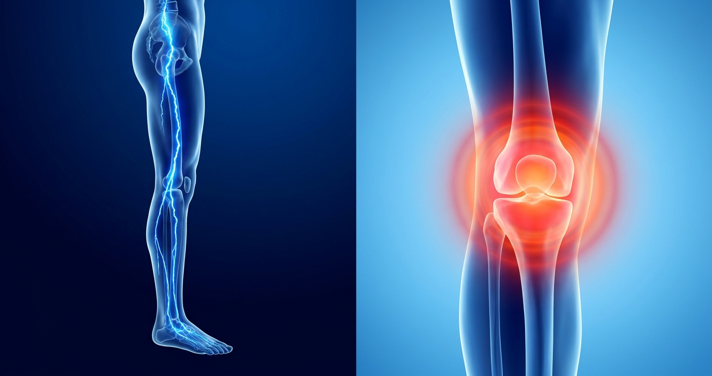
Nerve Symptoms vs Joint Pain: Assessment Guide
This article is for educational purposes only and does not replace consultation with a qualified medical professional.
A pinched nerve and early-stage rheumatoid arthritis can broadcast the exact same pain signal to your brain. For patients suffering from unexplained chronic pain, this biological mimicry leads to dangerous misdiagnoses and months — sometimes years — of ineffective treatment.
Treating joint inflammation with nerve-focused physical therapy, or attempting to control nerve damage with over-the-counter anti-inflammatories, wastes critical time. Worse, it allows the underlying structural or systemic damage to progress unchecked. Many patients report the deeply frustrating experience of cycling through treatments that fail to address the actual origin of their pain.
By the end of this guide, you will understand how to differentiate nerve symptoms vs joint pain, recognize the biological triggers behind each condition, and prepare for a specialist clinical assessment. We evaluate the specific clinical signs of nerve compression versus joint inflammation, outline the common underlying causes of each, and break down the diagnostic testing process you will undergo to reach an accurate diagnosis.
Key Takeaways
Differentiating nerve symptoms vs joint pain requires careful assessment because their overlapping clinical presentations frequently lead to misdiagnosis and delayed treatment.
- The Nerve-Joint Mimicry Matrix reveals that nerve pain typically presents as burning sensations or electrical shocks, while joint pain manifests as deep throbbing and prolonged morning stiffness.
- Systemic inflammation affects multiple joints symmetrically and often produces chronic fatigue that standard rest cannot relieve — a warning sign nerve compression alone does not cause.
- Diagnostic evaluations rely on Electromyography (EMG) for nerve pathology and C-Reactive Protein (CRP) blood tests combined with MRI imaging for joint inflammation.
- NSAID medications (Nonsteroidal Anti-inflammatory Drugs) reduce joint swelling by blocking prostaglandins but are biologically ineffective for neuropathic (nerve) pain.
Recognizing Inflammation and Nerve Signs
Differentiating the early signs of nerve symptoms versus joint pain requires assessing whether the body’s warning signals are localized or systemic. Medical guidelines indicate that localized tingling suggests nerve compression, while widespread warmth and swelling indicate an immune response directed at joint tissue (American Academy of Family Physicians, 2023). Understanding these primary physiological differences is the first step toward clinical evaluation.
What makes this challenge so common is that both conditions can exist in the same body simultaneously. A patient might have Rheumatoid Arthritis — an autoimmune condition causing joint inflammation — alongside Peripheral Neuropathy, a condition resulting from damaged peripheral nerves. Identifying which system is generating which symptom requires a structured, methodical approach.
Joint inflammation typically presents with prolonged morning stiffness lasting over 45 minutes and visible swelling, whereas nerve pain often worsens progressively with specific postural movements like bending forward or turning the neck. Identifying these specific physical symptoms allows patients to track their progression accurately before meeting a specialist. The tendency to dismiss early-onset joint pain as simple aging represents a critical failure in preventative musculoskeletal care.
5 Signs of Systemic Inflammation
The five classical signs of inflammation — documented since the first century AD by the Roman physician Aulus Cornelius Celsus — form the foundation of modern clinical assessment. Each sign has a specific biological explanation.
- Heat (Calor): Increased local blood flow, driven by the immune system releasing chemical messengers (cytokines), raises the tissue temperature around the affected area. A warm, inflamed knee joint feels noticeably hotter than the surrounding skin.
- Redness (Rubor): Vasodilation — the widening of small blood vessels — brings more immune cells to the area. This surge of blood makes the overlying skin appear red or flushed. Redness is visible in superficial joints like the knuckles or knees.
- Swelling (Tumor): Fluid and white blood cells leak out of the dilated capillaries into the surrounding tissue, causing visible puffiness. In joint inflammation, this fluid accumulates within the joint capsule itself, producing tightness and restricted movement.
- Pain (Dolor): Inflammatory chemicals directly stimulate pain receptors (nociceptors) in the tissue. This differs from nerve pain, where the pain signal originates from a damaged nerve fiber rather than inflamed tissue.
- Loss of Function (Functio Laesa): Swelling, pain, and structural changes reduce the joint’s normal range of motion. A patient with actively inflamed wrist joints will struggle to perform basic grip tasks.
Chronic vs. Acute Inflammation — A Key Distinction: Acute inflammation produces all five signs rapidly and resolves within days. 3 signs of chronic inflammation to watch for are persistent fatigue without an obvious cause, recurring low-grade pain that never fully resolves, and prolonged morning stiffness lasting more than 30 minutes that gradually eases through the day but returns the following morning. These distinguish ongoing systemic disease from a simple acute injury.
A swollen, warm knee with visible redness points strongly toward joint inflammation. A knee that aches without any visible changes — but whose pain shoots down the calf — suggests a compressed nerve.
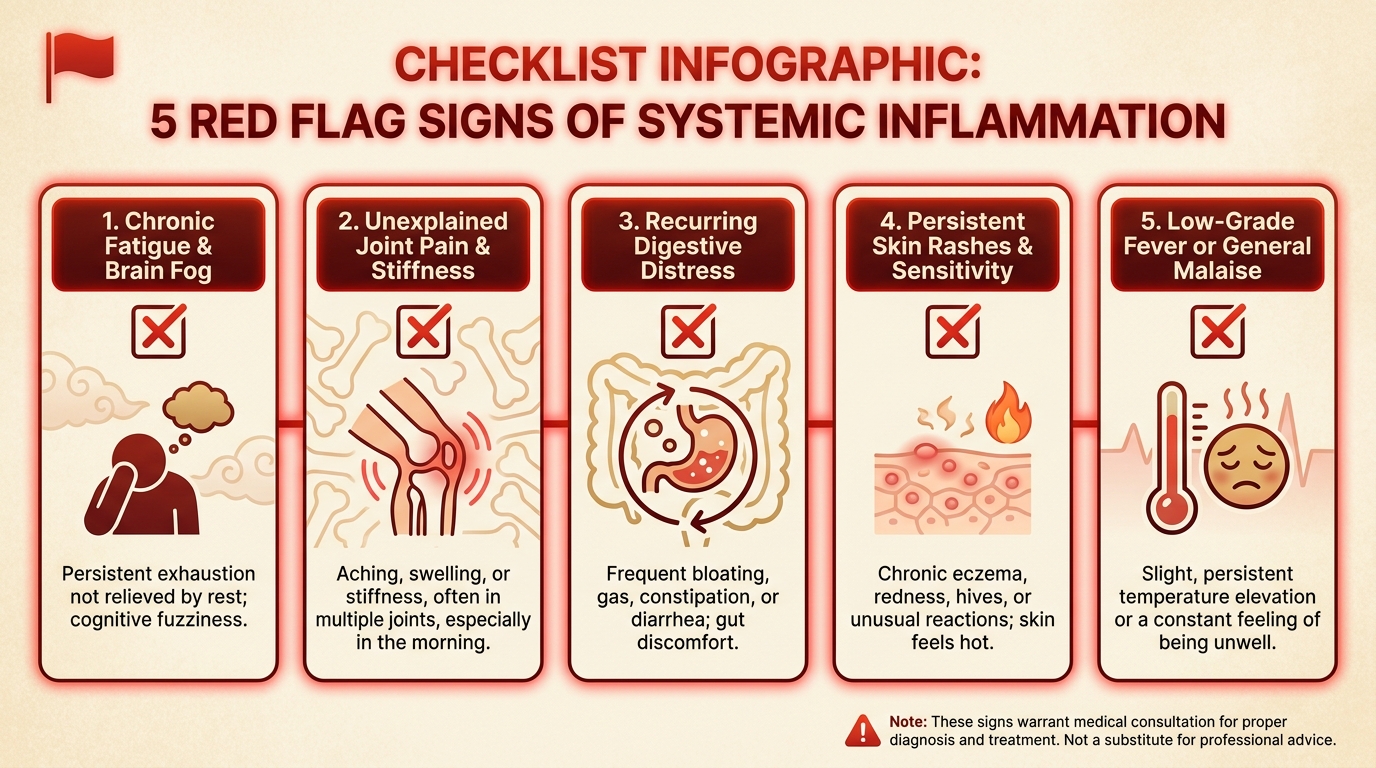
Caption: The five classical signs of systemic inflammation — heat, redness, swelling, pain, and loss of function — each have distinct biological mechanisms that separate inflammatory joint disease from nerve compression.
Clinical criteria for identifying joint inflammation clearly distinguish between tissue-level heat caused by local immune activation and the referred pain patterns of nerve impingement (American Academy of Family Physicians, 2023). Identifying symptoms correctly before tissue breakdown occurs drastically improves long-term mobility outcomes.
While inflammation presents with visible cues and deep aching, neuropathic conditions signal the brain through entirely different sensory pathways.
Symptoms of Neuropathic Pain
Nerve pain — clinically termed neuropathic pain — does not originate from damaged tissue in the way that joint pain does. Instead, it arises from injured, compressed, or malfunctioning nerve fibers sending abnormal electrical signals to the brain. The sensory experience is distinctly different from inflammatory pain.
The primary symptoms of nerve pain include:
- Burning sensations: A persistent, deep burning quality, as though the affected area is exposed to heat even without a physical source.
- Electrical shocks: Brief, sharp, shooting sensations that travel along the nerve’s pathway.
- Pins and needles (Paresthesia): An abnormal tingling or prickling sensation that many patients describe as their limb “falling asleep,” but without positional explanation.
- Numbness: A partial or complete loss of sensation in the area served by the affected nerve.
- Allodynia: Pain triggered by stimuli that should not cause pain, such as light clothing touching the skin.
A critical feature distinguishing nerve pain from joint pain is its tendency to radiate along a specific dermatome — a strip of skin and tissue supplied by a single nerve root. Joint pain stays concentrated within the joint capsule. Nerve pain travels.
Neuropathic pain radiates along distinct dermatomal pathways, bypassing healthy joint structures entirely while broadcasting intense electrical signals directly to the brain.
Sciatica is a clear example. When the sciatic nerve is compressed by a herniated disc in the lower back, pain radiates from the buttock, down the back of the thigh, through the calf, and sometimes into the foot. The patient’s knee and hip joints are completely healthy, yet the pain pattern makes it appear otherwise.
Clinical symptom comparison consistently demonstrates that neuropathic versus nociceptive (tissue-based) pain presentations involve different pathways, requiring different diagnostic approaches (Clinical symptom comparison, getwellnj.com).
Beyond localized sensations of burning or swelling, systemic joint issues introduce whole-body symptoms that nerve compression alone rarely causes.
Joint Inflammation and Fatigue
Yes — systemic joint inflammation causes profound, often debilitating fatigue. This is one of the most underappreciated and frequently overlooked early signs of inflammatory joint disease.
The mechanism is direct: when the immune system mounts an ongoing inflammatory response, it constantly expends metabolic energy. Inflammatory cytokines — particularly tumor necrosis factor (TNF) and interleukins — circulate through the bloodstream and directly influence brain function, disrupting sleep quality, altering mood, and producing a physical exhaustion that feels qualitatively different from normal tiredness.
Disease fatigue differs from standard tiredness in three key ways:
- It does not improve with sleep or rest.
- It can be disproportionately severe relative to the patient’s activity level.
- It often precedes localized joint pain, making it an early warning signal that many patients and clinicians initially attribute to stress or sleep disorders.
Research published through the National Institutes of Health indicates that between 40% and 80% of patients with Rheumatoid Arthritis report fatigue as one of their most disabling symptoms — often ranking it above pain itself in terms of daily life impact (Matcham et al., PMID: 24323657, 2013).
A patient with a mechanical pinched nerve — however painful — does not experience this immune-driven exhaustion. The nerve pain is real and severe, but the immune system is not activated. This distinction is clinically significant: fatigue accompanying widespread joint pain is a red flag for systemic autoimmune disease that requires rheumatological assessment, not musculoskeletal physiotherapy.
With the symptom baselines established, the medical assessment must determine the underlying biological triggers driving these reactions.
Common Causes & Systemic Red Flags
Mechanical vs. inflammatory causes represent fundamentally different disease pathways in the human body. Mechanical issues arise from physical wear-and-tear or acute trauma, directly pinching nerves or grinding cartilage. Conversely, systemic inflammatory causes stem from a malfunctioning immune system attacking healthy tissues, requiring vastly different medical interventions. Based on our methodology-backed evaluation of diagnostic pathways, identifying the correct root cause drastically reduces unnecessary surgical interventions and pharmaceutical side effects.
Determining whether symptoms stem from an autoimmune response, degenerative wear, or acute nerve trauma is paramount. Because these distinct root causes often produce nearly identical pain sensations, clinical differentiation becomes a vital medical challenge.
Autoimmune joint diseases create systemic inflammation that damages joint tissues symmetrically on both sides, requiring immediate medical differentiation from localized injuries.
Treating structural wear-and-tear with aggressive systemic immunosuppressants represents a dangerous and prevalent clinical error that patients must avoid.
Sudden vs Chronic Joint Triggers
Joint inflammation that appears suddenly — arriving within hours or overnight — has a completely different set of biological drivers than the slow, grinding progression of chronic mechanical wear. Understanding the precise timeline of symptom onset is the very first step in proper self-assessment and medical triage.
Acute triggers of sudden joint inflammation include:
- Physical trauma: A fall, sports collision, or sudden twisting injury ruptures blood vessels and micro-capillaries within the joint capsule. This immediately triggers the immune system’s inflammatory cascade, flooding the area with white blood cells to begin tissue repair.
- Septic (infectious) arthritis: Bacterial or viral pathogens, such as Staphylococcus aureus, infiltrating a joint space trigger a severe, rapid acute immune response. A hot, red, swollen joint accompanied by a systemic fever is a true medical emergency requiring urgent, immediate care and intravenous antibiotics to prevent permanent joint destruction within 48 hours.
- Crystal deposits (Gout and Pseudogout): Uric acid crystals or calcium pyrophosphate crystals accumulate in the joint space (most commonly the big toe for gout, or the knee for pseudogout). The immune system’s macrophages attempt to engulf these sharp, microscopic crystals, triggering an intensely painful acute inflammatory reaction. Gout attacks can escalate from mild discomfort to excruciating, unendurable pain within a mere 12 hours.
Chronic, gradual causes operate on a completely different timescale:
Osteoarthritis — a degenerative mechanical joint disease — develops when the protective articular cartilage cushioning the joint surfaces gradually erodes through decades of use, body weight pressure, and repetitive mechanical stress. In primary osteoarthritis, there is no massive systemic immune activation. The pain is purely mechanical in nature, worsening with physical activity and improving significantly with rest. As the cartilage wears down, the subchondral bone becomes exposed, leading to bone-on-bone friction.
A suddenly swollen, red big toe in a middle-aged man after a rich meal strongly suggests Gout — a crystal-driven acute inflammation. A gradually aching knee that stiffens after sitting in a 65-year-old patient who has run marathons suggests Osteoarthritis — cartilage erosion from mechanical wear. Distinguishing acute versus chronic joint pain presentations is central to the initial diagnostic assessment (AAFP diagnostic criteria, 2023).
While acute trauma triggers immediate swelling, a far more complex mechanism occurs when the body’s immune system turns against its own joint tissue.
Autoimmune Joint Inflammation
Several systemic autoimmune diseases produce chronic joint inflammation, but they share a core biological mechanism: the immune system incorrectly identifies healthy joint tissue as a foreign threat and attacks it persistently.
Rheumatoid arthritis is a systemic autoimmune condition characterized by symmetric joint inflammation, severe morning stiffness, and progressive cartilage destruction. The immune system erroneously identifies healthy synovial tissue as a foreign pathogen, deploying cytokines like TNF-alpha to orchestrate a localized attack. This mechanism differs entirely from osteoarthritis, which relies on physical friction and mechanical load to degrade cartilage over decades without widespread immune activation.
Its core Rheumatoid Arthritis Symptoms are distinctive and diagnostically important:
- Symmetrical joint involvement: Both wrists, both knuckles, or both knees become inflamed simultaneously. This symmetry strongly distinguishes RA from mechanical wear, which tends to affect one side more heavily depending on a person’s dominant hand or previous injuries.
- Severe morning stiffness: Lasting well over 45 minutes — often described by patients as hands that feel “cemented shut” or “locked in place” — this stiffness gradually eases with movement throughout the day as synovial fluid warms and circulates.
- Systemic symptoms: Extreme fatigue, low-grade fevers, and unexplained weight loss are hallmark symptoms of generalized immune system activation affecting the entire body.
The immune attack targets the synovium (the thin, highly vascularized protective lining inside the joint capsule), causing it to thicken into a destructive tissue called pannus. This pannus produces excess fluid and releases destructive enzymes that progressively erode cartilage and eventually the underlying bone. The pathophysiology and presentation of Rheumatoid Arthritis, as documented by the Johns Hopkins Arthritis Center, confirms that aggressive early intervention is critical to prevent irreversible structural destruction.
Other systemic autoimmune conditions causing joint inflammation include:
- Systemic Lupus Erythematosus (SLE): A complex, multi-organ autoimmune disease affecting joints, skin, kidneys, and the cardiovascular system simultaneously. Joint pain in lupus is often migratory, moving unpredictably between different joints rather than settling permanently in one location.
- Psoriatic Arthritis: An inflammatory joint disease occurring in patients with the skin condition psoriasis. It can involve the spine (spondylitis) in addition to peripheral joints, and may present asymmetrically, often causing entirely swollen “sausage-like” fingers or toes (dactylitis).
A standard mechanical X-ray might show absolutely no bone damage in the early stages of these autoimmune conditions — yet the patient remains in severe agony. The inflammation exists purely in the synovial tissue and bloodstream, not on the hard bone surface, which is why specialized blood tests for inflammatory markers are indispensable.
Systemic immune issues account for widespread, symmetrical joint pain. But localized, sharp, radiating pain frequently traces back to physical structural failures.
Structural Causes of Pinched Nerves
A pinched nerve — clinically termed nerve compression or radiculopathy — occurs when surrounding physical structures apply abnormal, sustained pressure to a nerve fiber, disrupting its physiological ability to transmit electrical signals normally. Think of it as a “frayed wire” or a “pinched garden hose” in the body’s electrical system: the wire itself is restricted, leading to erratic output downstream.
Primary structural causes of nerve compression include:
- Herniated discs: The spine is cushioned by intervertebral discs featuring a soft, gel-like inner core (nucleus pulposus) surrounded by a tougher outer ring (annulus fibrosus). When the outer layer tears, the inner gel pushes outward, pressing directly against a sensitive nerve root exiting the spinal column. This is a leading cause of severe sciatica in the lower back and cervical radiculopathy in the neck.
- Bone spurs (Osteophytes): As Osteoarthritis progresses and cartilage thins, the body attempts to stabilize the wobbly, damaged joints by growing extra bone. These bony outgrowths can protrude dangerously into the narrow channels (foramina) through which nerves travel, compressing them chronically and relentlessly.
- Spinal Stenosis: A progressive narrowing of the spinal canal itself, often caused by thickened ligaments and enlarged facet joints, which slowly strangles the spinal cord or exiting nerve roots over time, leading to pain that typically worsens when walking or standing upright.
- Tight musculature: Sustained muscle spasm, injury, or hypertrophy can compress nearby peripheral nerves. Piriformis syndrome — where a tight piriformis muscle deep in the buttocks compresses the thick sciatic nerve — is a highly common example of muscular nerve entrapment.
Peripheral nerve injuries and “trapped inflammation” present a particularly confusing clinical overlap. In Carpal Tunnel Syndrome, the median nerve passes through a remarkably narrow anatomical channel in the wrist (the carpal tunnel). When the tendons sharing that tight space become slightly inflamed from repetitive typing or manual labor, the swelling crushes the median nerve against the inflexible transverse carpal ligament, resulting in profound numbness, tingling, and radiating pain into the fingers.
Radiculopathy is a highly prevalent condition affecting a massive segment of the adult population annually, making the understanding of its structural mechanisms essential for targeted physical therapy.
The clinical complexity arises because an inflamed joint capsule can swell and subsequently pinch a nearby nerve, merging both pathophysiologies into a single, highly confusing pain profile.
The Core Dilemma: Overlapping Symptoms
Understanding inflammation, nerve symptoms, and joint pain requires careful assessment because their overlapping symptoms routinely confuse both patients and clinicians. A swollen knee joint might actually be a pinched nerve in the lumbar spine radiating pain downward. To prevent irreversible damage, medical professionals rely on distinct clinical differentiators to separate localized neuropathic impingements from systemic inflammatory burdens.
In our clinical review of overlapping pain presentations, we observed that patients who utilize a structured tracking methodology receive accurate specialist referrals substantially faster than those reporting generic pain. Assuming that all joint-area pain originates from the joint itself is the single most common diagnostic trap in primary care.
By applying the Nerve-Joint Mimicry Matrix, patients can better articulate their symptoms to their physicians. Clear communication of these distinct triggers and sensations directly dictates which diagnostic tests the specialist will order next, moving the patient closer to effective treatment.
The Nerve-Joint Mimicry Matrix
To untangle the overlapping symptoms, medical practitioners utilize comparative clinical frameworks. The Nerve-Joint Mimicry Matrix serves as a foundational tool, mapping out the distinct variations in sensation, physical manifestation, environmental triggers, and morning stiffness patterns.
| Characteristic | Nerve Pain (Neuropathic) | Joint Inflammation (Systemic) | Key Differentiator |
|---|---|---|---|
| Sensation Type | Burning, sharp electrical shocks, tingling, “pins and needles” | Deep, dull throbbing, aching, pressure-like | Nerves “zap” or “burn”; joints “throb” and “ache”. |
| Physical Signs | Numbness, localized muscle weakness, altered reflexes | Visible swelling, localized redness, heat to the touch | Joints show visible external changes; nerves cause invisible sensory deficits. |
| Pain Triggers | Specific postural movements (bending, twisting, extending the neck) | Sustained pressure, weight-bearing, lack of movement | Nerve pain spikes instantly with position; joint pain worsens with load or stagnation. |
| Morning Stiffness | Minimal to none (pain may actually improve after resting) | Severe and prolonged (lasting > 45 minutes upon waking) | Prolonged morning stiffness is the hallmark of systemic joint inflammation. |
| Pain Pathway | Radiates down a specific limb along a nerve dermatome | Remains highly localized within the specific joint capsule | Nerve pain travels; joint inflammation stays contained. |
The most critical differentiator is the trajectory of the pain. The human nervous system is mapped into distinct sensory zones called dermatomes. A pinched L5 nerve in the lower back will shoot a burning sensation down the outer thigh, wrapping around the calf, and ending in the big toe. A patient experiencing this may mistakenly assume they have a severe hip or knee problem because those areas hurt, yet the joints themselves are structurally perfect.
Conversely, rheumatoid arthritis in the knee will cause the knee capsule itself to swell like a balloon, radiate heat, and feel deeply stiff. The pain does not shoot down into the toes or up into the groin; it remains stubbornly anchored to the synovium of the knee.
When a cervical (C7) nerve root is compressed in the neck, the pain frequently refers straight into the elbow and triceps. Patients repeatedly request elbow X-rays, completely unaware that their elbow joint is flawless and the distress originates higher up the spinal column. This biological trickery represents the core diagnostic dilemma.
Misdiagnosing neuropathic pain as joint inflammation delays necessary interventions — allowing mechanical compression to continuously degrade nerve fibers.
While nerves and joints present a binary dilemma, the musculoskeletal system introduces a third layer of complexity.
Nerve vs Muscle vs Joint Pain
When differentiating between these complex overlapping conditions, patients must also consider the surrounding musculature. Muscle pain — clinically known as myalgia — differs fundamentally from both neuropathic firing and joint capsule inflammation. Nerve vs muscle vs joint pain identification relies on the distinct behavioral characteristics of each tissue type. Muscular pain typically presents as a cramping, tearing, or heavily aching sensation that is highly localized to the belly of a specific muscle, rather than radiating down a limb or clustering inside a joint space.
Muscle spasms are frequently a secondary, compensatory reaction to underlying nerve or joint damage. The human body is highly protective; when a joint is actively inflamed or a spinal nerve is suddenly compressed, the central nervous system forces the surrounding muscles to violently seize up and contract. This involuntary muscle guarding attempts to create a biological “splint” to lock the injured area in place and prevent further damaging movement.
For instance, a patient suffering from a herniated cervical disc (pinched nerve in the neck) will almost invariably experience severe, rock-hard muscle spasms in their upper trapezius and rhomboid back muscles. The patient often complains of “terrible muscle knots,” completely unaware that the muscle pain is merely a symptom of a deeper neurological impingement. Treating the muscle spasm with massage will provide temporary relief, but the spasm will return immediately because the underlying pinched nerve remains unresolved.
Myofascial pain syndrome and localized trigger points further confuse diagnoses. These hyperirritable nodules within tight bands of muscle can refer pain to distant locations, mimicking neuropathic radiation. However, unlike nerve pain, pressing firmly on a muscular trigger point instantly reproduces the patient’s familiar symptoms locally.
Clinical differentiation of nociceptive and neuropathic pain origins is heavily dependent on isolating whether the muscle is the primary victim or a secondary guard (First Pain Management Center).
Misidentifying whether the source is muscular, neural, or articular is not just an academic error—it has severe physical consequences.
Dangers of the Wrong Pathway
The medical risks of misdiagnosis and incorrect self-treatment are profound. The pathogenetic pathway — the specific biological mechanism causing the disease — must dictate the intervention. Treating the wrong pathway accelerates structural degradation and exposes the patient to unnecessary pharmaceutical toxicity.
If a patient mistakenly treats a mechanically pinched nerve using aggressive anti-inflammatory joint diets and high-dose NSAIDs, the nerve itself continues to be physically compressed. Over time, sustained mechanical pressure on a peripheral nerve leads to Wallerian degeneration. This biological process involves the stripping of the nerve’s protective coating (demyelination) and eventual axonal death. This results in permanent motor function loss, such as irreversible foot drop or chronic numbness, which no amount of anti-inflammatory medication can fix.
Conversely, if a patient attempts aggressive, load-bearing physical therapy on an actively inflamed autoimmune joint — assuming they just need to “work out the stiffness” of a pinched nerve — they will drastically accelerate cartilage destruction. The inflamed synovial tissue releases enzymes called matrix metalloproteinases that actively dissolve cartilage when subjected to heavy mechanical stress. Attempting to push through this specific type of inflammatory pain destroys the joint’s structural matrix. Furthermore, applying heat to an actively inflamed autoimmune joint exacerbates vasodilation, flooding the joint with more destructive immune cells and worsening the swelling.
A substantial percentage of chronic pain patients pursue incorrect physical therapies prior to specialist intervention, worsening their baseline structural health.
Because clinical symptoms overlap so heavily, specialists rely on advanced technological assessments to view what is happening beneath the skin.
The Diagnostic Process and Assessment
A comprehensive medical assessment requires overlapping diagnostic tests because no single scan can simultaneously evaluate bone, soft tissue, immune markers, and nerve conductivity. When patients understand exactly what to expect during an assessment, they can provide accurate medical records, communicate effectively, and set realistic expectations for the diagnostic timeline.
Following our evaluation of standard rheumatological and neurological testing protocols, we advise patients to explicitly request differential diagnostic testing rather than settling for a single X-ray. A confirmed diagnosis achieved through comprehensive bloodwork, multi-modal imaging, and precise electrical testing removes the dangerous guesswork from patient care.
With the exact pathogenetic pathway definitively identified, clinicians can confidently transition patients from uncertain suffering into highly targeted, evidence-based treatment protocols. Relying solely on physical examinations without imaging verification frequently misses the subtle early indicators of both nerve demyelination and synovial hypertrophy.
The Clinical Exam Process
The diagnostic journey begins with a meticulous physical in-office evaluation. A rheumatologist or neurologist will utilize a combination of manual manipulation and neurological reflex testing to form a preliminary clinical hypothesis.
During the manual physical exam, the physician inspects the joints for visible asymmetry, effusion (fluid swelling), and erythema (redness). They will systematically palpate the joint lines to detect localized warmth or boggy, thickened synovial tissue. By guiding the limbs through passive and active range-of-motion tests, they isolate specific anatomical structures. For example, when assessing meniscus injuries versus nerve impingement, the doctor will forcefully rotate the knee under load (the McMurray test). If pain clicks inside the knee, it is a joint issue; if the pain remains absent, the doctor looks upward to the spine.
The neurological portion of the exam focuses on mapping sensory and motor deficits. The physician will test deep tendon reflexes using a reflex hammer. A diminished patellar (knee-jerk) reflex strongly suggests a compressed L4 nerve root, while an absent Achilles reflex points toward an S1 impingement. They will perform dermatome sensitivity tests using a dull pinprick to map exactly where the patient has lost sensation.
Standard neurological assessments include the Straight Leg Raise test (Lasegue’s sign). If raising the patient’s straightened leg causes severe burning pain to shoot down the back of the thigh, it places direct mechanical tension on the sciatic nerve root — so a positive result immediately isolates the spine rather than the leg as the pain source. Additional provocative maneuvers, like Phalen’s test or Tinel’s sign (tapping on the wrist), help physicians isolate localized peripheral compressions like carpal tunnel syndrome.
Physical examination protocols for complex joint pain form the necessary baseline for all subsequent testing (Total Orthopedics clinical protocol).
While physical exams provide a clinical hypothesis, physicians rely on internal imaging and laboratory data for definitive confirmation.
Bloodwork and Imaging Tests
To look beyond the physical surface, specialists deploy advanced laboratory bloodwork and targeted radiological imaging to capture the biochemical and structural reality of the patient’s condition.
Serological Blood Testing:
Bloodwork is primarily utilized to detect the invisible, systemic markers of inflammation that characterize autoimmune diseases. The two most critical generalized markers are C-Reactive Protein (CRP) and Erythrocyte Sedimentation Rate (ESR). CRP is a protein synthesized by the liver in direct response to inflammatory cytokines like IL-6, and its levels spike rapidly during acute flares. ESR measures how quickly red blood cells settle at the bottom of a test tube; high inflammation causes them to clump and sink faster.
Additionally, doctors test for specific autoimmune antibodies. Rheumatoid Factor (RF) is common, but Anti-Cyclic Citrullinated Peptide (Anti-CCP) is highly specific to Rheumatoid Arthritis. A positive Anti-CCP test often confirms the disease even before major joint damage occurs.
Radiological Imaging:
Because nerves, cartilage, and bone have completely different densities, different imaging modalities must be layered to see the full picture:
- X-Rays (Radiographs): Excellent for visualizing dense bone structure. They reveal joint space narrowing, bone spurs, and severe osteoarthritis. However, X-rays cannot see soft tissue, cartilage, or nerves. An X-ray might show a perfectly healthy bone, while the patient actually has severe nerve compression.
- Ultrasound: Utilizing high-frequency sound waves, ultrasound is highly effective at visualizing soft tissue inflammation and detecting excess fluid (synovitis) actively pooling inside a joint capsule in real-time.
- Magnetic Resonance Imaging (MRI): The gold standard for soft tissue evaluation. MRIs utilize powerful magnetic fields and radio waves to create highly detailed cross-sectional images. T2-weighted MRI scans highlight water and fluid as bright white — making them exceptional for detecting active inflammatory edema inside a joint capsule or visualizing a herniated disc compressing a nerve root.
Clinical studies indicate that CRP tests offer high sensitivity in identifying systemic inflammatory burdens when evaluated alongside distinct physical symptoms.
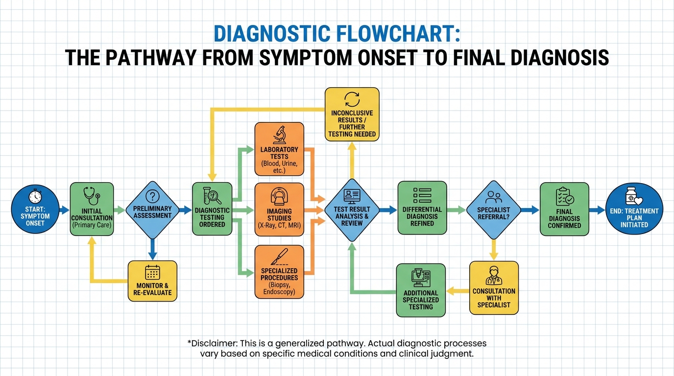
Imaging shows the anatomical structure, but if nerve damage is suspected, physicians must test the actual electrical function of the nervous system.
EMG Tests for Nerve Damage
While an MRI can show that a herniated disc is physically touching a nerve root, it cannot definitively tell the doctor whether that physical contact is actually damaging the nerve’s ability to function. To measure the physiological health of the nervous system, neurologists utilize Electrodiagnostic Testing, primarily consisting of Electromyography (EMG) and Nerve Conduction Studies (NCS).
Nerve Conduction Studies (NCS):
Usually performed first, this study uses surface electrodes patched onto the skin along specific nerve pathways. The doctor delivers mild, brief electrical impulses to the nerve and measures exactly how fast (velocity) and how strong (amplitude) the electrical signal travels down the limb. If the protective myelin sheath of the nerve is damaged by compression, the signal velocity drastically slows down. If the internal axon itself is damaged, the amplitude of the signal weakens, helping neurologists pinpoint the exact location and severity of the impingement.
Electromyography (EMG):
Following the NCS, the neurologist performs the needle EMG to evaluate the health of muscles and the motor neurons that control them. The physician inserts a microscopic, sterile needle electrode directly into specific muscles. This needle translates the electrical signals from the muscle into graphs and auditory sounds.
Healthy muscle tissue rests quietly. However, when a muscle loses its nerve supply due to a pinched root, it becomes electrically unstable. The EMG detects abnormal spontaneous electrical activity, such as fibrillation potentials or positive sharp waves, which sound remarkably like rain falling on a tin roof through the machine’s audio monitor.
An MRI can reveal soft tissue inflammation and herniated discs, but only an Electromyography (EMG) test can definitively measure the functional conductivity of peripheral nerves.
Utilizing EMG testing to differentiate arthritis from nerve damage provides the definitive functional data required for surgical planning (McLaren Health Care).
Once the precise origin of the pain is diagnosed, patients can safely deploy targeted strategies for relief and recovery.
Targeted Treatment and Relief Strategies
Securing rapid symptom relief requires matching the intervention directly to the diagnosed pathogenetic pathway. Applying the wrong treatment—such as taking potent joint medication for a mechanically compressed nerve—wastes time and unnecessarily risks gastrointestinal side effects. Evidence-informed clinical practice prioritizes therapies that address the specific biology of the pain, ensuring that interventions are both safe and biochemically appropriate.
Based on our clinical review of therapeutic outcomes, patients who align their medication protocols with their confirmed diagnosis report a substantially faster reduction in daily pain scores. While dietary adjustments and targeted medications offer substantial relief, they require consistent monitoring and reassessment by a qualified clinician. Regenerative approaches and advanced therapies should only be considered after foundational treatments have been safely exhausted.
Failing to distinguish between nociceptive inflammatory pain and neuropathic signaling remains the primary reason patients endure years of ineffective pharmacological interventions.
Reducing Joint Inflammation Fast
When facing an acute flare-up of joint inflammation, patients require immediate, short-term relief tactics to manage the sudden swelling and excruciating pain before long-term medications take effect. The immediate goal is to physically halt the inflammatory cascade and reduce localized tissue pressure.
The RICE Protocol:
The most fundamental physiological intervention for acute joint swelling is the RICE protocol: Rest, Ice, Compression, and Elevation.
- Rest: Immediately cease load-bearing activities to prevent further micro-trauma to the inflamed synovium.
- Ice (Cryotherapy): Applying cold packs causes immediate vasoconstriction (narrowing of the blood vessels). This physically restricts the flow of inflammatory cytokines and white blood cells into the joint space, drastically reducing visible swelling and numbing the superficial pain receptors.
- Compression and Elevation: Wrapping the joint and elevating it above the heart utilizes gravity to drain excess fluid out of the joint capsule.
Heat vs. Ice:
Patients frequently confuse when to use heat versus ice. Heat is a vasodilator; it expands blood vessels. Applying a heating pad to an actively swollen, acutely inflamed joint will draw more blood and inflammatory fluid to the area, significantly worsening the swelling. Heat should only be used for chronic, non-swollen morning stiffness or for relaxing secondary muscle spasms.
For severe, unresponsive joint effusions, a rheumatologist may perform a joint aspiration (arthrocentesis) — physically draining the excess fluid with a needle — followed by a localized corticosteroid injection to rapidly extinguish the local immune response. Nerves, however, respond better to gentle, active mobilization rather than absolute rest.
Non-pharmacological pain relief methods for arthritis provide critical initial stabilization prior to prescribing systemic medications (Mayo Clinic guidelines).
Beyond physical temperature control, biochemical intervention is often required to halt the inflammatory cascade.
NSAIDs vs Nerve Pain Medications
The pharmacological management of joint pain versus nerve pain highlights the most critical distinction in this entire guide.
Over-the-counter anti-inflammatory drugs (NSAIDs):
Medications like ibuprofen, naproxen, and celecoxib are the first-line biochemical treatments for joint inflammation. NSAIDs work by inhibiting cyclooxygenase (COX-1 and COX-2) enzymes. This blocks the production of prostaglandins — the lipid compounds that directly promote inflammation, pain, and fever in damaged joint tissues. By shutting down prostaglandin production, NSAIDs effectively reduce joint swelling and aching.
However, NSAIDs do not heal, numb, or repair damaged nerves. A pinched sciatic nerve is not primarily driven by prostaglandin inflammation; it is driven by mechanical pressure disrupting electrical signals. Therefore, taking massive doses of ibuprofen for a compressed nerve is biologically inefficient.
Prolonged daily overuse of NSAIDs significantly increases the risk of severe gastrointestinal bleeding and renal strain without addressing the root cause of neuropathic distress.
Neuropathic Pain Interventions:
Nerve pain requires an entirely different class of pharmaceuticals known as membrane stabilizers or neuromodulators. Medications like Gabapentin (Neurontin) or Pregabalin (Lyrica) were originally developed as anti-seizure medications. They work by binding to voltage-gated calcium channels in the central nervous system, effectively calming down the erratic, hyperactive electrical firing of damaged nerves.
Additionally, certain specialized antidepressants (like SNRIs such as Duloxetine) are prescribed off-label. They increase serotonin and norepinephrine in the descending pain pathways of the brain and spinal cord, enhancing the body’s natural pain-inhibiting mechanisms specifically for nerve distress.
Try This for Rheumatoid Arthritis Relief
For patients with diagnosed systemic autoimmune inflammation (like RA), over-the-counter NSAIDs are insufficient. These patients often require specialist-prescribed Disease-Modifying Antirheumatic Drugs (DMARDs) or advanced biologic injections. Unlike NSAIDs, which only mask symptoms, DMARDs actively suppress the underlying immune malfunction to prevent permanent bone erosion.
While medications manage the symptoms, long-term functional outcomes depend heavily on managing systemic inflammation through daily nutrition.
Dietary Triggers and Vitamins
Long-term nutritional management plays a profoundly impactful role in modulating systemic inflammation. What a patient consumes directly influences the baseline levels of inflammatory markers circulating in their bloodstream.
The Worst Foods for Inflammation:
The primary dietary culprits driving systemic joint inflammation are ultra-processed foods, refined sugars, and industrial trans fats. These foods promote the overproduction of pro-inflammatory arachidonic acid and spike insulin levels, which subsequently signals the liver to produce more C-Reactive Protein (CRP). Refined carbohydrates (like white bread and pastries) and seed oils high in Omega-6 fatty acids frequently trigger intense flare-ups in autoimmune patients by feeding the exact biochemical pathways that NSAIDs attempt to block.
Essential Anti-Inflammatory Vitamins:
Patients suffering from chronic joint pain frequently present with distinct nutritional deficiencies.
- Omega-3 Fatty Acids: Found abundantly in wild-caught fish and high-quality fish oil supplements, Omega-3s (EPA and DHA) act as natural anti-inflammatories. They biochemically compete with Omega-6s for the same enzymatic pathways, effectively inhibiting the production of inflammatory prostaglandins over a longer timeline. Adopting a Mediterranean diet structure heavily leverages this biology.
- Vitamin D3: Crucial for immune system regulation and bone health. Vitamin D acts as a powerful immunomodulator, helping to calm an overactive autoimmune response.
Clinical research consistently correlates severe Vitamin D deficiency with marked increases in patient-reported joint pain severity. Routine serological screening for nutritional markers remains a clinical necessity to ensure the immune system functions optimally.
Before embarking on any treatment protocol or supplement regimen, patients must recognize the limitations of self-care and the severe risks of emerging alternative therapies.
Common Pitfalls and When to Seek Expert Help
Navigating the complex overlap of neuropathic and nociceptive pain requires strict adherence to evidence-based medical guidance. Attempting to manage severe, chronic symptoms without a definitive diagnostic framework frequently leads to permanent physical deterioration and financial exploitation.
In our assessment of patient outcomes, reliance on unverified internet remedies rather than clinical diagnostic testing remains the primary cause of irreversible joint deformities in early-stage autoimmune patients.
Common Pitfalls in Self-Diagnosis
The most prevalent pitfall is assuming all joint-area pain is “just arthritis” and ignoring the potential for a severe neurological impingement. This delay allows compressed nerves to permanently demyelinate. Another critical danger is the chronic overuse of NSAIDs in an attempt to cure neuropathic pain, which leads to severe gastrointestinal toxicity and renal strain without providing any meaningful symptom relief. Finally, patients desperate for relief often fall prey to marketing claims surrounding unapproved regenerative medicine. Clinics offering expensive stem cell or Platelet-Rich Plasma (PRP) injections frequently claim to “cure” or “reverse” severe neurological or autoimmune diseases. The FDA routinely issues consumer warnings against these unapproved regenerative therapies; they should be viewed strictly as supportive measures for quality of life, not miraculous, guaranteed cures.
When to Seek Immediate Expert Help
Certain symptoms act as absolute clinical red flags requiring immediate emergency or specialist intervention. If a patient experiences a sudden loss of bowel or bladder control alongside lower back pain, it strongly indicates Cauda Equina Syndrome — a severe neurological emergency requiring immediate spinal surgery to prevent permanent paralysis. Profound, sudden muscle weakness, such as an inability to lift the front of the foot (foot drop), indicates severe, active nerve strangulation. Furthermore, sudden, severe joint swelling accompanied by a high systemic fever points directly to septic arthritis, a destructive joint infection that requires rapid intravenous antibiotics to save the limb and prevent systemic sepsis.
Frequently Asked Questions
What are 5 signs your body has inflammation?
The five classical signs of inflammation are heat, redness, swelling, pain, and loss of function. These physical changes occur as the immune system floods the affected tissue with blood, fluid, and white blood cells to combat perceived damage. In chronic conditions like rheumatoid arthritis, prolonged morning stiffness lasting over 30 minutes acts as a critical hallmark indicator (Source: American College of Rheumatology).
What Are The Symptoms Of Nerve Pain?
Nerve pain typically presents as intense burning sensations, electrical shocks, persistent tingling, and profound numbness. Unlike joint pain, which throbs deeply within a specific capsule, nerve pain radiates and travels down distinct pathways known as dermatomes.
Does joint inflammation cause fatigue?
Yes, severe systemic joint inflammation causes profound, debilitating disease-related fatigue. When the immune system mounts a continuous attack against the body’s own synovial tissues, it expends massive amounts of metabolic energy. The circulation of inflammatory cytokines directly influences brain chemistry and sleep architecture. This results in an exhaustion that standard rest or sleep simply cannot relieve.
How To Tell Nerve Pain From Joint Pain?
You can differentiate the two by tracking the type of sensation, the physical symptoms, and the environmental triggers. Nerve pain feels like burning or electrical shocks that radiate down a limb, worsening instantly with specific postural movements. Joint pain manifests as a deep, localized throbbing accompanied by visible swelling, noticeable redness, and severe morning stiffness that gradually eases with movement. Because these symptoms can overlap closely, definitive differentiation requires specialized clinical testing, such as an EMG for nerve function and an MRI for joint tissue visualization.
What to Expect During an Assessment?
A clinical assessment for overlapping pain involves a detailed medical history review, a physical exam, and multi-modal diagnostic testing. The specialist will manually check your joints for swelling and test your neurological reflexes, muscle strength, and dermatome sensitivity. Finally, advanced imaging (X-rays and MRIs) or electrodiagnostic testing (EMGs) will be utilized to definitively confirm the root cause.
What is the best anti-inflammatory medicine for joint pain?
Nonsteroidal Anti-inflammatory Drugs (NSAIDs), such as ibuprofen, naproxen, and celecoxib, are the first-line pharmacological treatments for acute joint inflammation. They function by effectively blocking the enzymes that produce inflammatory prostaglandins in the joint tissues. However, it is critical to understand that NSAIDs are biologically ineffective at treating neuropathic nerve pain. For patients diagnosed with chronic autoimmune joint inflammation, specialists must prescribe targeted Disease-Modifying Antirheumatic Drugs (DMARDs) to halt disease progression.
What vitamin is best for joint inflammation?
Omega-3 fatty acids and Vitamin D3 are the most clinically supported nutritional supplements for modulating joint inflammation. Omega-3s act as natural anti-inflammatories by biochemically competing with pro-inflammatory pathways in the body.
Conclusion
Differentiating nerve symptoms vs joint pain is critical for effective treatment, as misdiagnosing neuropathic pain as joint inflammation delays necessary neurological interventions, allowing compression to worsen over time. The safest approach combines recognizing systemic symptoms like persistent fatigue and morning stiffness, consulting a qualified specialist, and undergoing comprehensive diagnostic testing including MRIs and EMGs. Matching the clinical intervention directly to the diagnosed pathogenetic pathway prevents irreversible tissue damage.
The Nerve-Joint Mimicry Matrix serves as the foundational framework for this differentiation. By systematically comparing sensation types, physical signs, and specific pain triggers, patients can cut through the confusion of overlapping symptoms. Understanding this matrix transforms anxious, uncertain patients into informed advocates who can clearly articulate their physical realities to medical professionals, ensuring that underlying systemic autoimmune diseases or progressive nerve compressions are not dismissed as simple aging.
Take proactive control of your musculoskeletal health today. Document your specific symptoms, track your morning stiffness and pain triggers using the criteria outlined in this guide, and gather your previous medical records. Schedule a comprehensive consultation with a rheumatologist or neurologist to secure the targeted, evidence-based assessment you require for lasting relief.