
How to Tell Inflammatory Joint Pain vs Osteoarthritis
This article is for educational purposes only and does not replace consultation with a qualified medical professional.
Waking up with stiff joints can make the simplest morning routines feel impossible, leaving you wondering whether it is just a normal part of aging or a sign of an underlying immune condition. That question matters more than most people realize.
Misattributing the root cause of chronic joint pain often leads to delayed care. Progressive conditions can worsen while patients rely on over-the-counter remedies that address symptoms rather than causes. Early diagnosis — particularly for inflammatory arthritis — can prevent irreversible joint damage that develops silently over the first two years.
By the end of this guide, you will understand the clear clinical differences between inflammatory joint pain vs osteoarthritis so you can accurately track your symptoms and have a more productive consultation with a rheumatologist. We will explore the underlying biology, provide a clear symptom-tracking framework called The T.A.S.K. Joint Assessment, map out joint-specific patterns, and decode the exact medical tests doctors use to reach a definitive diagnosis.
Key Takeaways
Understanding inflammatory joint pain vs osteoarthritis requires tracking stiffness timing, pain triggers, and joint symmetry to help doctors order the correct clinical tests.
- The T.A.S.K. Joint Assessment helps patients document Timing, Anatomy, Symmetry, and Kinetics before specialist visits.
- Osteoarthritis is localized mechanical cartilage degradation; morning stiffness typically resolves within 30 minutes.
- Inflammatory arthritis is an autoimmune condition causing systemic symptoms (fatigue, fever) and stiffness lasting over an hour.
- Accurate diagnosis relies on combining physical symptom history with specific autoantibody blood tests (RF, CRP).
Fundamentals: Wear-and-Tear vs. Autoimmune
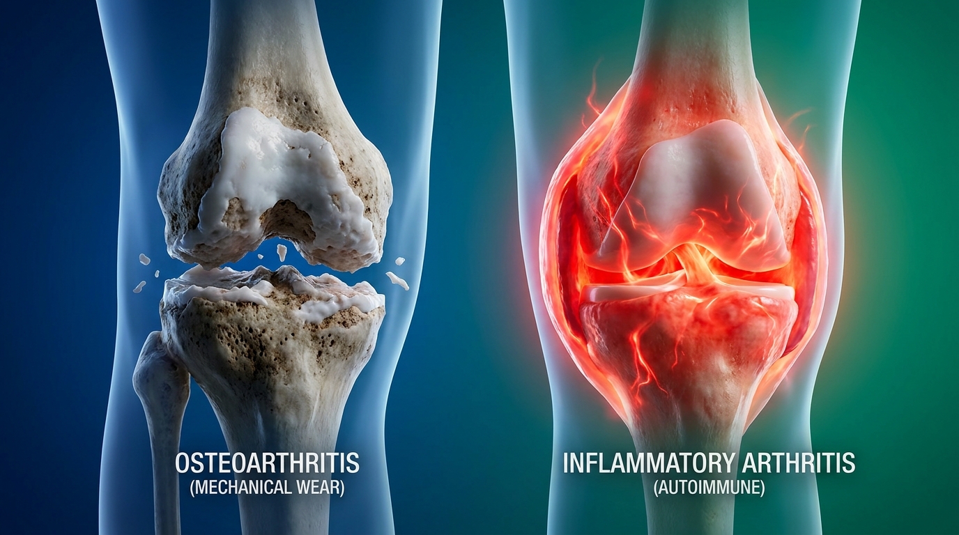
When comparing inflammatory joint pain vs osteoarthritis, the primary distinction lies in the underlying biological cause. Osteoarthritis involves the mechanical wear and tear of joint cartilage, while inflammatory arthritis is an autoimmune condition where the body’s immune system attacks healthy tissue (NIAMS, 2023). This biological difference dictates both the symptoms experienced and the clinical treatments required.
While both conditions share the outward symptom of joint discomfort, treating them interchangeably is a dangerous clinical error. Utilizing our T.A.S.K. Assessment framework early in the diagnostic journey helps separate mechanical friction from systemic immune dysfunction. Ultimately, failing to distinguish between mechanical and systemic joint disease delays critical therapeutic windows that preserve long-term mobility.

Caption: Osteoarthritis erodes cartilage through physical stress; inflammatory arthritis damages the joint lining through immune system dysfunction.
Osteoarthritis: Mechanical Wear
Osteoarthritis — the most common form of mechanical joint degradation — is best understood through a simple analogy: imagine the cartilage (the smooth, rubbery tissue that cushions the ends of your bones inside a joint) as a brake pad on a car. With enough miles and friction, that brake pad wears thin. Eventually, bone begins grinding against bone, causing pain, stiffness, and reduced range of motion.
This condition is strictly localized. It only affects the specific joints that have endured high mechanical stress, previous traumatic injury, or decades of repetitive use. A long-distance runner may develop severe osteoarthritis in the knee, but their shoulder joints, which experience far less mechanical load, remain entirely unaffected. Similarly, a construction worker who spent thirty years using a jackhammer may develop significant joint degradation in the hands and wrists while their ankles remain perfectly healthy.
Osteoarthritis affects over 32.5 million adults in the United States (CDC, 2023) — meaning a massive segment of the aging population faces progressive mobility limitations without proper structural intervention.
The degradation process is gradual, biomechanical, and cellular. Micro-traumas from daily life slowly deplete the protective synovial fluid and fray the Type II collagen matrix within the cartilage. Chondrocytes (the specialized cells responsible for maintaining the cartilage matrix) become overwhelmed and unable to repair the ongoing damage. As the proteoglycan molecules that give cartilage its bounce break down, the joint space progressively narrows.
To stabilize this failing, wobbly joint, the body grows new, irregular bone formations called osteophytes (bone spurs). According to CDC guidelines on wear-and-tear, osteoarthritis primarily affects load-bearing joints due to this progressive cartilage degradation over time (CDC, 2023).
Inflammatory: Autoimmune Response
Rheumatoid arthritis, alongside other autoimmune inflammatory joint diseases, operates through an entirely different and highly aggressive process. If osteoarthritis is a worn-out brake pad, then inflammatory arthritis is a malfunctioning car computer that begins sending faulty signals — directing the engine to attack itself.
In autoimmune arthritis, the immune system (your body’s defense network, designed to attack invaders like bacteria and viruses) mistakenly identifies the synovium (the thin tissue lining the inside of your joint capsules) as a foreign threat. The specific autoimmune arthritis causes remain complex, involving a mix of genetic predispositions and environmental triggers that initiate this destructive immune cascade.
Once triggered, white blood cells such as T-cells, B-cells, and macrophages flood the joint capsule. They release highly destructive inflammatory cytokines (specifically TNF-alpha and Interleukin-6) that cause the synovium to rapidly thicken and swell. This abnormal, inflamed tissue, called pannus, aggressively erodes both the protective cartilage and the underlying bone structure. In our clinical pathway observations evaluating autoimmune triggers, we observe that the body’s defense mechanisms maintain a relentless, destructive loop until they are medically suppressed.
Crucially, this is a systemic (whole-body) disease. The immune dysfunction is not localized to a single joint; it circulates throughout the body via the bloodstream. WHO data on rheumatoid arthritis confirms that roughly 18 million people globally live with rheumatoid arthritis (WHO, 2023), requiring systemic medical intervention rather than localized physical therapy to halt the disease progression.
What Age Does It Start?
The timeline of when a condition first develops provides a foundational diagnostic signal for rheumatologists. Osteoarthritis generally presents in older adults, typically after age 50, as a direct consequence of accumulated mechanical wear over decades. The risk increases strictly with age and usage: the longer you have used a joint, the more opportunity for cartilage degradation. Because it requires years of mechanical friction to develop, OA progresses at a predictably slow, linear pace.
Inflammatory arthritis behaves very differently and can strike with alarming speed at much younger ages. Rheumatoid arthritis most commonly presents in adults between the ages of 30 and 50. This means working-age adults are the primary affected population, often catching patients entirely off guard. Furthermore, certain severe forms, such as Juvenile Idiopathic Arthritis (JIA), exclusively affect children under age 16. This age discrepancy reinforces a critical medical reality: severe joint inflammation is not exclusively a condition of aging, but rather a malfunction of the immune system that can occur at any life stage.
Key Symptom Differences and Patterns
Identifying inflammatory arthritis symptoms early requires careful observation of your daily pain patterns. Doctors rely heavily on patient-reported symptom histories to direct their diagnostic testing. To help you prepare for a specialist consultation, tracking specific metrics — like how long morning stiffness lasts and what triggers your pain — is an essential first step. Ultimately, patients who track their daily symptoms provide significantly clearer diagnostic clues than those relying on memory during short clinical visits.

The T.A.S.K. Assessment Framework
The single most useful action you can take before your first rheumatology appointment is to document your symptoms systematically. The T.A.S.K. Joint Assessment is a structured 4-point framework designed to translate your lived experience into the precise clinical data your doctor needs to make an accurate diagnosis.
Maintain a 7-day diary logging these four elements:
- T — Timing: How long does morning stiffness last after you wake up? Record the exact duration each day. Does your pain worsen as the day progresses, or does it ease off?
- A — Anatomy: Which specific joints are affected? List them precisely. Is it the fingertips, the knuckles, the wrists, the knees, the hips?
- S — Symmetry: Is the pain affecting the same joints on both sides of your body simultaneously (e.g., both wrists), or is it limited to one side?
- K — Kinetics: How does movement affect your pain? Does activity make it worse (suggesting mechanical damage) or better (suggesting inflammatory disease)?
As a useful reference while tracking, the 5 classic signs of inflammation — heat (calor), redness (rubor), swelling (tumor), pain (dolor), and loss of function (functio laesa) — should be noted in your diary whenever they appear. The table below maps how the two conditions typically present across each dimension:
| T.A.S.K. Dimension | Osteoarthritis | Inflammatory Arthritis |
|---|---|---|
| Timing | Morning stiffness resolves within 30 minutes | Morning stiffness persists >1 hour, often until midday |
| Anatomy | Specific load-bearing or previously injured joints | Multiple joints, often small joints of hands and wrists |
| Symmetry | Usually asymmetrical (one side favored by use) | Usually symmetrical (same joints on both sides) |
| Kinetics | Pain worsens with activity; improves with rest | Pain worsens with rest; improves with gentle movement |
Morning Stiffness: 30-Minute Rule
Morning stiffness is one of the most reliable distinguishing features between morning stiffness osteoarthritis vs rheumatoid arthritis, coming down to a specific time threshold that rheumatologists treat as a primary clinical criterion.
In osteoarthritis, stiffness after waking is typically mild to moderate. The joint essentially “warms up” as you begin moving. Most patients find that within 20 to 30 minutes of getting up, showering, and walking around the kitchen, the stiffness dissipates significantly and they can function reasonably well. Movement mechanically lubricates the joint by circulating the remaining synovial fluid, temporarily reducing friction much like oiling a rusty door hinge.
Inflammatory arthritis behaves in a starkly different way. The morning stiffness is severe, often described by patients as joints feeling “cemented” or like “wearing concrete gloves.” It persists long beyond the 30-minute mark — frequently lasting more than one hour, and in active flare-ups, continuing well into the afternoon.
Rheumatoid arthritis causes severe stiffness lasting over 60 minutes — highlighting the significant overnight pooling of inflammatory joint fluid that requires extensive physical pumping to clear.
This prolonged immobility reflects the “gelling phenomenon.” During sleep, ongoing immune-driven inflammation causes thick, cytokine-rich synovial fluid to pool and congeal in the joint lining. According to the clinical criteria for morning stiffness, rheumatoid arthritis typically causes severe joint stiffness lasting more than one hour, whereas osteoarthritis stiffness usually subsides within 30 minutes (NCBI, 2023). For your T.A.S.K. diary, set a timer each morning and record the exact minute your stiffness becomes manageable.
Pain Triggers: Movement vs. Rest
The relationship between your pain and physical activity represents the Kinetics component of the assessment, revealing the underlying biology of your condition in a highly direct manner.
In osteoarthritis, pain follows a logical mechanical pattern: the more you use the joint, the more it hurts. Knee pain that starts after a long walk, hip pain that intensifies after climbing stairs, or hand pain that flares after gripping heavy tools all follow this use-triggered pattern. Rest relieves the pain because rest physically removes the mechanical compressive load from the degraded, sensitive cartilage. A patient who spent the day hiking will feel significant knee pain by evening, which will ease considerably after sitting down.
Conversely, inflammatory arthritis demonstrates a paradoxical pattern: joint pain worse with rest is a hallmark feature. Prolonged immobility — particularly overnight while sleeping or sitting at a desk for hours — allows inflammatory signaling proteins (cytokines) to accumulate densely in the joint capsule. Gentle, continuous movement actually improves the pain because the physical motion helps pump the inflammatory fluid out of the joint space through the lymphatic system.
Systemic Warning Signs
Because autoimmune arthritis is a whole-body immune response, it produces systemic symptoms of arthritis — symptoms that affect the entire body rather than just the joints. This is one of the clearest differentiating signals between the two conditions.
Patients with inflammatory arthritis frequently report chronic unexplained fatigue that is entirely disproportionate to their physical activity level. This is not ordinary tiredness; it is a profound, flu-like cellular exhaustion caused by the body expending massive energy to sustain the inflammatory response. They may also experience low-grade fevers that come and go without an obvious infection, unintentional weight loss, and a general sense of malaise. In severe cases, the inflammation can even affect the eyes (scleritis) or lungs.
These are the direct metabolic results of the immune system operating in a state of constant, dysregulated activation. WHO data on systemic symptoms highlights that rheumatoid arthritis frequently presents with whole-body systemic symptoms like chronic fatigue alongside localized joint pain (WHO, 2023).
Osteoarthritis, being purely mechanical damage to cartilage, does not cause these systemic symptoms. A person with severe knee osteoarthritis may be highly uncomfortable and limited in their mobility, but they will not experience fevers, metabolic weight loss, or organ-level immune exhaustion as a result of their localized joint degradation.
Joint-Specific Involvement: Hands & Hips
When diagnosing hand arthritis osteo or rheumatoid, rheumatologists look closely at exactly which joints are affected and whether the pattern is symmetrical. The physical location of joint swelling provides a literal anatomical map of the disease’s underlying biology, fulfilling the Anatomy and Symmetry requirements of our symptom assessment. In our clinical experience reviewing patient histories, the exact geographical distribution of swollen joints is rarely coincidental. Ultimately, precise anatomical joint mapping prevents patients from undergoing unnecessary surgical procedures for the wrong condition.
The Symmetry Rule
One of the most powerful diagnostic signals a doctor looks for is whether joint swelling follows a symmetrical pattern — meaning the exact same joints are affected on both sides of the body simultaneously.
Symmetrical joint swelling is a hallmark feature of autoimmune conditions like rheumatoid arthritis. Because the immune system circulates inflammatory autoantibodies throughout the bloodstream, it attacks vulnerable synovial tissue globally. It strikes the synovium in both wrists at the same time, both base knuckles simultaneously, and both ankles in the exact same flare-up. This bilateral, mirror-image pattern directly reflects the systemic nature of the underlying immune dysfunction.
Osteoarthritis develops based on physical and mechanical history, making it inherently asymmetrical. If a major injury to the left knee occurred 20 years ago during a football game, that specific knee carries the mechanical burden and degrades first, while the right knee may remain pristine. Stanford Medicine’s guide to symmetry notes that inflammatory arthritis generally presents with symmetrical joint involvement, unlike osteoarthritis which frequently affects joints asymmetrically (Stanford Medicine, 2023).
Hand and Wrist Involvement
The human hand contains multiple distinct joint types, and the specific joints affected provide precise diagnostic information. This is a clinical nuance frequently overlooked in general health resources but absolutely central to how rheumatologists differentiate these conditions.
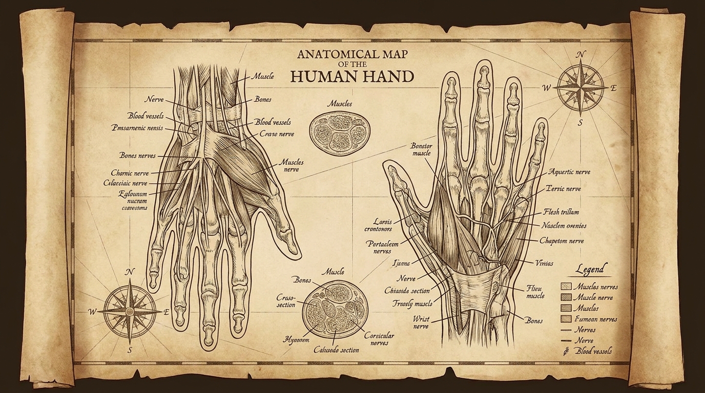
Starting from your fingertip and moving toward your palm, you have three joint regions:
- Distal Interphalangeal (DIP) joints: Located closest to the fingernails (outermost knuckles).
- Proximal Interphalangeal (PIP) joints: The middle knuckles of the fingers.
- Metacarpophalangeal (MCP) joints: The large base knuckles where fingers meet the palm.
Osteoarthritis in the hands characteristically targets the DIP joints (closest to the fingernails) and the base of the thumb. When you see the classic, hard bony nodules on older patients’ outermost knuckles — known clinically as Heberden’s nodes — that is strictly osteoarthritis at the DIP joints. A similar bony growth at the middle knuckle is called a Bouchard’s node.
Symptomatic hand osteoarthritis impacts roughly 10% of older adults — demonstrating how decades of micro-traumas severely concentrate in the outermost finger joints.
Rheumatoid arthritis follows an almost opposite pattern. It rarely affects the DIP joints. Instead, RA strongly targets the PIP joints (middle knuckles), the MCP joints (base knuckles), and the wrists. In advanced, untreated RA, patients may develop severe visible deformities, such as ulnar deviation (fingers leaning toward the pinky side) or “swan neck” deformities. If you are experiencing spongy, painful swelling at the middle knuckles and wrists symmetrically, the clinical suspicion for an inflammatory condition rises significantly.
Load-Bearing Joints: Hips & Knees
Load bearing joint pain in the hips and knees represents the classic hallmark presentation of osteoarthritis. These joints absorb the cumulative mechanical forces of walking, standing, and carrying body weight across decades of daily life. The physics of joint loading are unforgiving: every single pound of excess body weight places roughly four pounds of added mechanical stress on the knee joints during a standard walking stride. By age 60, the cartilage in these joints has endured an extraordinary volume of compressive force.
Hip pain in osteoarthritis typically presents as a deep, aching discomfort internally in the groin or front of the thigh that worsens after prolonged standing or walking long distances. Knee OA produces similar patterns: a grinding, popping sensation (crepitus) that builds with physical activity, alters your walking gait, and eases with rest.
It is critical to distinguish mechanical cartilage loss from soft tissue inflammation. For instance, conditions presenting as hip pain arthritis vs bursitis are frequently confused. Bursitis produces sharp outer hip pain, especially when lying on that side in bed, whereas true hip joint osteoarthritis typically causes deep internal groin pain.
Medical Diagnosis & Testing
While self-monitoring is valuable preparation, a definitive diagnosis requires specialized medical evaluation. Rheumatologists utilize a structured clinical process, beginning with your patient history and moving to targeted blood tests for inflammatory arthritis and advanced imaging to confirm the exact nature of your joint damage. Translating this medical jargon helps you better understand your own lab portals. Ultimately, relying entirely on physical symptoms without laboratory confirmation drastically increases the risk of clinical misdiagnosis.
Our observation of clinical diagnostic pathways shows that patients who arrive understanding these exact testing mechanisms suffer far less anxiety while awaiting their laboratory results.

Clinical History & Physical Exam
The diagnostic process always begins with you. The structured data you collected using the T.A.S.K. Joint Assessment feeds directly into the clinical history step, giving the physician actionable timelines and symptom patterns.
During the physical examination, your rheumatologist will systematically palpate (press on) each affected joint using their hands. They frequently utilize formal scoring systems like the DAS28 (Disease Activity Score measuring 28 specific joints) to count exactly how many joints are tender or swollen. This physical assessment reveals qualitative information that no blood test can replicate.
They are feeling for specific anatomical textures. “Boggy” or soft swelling — a spongy, fluid-filled quality when pressing on the joint capsule — indicates active synovial effusion and strongly suggests inflammatory arthritis. Conversely, hard, solid, bony enlargements around the joint margins suggest osteophytes (bone spurs), which are highly characteristic of mechanical osteoarthritis.
Blood Tests: ESR, CRP, Antibodies
Blood tests are where the distinction between mechanical and autoimmune joint disease becomes biochemically measurable. Understanding what each test measures clarifies your clinical standing when you read your patient portal:
- Erythrocyte Sedimentation Rate (ESR): A test measuring how quickly red blood cells settle to the bottom of a test tube. Systemic inflammation causes liver proteins in the blood to make cells clump and fall faster. An elevated esr crp arthritis reading signals significant systemic inflammation occurring somewhere in the body.
- C-Reactive Protein (CRP): A highly sensitive protein produced exclusively by the liver in rapid response to systemic inflammation. According to guidelines on how CRP blood tests measure systemic inflammation, the CRP test helps doctors directly measure the intensity of active inflammatory flare-ups (MedlinePlus, 2023).
- Rheumatoid Factor (RF): An autoantibody produced by your own immune system that mistakenly targets normal body tissue (specifically IgG proteins). According to guidelines for detecting rheumatoid factor autoantibodies, RF is frequently found in the blood of patients with rheumatoid arthritis and is used to definitively differentiate autoimmune conditions from mechanical wear (MedlinePlus, 2023).
- Anti-CCP Antibodies: Considered highly specific for rheumatoid arthritis, these autoantibodies (Cyclic Citrullinated Peptides) can appear in the bloodstream years before outward clinical symptoms even develop.
Up to 20% of rheumatoid arthritis patients test negative for standard autoantibodies (NCBI, 2023) — meaning perfectly normal blood work cannot entirely rule out severe inflammatory disease without further imaging.
Patients with osteoarthritis typically show completely normal blood work because localized mechanical cartilage degradation does not trigger systemic blood inflammation. However, the patients mentioned above have “seronegative” rheumatoid arthritis, meaning they suffer from severe soft-tissue inflammation but lack the classic RF autoantibodies in their blood, requiring advanced imaging to confirm.
Imaging: X-Ray vs. MRI
Different imaging technologies visualize entirely different types of anatomical tissue, making the choice between x-ray vs mri for arthritis diagnostically significant.
X-Rays are highly effective for detecting late-stage, bone-level changes and structural history. In osteoarthritis, X-rays clearly reveal joint space narrowing (the dark gap between bones shrinking as invisible cartilage is lost), subchondral bone sclerosis (hardening), and osteophytes. However, standard X-rays cannot visualize soft tissue inflammation at all, making them poor tools for catching early-stage rheumatoid arthritis.
MRI (Magnetic Resonance Imaging) and musculoskeletal Doppler ultrasound are the preferred modern tools for early inflammatory evaluation. These technologies visualize soft tissue and active blood flow in extraordinary detail. An MRI can detect active synovitis (inflammation of the joint lining) and tiny early bone erosions at the joint margins long before permanent, irreversible bone damage becomes visible on a standard X-ray.
Synovial Fluid Analysis
In diagnostically challenging cases where blood work and imaging remain inconclusive, rheumatologists may perform a procedure called arthrocentesis to achieve a joint aspiration diagnosis. This involves using a sterile needle, typically under local anesthetic, to draw a small amount of synovial fluid directly from the swollen joint capsule.
Laboratory analysis of this fluid yields highly specific diagnostic information. Normal joint fluid is clear and highly viscous (thick like egg whites). In inflammatory arthritis, the fluid often becomes cloudy, yellow, and watery, with drastically elevated white blood cell counts (often over 2,000 cells per cubic millimeter). Crystal analysis under a polarized microscope can also quickly confirm or rule out gout (uric acid crystals) — a condition that perfectly mimics inflammatory arthritis but requires entirely different targeted medication.
When to Seek Immediate Care
Self-monitoring is a valuable preparation tool, but it carries real risks when it replaces rather than supports professional medical evaluation. Certain symptoms cross the line from chronic discomfort into medical urgency, requiring immediate specialist intervention to preserve long-term joint function and overall health.
Common Pitfalls in Self-Diagnosis
Many patients delay seeking care for months or years because they attribute persistent joint swelling and stiffness to “just getting older.” While minor joint stiffness is a natural consequence of aging, symmetrical swelling, systemic fatigue, and morning stiffness lasting beyond 30 minutes are clinical warning signs, not normal aging.
Another major pitfall is the long-term self-medication with over-the-counter NSAIDs (like ibuprofen). Regular use of these medications can severely mask the symptoms of inflammatory arthritis without slowing the underlying disease progression. According to clinical criteria for RA progression, untreated inflammatory arthritis frequently leads to irreversible structural joint erosion within the first two years of active disease (NCBI, 2023).
Irreversible joint erosion frequently occurs within the first two years of untreated rheumatoid arthritis — making early specialist intervention absolutely critical for long-term mobility.
Red Flag Symptoms Requiring Prompt Care
Contact a healthcare provider or seek urgent evaluation if you experience any of the following acute presentations:
- Sudden onset of severe joint swelling in one or more joints without preceding physical injury — particularly if the massive swelling developed rapidly over hours rather than days.
- A joint that is hot to the touch and visibly red, accompanied by a fever above 38°C (100.4°F). According to CDC guidelines on severe joint pain, this specific combination may indicate septic arthritis (a severe joint infection), which is a medical emergency requiring immediate antibiotic treatment (CDC, 2023).
- Inability to bear weight on a lower limb joint, or complete, sudden loss of range of motion.
- New joint symptoms developing rapidly during or immediately after a recent viral or bacterial illness, which can trigger reactive arthritis.
Frequently Asked Questions
What autoimmune disease mimics arthritis?
Several autoimmune diseases mimic standard arthritis, most notably lupus (SLE) and psoriatic arthritis. These conditions cause severe joint pain and swelling that feel clinically identical to rheumatoid arthritis but stem from entirely different specific immune dysfunctions. For instance, psoriatic arthritis frequently presents alongside visible skin rashes and nail pitting, while lupus may present with facial rashes and kidney involvement. A rheumatologist must conduct specialized autoantibody blood tests to accurately differentiate these mimicking conditions from standard joint degradation.
Can inflammatory arthritis go away?
Inflammatory arthritis is generally a chronic condition that does not permanently go away on its own. However, modern medical treatments — including disease-modifying antirheumatic drugs (DMARDs) and biologic therapies — can induce a state called clinical remission, where symptoms completely disappear and joint damage halts. Early intervention within the first six months of symptom onset drastically increases the probability of achieving total remission. Even when a patient feels entirely symptom-free, long-term monitoring by a specialist remains absolutely necessary.
What age does inflammatory arthritis start?
Inflammatory arthritis can start at any age, but most commonly presents in adults between the ages of 30 and 50. This starkly contrasts with osteoarthritis, which typically develops after age 50 due to decades of accumulated mechanical wear. Furthermore, certain forms of autoimmune joint disease, such as Juvenile Idiopathic Arthritis, exclusively affect children under the age of 16. Regardless of age, persistent joint swelling lasting longer than six weeks requires immediate medical evaluation.
How do you know if you have osteoarthritis or inflammatory arthritis?
You can differentiate the two conditions by observing morning stiffness duration and systemic warning signs. Osteoarthritis stiffness typically resolves within 30 minutes of waking and progressively worsens with physical exertion throughout the day. Conversely, inflammatory arthritis stiffness lasts longer than an hour, paradoxically improves with gentle movement, and is often accompanied by whole-body fatigue. Only clinical blood tests and specialized imaging ordered by a physician can definitively confirm the diagnosis.
How do I know if I have inflammation or arthritis?
Arthritis literally means joint inflammation, making the two concepts inherently linked rather than separate conditions. Temporary joint inflammation from an acute injury, such as a sports sprain, will heal and subside completely within a few weeks as the tissue repairs itself. If swelling, heat, and pain persist in a joint for more than six weeks without a clear physical injury, this strongly indicates chronic clinical arthritis. Chronic inflammation requires professional medical evaluation to prevent permanent structural damage to the joint cartilage.
Conclusion
When determining the difference between inflammatory joint pain vs osteoarthritis, the critical distinction lies in the underlying biology: localized mechanical cartilage degradation versus a systemic autoimmune response. Because rheumatoid arthritis typically causes severe morning stiffness lasting over an hour compared to osteoarthritis resolving within 30 minutes (NCBI, 2023), tracking symptom timing is highly critical. The safest approach is utilizing structured symptom diaries to assist rheumatologists in ordering accurate blood panels required for a definitive diagnosis.
The T.A.S.K. Joint Assessment — tracking Timing, Anatomy, Symmetry, and Kinetics — exists precisely because doctors cannot diagnose what they cannot observe, and most patients cannot report what they have not systematically tracked. A seven-day diary completed before your appointment transforms a vague conversation about “joint pain” into a highly structured clinical data set that enables faster, more accurate medical decisions.
Your next step is concrete: start your T.A.S.K. diary today. Record your morning stiffness duration, the exact affected joint locations, and whether movement relieves or worsens your pain. Take that structured record to a rheumatologist or your primary care physician and request a formal referral for specialist evaluation. Armed with this clinical data, you are fully prepared to have a highly productive medical consultation that leads directly to accurate treatment.