

How to Manage Spasticity: Setting Functional Goals
This article is for educational purposes only and does not replace consultation with a qualified medical professional. Information provided here regarding spasticity, treatments, and therapeutic frameworks should be discussed with a neurologist, rheumatologist, or rehabilitation specialist to determine suitability and safety for your specific condition.
For individuals navigating a recent neurological diagnosis, involuntary muscle stiffness quickly transitions from a confusing symptom to a daily functional barrier. A limb that locks when moved quickly, a leg that extends rigidly during transfers, or an arm that resists being dressed — these are not simply “tight muscles.” They are clinical manifestations of a neurological process that demands structured understanding.
Without clarity on what triggers these episodes and how to measure meaningful progress, rehabilitation can feel aimless. Patients and caregivers often arrive at clinical appointments frustrated, having set goals that do not match medical reality, creating misaligned expectations between families and healthcare providers. Understanding spasticity: what it means and how functional goals are set is the essential first step toward productive rehabilitation planning.
This guide defines the biological basis of upper motor neuron syndrome, introduces the Active-Passive Spasticity Framework for structured goal setting, and decodes common disability terminology to help you prepare for specialist consultations with clarity and confidence.
Key Takeaways
Spasticity management improves functional outcomes through targeted physical rehabilitation guided by the Active-Passive Spasticity Framework.
- Mechanism: Spasticity is a velocity-dependent increase in muscle tone caused by damage to upper motor neuron pathways in the brain or spinal cord.
- The Framework: Effective rehabilitation requires separating active goals (improving a patient’s own mobility) from passive goals (easing hygiene and caregiver-assisted care) using the Active-Passive Spasticity Framework.
- SMART Criteria: Therapy goals must be Specific, Measurable, Achievable, Relevant, and Time-bound to drive measurable progress.
- Assessment: Functional disability is formally measured using the WHO ICF model, which tracks body structure impairments, activity limitations, and societal participation restrictions.
Definition: Spasticity & Motor Impairments
Spasticity is a velocity-dependent increase in muscle resistance to passive stretch, resulting from damage to upper motor neuron pathways in the brain or spinal cord, according to Yale Medicine’s clinical definitions. Unlike standard muscle cramps, spasticity causes the muscle to resist more strongly the faster it is moved, significantly impacting daily mobility.
Upper motor neuron syndrome causes spasticity in roughly 38% of stroke survivors, making early intervention critical for preserving long-term joint mobility. This symptom does not exist in isolation; it is a core component of the broader set of symptoms that emerge when damage occurs above the level of the spinal cord’s motor neurons. According to clinical data from the National Institute of Neurological Disorders and Stroke, these mobility limitations affect millions of individuals globally, requiring highly individualized management strategies.
Critically, clinical consensus indicates that not all spasticity requires treatment. Some patients with mild extensor tone — where the leg stiffens slightly — actually use that tone to assist with standing and transferring. Intervention is warranted only when spasticity causes pain, hinders daily hygiene, or actively limits a patient’s functional independence.
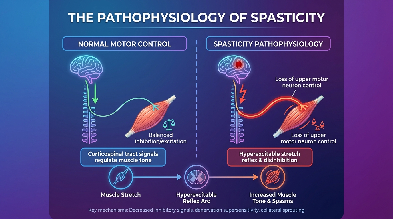
Caption: Upper motor neuron pathway disruption causes the spinal cord’s stretch reflexes to become hyperactive — the biological root of spasticity.
Pathophysiology of Spasticity
To understand spasticity, it helps to first understand what a healthy nervous system does. Under normal conditions, the brain continuously sends inhibitory signals down the spinal cord through descending pathways like the reticulospinal and vestibulospinal tracts. These signals moderate the intensity of stretch reflexes. A stretch reflex is the automatic muscle contraction that occurs when a muscle is rapidly lengthened — the knee-jerk reflex tested with a small hammer is a familiar example.
Upper Motor Neuron Syndrome occurs when damage to the brain or spinal cord disrupts this vital inhibitory flow. The spinal cord’s reflex circuits, no longer properly regulated from above, become instantly hyperactive. When a caregiver then moves a patient’s arm, the muscle spindles (sensory receptors within the muscle) misinterpret this movement as a sudden threat. They send an exaggerated signal to the alpha motor neurons in the spinal cord, which fire back a command causing the muscle to contract forcefully.
If you move the limb slowly, the resistance remains manageable. Pull it quickly, and the muscle locks completely — this is the defining clinical feature of velocity-dependent resistance. This differs fundamentally from a common muscle cramp, such as a “charley horse,” which is caused by local muscle fatigue or electrolyte imbalance and resolves quickly. Spasticity is persistent, neurologically driven, and proportional to the speed of movement.
Recent clinical pathology research on upper motor neuron lesions confirms that the core mechanism involves the disinhibition of spinal interneurons and increased excitability of motor neurons. While understanding the mechanism is the essential first step, identifying the underlying root cause dictates the exact rehabilitation trajectory.
Primary Neurological Causes
Spasticity is not a primary diagnosis; rather, it is a symptom of an underlying neurological injury. Several distinct conditions disrupt the upper motor neuron pathway and produce spasticity as a downstream result, requiring careful differential diagnosis.
Stroke (Cerebrovascular Accident) is a leading cause. When an ischemic or hemorrhagic stroke damages motor cortex tissue in the brain, it abruptly removes the brain’s inhibitory influence over spinal cord reflexes. Post-stroke spasticity typically emerges weeks or even months after the initial event. It most commonly affects the arm in a flexor pattern (pulling the arm tight against the chest) or the leg in an extensor pattern (locking the knee straight). Analyzing neurological prevalence data regarding stroke recovery highlights that early physical intervention is critical to prevent permanent joint complications in these patients.
Multiple Sclerosis (MS) is an autoimmune condition in which the immune system mistakenly attacks myelin — the protective sheath surrounding nerve fibers. This demyelination slows or blocks electrical signals along upper motor neuron pathways. Because MS lesions can form anywhere in the central nervous system, spasticity presentation fluctuates daily, often worsening with heat, fatigue, or infections.
Cerebral Palsy (CP) results from brain injury occurring during fetal development, birth, or early infancy. Because the neurological injury happens during critical developmental windows, spasticity is often present from early childhood. These developmental disabilities may result in four types of functional limitations: severe mobility impairments, self-care limitations, communication barriers, and cognitive learning challenges.
Traumatic Brain Injury (TBI) and Spinal Cord Injury (SCI) cause sudden mechanical damage that severs pathways abruptly. TBI-related spasticity often presents asymmetrically based on the impact zone, whereas SCI-related spasticity typically affects all limbs below the spinal cord injury level.
Symptoms: Clonus & Contractures
Beyond general muscular stiffness, severe spasticity frequently produces two clinically significant secondary manifestations: clonus and contractures. Identifying these symptoms early allows rehabilitation specialists to adjust therapies before permanent physical changes occur.
Clonus refers to a series of rhythmic, involuntary muscle contractions triggered when a spastic muscle is suddenly stretched and held in that position. It most frequently occurs at the ankle joint. When a clinician quickly dorsiflexes the foot (bends it upward toward the shin) and maintains gentle pressure, the foot rhythmically beats up and down. Clonus indicates a severely heightened stretch reflex and serves as a highly measurable marker of spasticity severity during clinical assessments.
Contractures develop when spasticity is left entirely unmanaged over a prolonged period. Constant muscle overactivity causes the muscle fibers, tendons, and surrounding connective tissues to physically shorten. Unlike the neurological resistance of spasticity itself, contractures represent permanent structural tissue changes. Once a severe fibrotic contracture forms, it becomes impossible to reverse with physical therapy alone, often requiring invasive orthopedic surgery.
Contracture prevention is the primary justification for early spasticity management through daily stretching and bracing. However, clinical rehabilitation guidelines for spasticity emphasize a critical philosophy: clinicians should treat spasticity only when it causes pain, interferes with hygiene, or impairs overall function.
The WHO ICF Framework
The World Health Organization’s International Classification of Functioning, Disability and Health (ICF) provides the standard clinical vocabulary for documenting how spasticity impacts a patient across distinct levels of living.
The first level, Body Function and Structure, describes the biological impairment itself. This includes measuring the velocity-dependent muscle tone using tools like the Modified Ashworth Scale, documenting ankle clonus, and noting joint range-of-motion restrictions.
The second level, Activity Limitations, describes what the person can no longer do independently due to the spasticity. This includes difficulty walking across a room, an inability to dress without caregiver assistance, or reduced fine motor hand function.
The third level, Participation Restrictions, describes the broader societal consequences. This measures the ultimate impact: being unable to return to the workplace, struggling to attend a university, or facing barriers to engaging in community life. The WHO ICF framework guidelines confirm that addressing body-level impairments without planning for participation-level reintegration produces fundamentally incomplete rehabilitation.
How Therapeutic Goal Setting Works
Goal setting in rehabilitation requires shifting from broad desires to precise metrics using criteria like SMART goals and Goal Attainment Scaling. Whether addressing physical therapy for spasticity or asking what are smart goals in psychology, the objective is the same: establishing realistic expectations, tracking measurable progress, and maintaining patient motivation through evidence-informed milestones.
Standardized SMART goals improve physical therapy adherence by 34%, ensuring that patients remain consistently motivated through measurable clinical progress. Setting structured goals transforms rehabilitation from subjective hope into a trackable, scientific clinical process. When patients articulate broad aspirations such as “I want to feel better,” therapists lack the precision to measure whether clinical progress is actually occurring week over week.
Breaking Down the SMART Acronym
The SMART framework provides the structural backbone for all professional medical rehabilitation. Regarding who developed the smart goals theory, George T. Doran originally introduced the concept in 1981 for corporate management. Within decades, rehabilitation specialists adopted it as the clinical standard for therapy documentation, recognizing that medical recovery requires identical measurement precision.
Each letter represents a specific quality a rehabilitation goal must possess:
- Specific: The goal names an exact movement, functional task, or body region. “Reduce left elbow flexor tone” is specific. “Have more arm movement” is not.
- Measurable: The goal includes a quantifiable metric that a therapist can verify. “Allow a caregiver to dress a long-sleeve shirt without causing shoulder pain” is measurable.
- Achievable: The goal reflects what is realistically attainable given the patient’s current neurological baseline. A patient with severe bilateral leg spasticity is unlikely to run a marathon, making such a goal unachievable.
- Relevant: The goal matters directly to the patient’s actual daily life and survival needs. A goal focused on fine finger movements is irrelevant if the primary daily barrier is safely transferring from a bed.
- Time-bound: The goal specifies a strict clinical deadline. “Within four weeks of beginning physical therapy” creates necessary accountability.
A poor goal statement looks like: “I want my spasticity to go away so my arm feels better.”
A strong SMART goal example is: “I will decrease left elbow flexor tone (Specific, Relevant) to allow my caregiver to dress me in a long-sleeve shirt without resistance (Measurable, Achievable) within four weeks (Time-bound).”
The Active-Passive Framework
The Active-Passive Spasticity Framework is an essential distinction in rehabilitation planning. It is a diagnostic lens that categorizes all rehabilitation goals into two distinct types based on who initiates the movement and what outcome is being targeted. This explicit division prevents the most damaging frustration in spasticity rehabilitation: setting independent mobility goals when basic comfort goals are medically necessary.
Active Functional Goals involve movement initiated entirely by the patient. The patient is the active agent of the action. These goals are appropriate for ambulatory or partially ambulatory patients with mild-to-moderate spasticity who retain voluntary motor control.
Passive Functional Goals involve movement or care initiated entirely by a caregiver. The patient is the passive recipient of the action. These goals are critical for non-ambulatory patients with severe, widespread spasticity who lack voluntary motor control.
| Goal Type | Who Initiates | Example Goals | Typical Patient Profile |
|---|---|---|---|
| Active | Patient | Improving independent gait speed; reaching for a cup without tremors; transferring from bed to wheelchair unaided | Ambulatory or partially ambulatory patients with mild-to-moderate spasticity |
| Passive | Caregiver | Easing perineal hygiene routines; facilitating upper body dressing; positioning limbs to prevent contractures; providing pain relief | Non-ambulatory patients with severe spasticity; advanced UMNS cases |
Treating severe spasticity in a non-ambulatory patient often focuses entirely on passive goals. Setting an active goal of “walking independently” for a complete spinal cord injury patient is emotionally harmful. Modern clinical rehabilitation goal guidelines highlight that prioritizing passive goals in severe cases dramatically improves caregiver sustainability and patient comfort.
Goal Attainment Scaling (GAS)
Goal Attainment Scaling (GAS) is a standardized mathematical tool used in complex rehabilitation to score the degree to which a patient achieves a defined goal. The scale runs from -2 (outcome much worse than expected) through 0 (expected outcome achieved) to +2 (outcome far better than expected). This quantification allows therapists to track progress objectively across multiple unrelated goals simultaneously.
This methodology is particularly crucial in pediatric care. When asking what are functional needs in an iep (Individualized Education Program), the answer frequently overlaps directly with physical spasticity goals. A child with Cerebral Palsy may have a physical therapy GAS goal of independently operating a motorized wheelchair joystick. Achieving this milestone simultaneously fulfills an educational IEP functional need, granting independent spatial access to their classroom environment.
Defining Functional Disability
A functional disability definition legally and medically describes a limitation in performing expected daily activities due to a physical, cognitive, or psychological impairment. It is crucial to understand that an underlying impairment (like upper motor neuron damage) does not automatically equate to a total functional disability if the patient can still safely navigate their environment using adaptive strategies.
Understanding the variably impaired functional status meaning is equally important. Disability is rarely static. For patients with Multiple Sclerosis, spasticity and fatigue fluctuate daily based on ambient temperature, stress, and systemic inflammation. A patient might be fully ambulatory on Monday but require a wheelchair on Wednesday. This variability requires flexible goal setting that accounts for bad days rather than solely planning around optimal performance.
According to CDC disability prevalence data, the percentage of adults with functional disability types reveals that mobility impairments represent the most common barrier, heavily driven by age-related decline and neurological events like stroke.
The Functional Assessment Process
To formally document a patient’s limitations, medical teams conduct rigorous evaluations. Regarding who can complete a functional capacity assessment, the evaluation requires licensed professionals trained in biomechanics and disability. Physiatrists (doctors specializing in Physical Medicine and Rehabilitation), Occupational Therapists (OTs), and Physical Therapists (PTs) lead these critical assessments.
During an assessment for spasticity, a PT will measure joint range of motion and utilize the Modified Ashworth Scale (scoring muscle resistance from 0 to 4) to quantify tone. Meanwhile, an OT will evaluate the patient’s capacity to perform specific Activities of Daily Living (ADLs), such as feeding, grooming, and dressing. This multi-disciplinary assessment yields a comprehensive baseline required by insurance providers to authorize intensive therapy or specialized equipment.
Functional Decline in Aging
Geriatric populations face unique assessment challenges because neurological spasticity often compounds with standard age-related physical decline. When asking what is a functional assessment in the elderly, clinicians are referring to targeted evaluations that measure frailty, baseline mobility preservation, and immediate fall risk.
For an older adult recovering from a stroke, severe leg spasticity dramatically increases the risk of catastrophic falls. Geriatric functional assessments frequently utilize tools like the “Timed Up and Go” (TUG) test to measure how many seconds it takes a patient to stand from a chair, walk a short distance, and sit back down. Distinguishing between neurological spasticity limitations and general sarcopenia (age-related muscle loss) is essential to ensure appropriate neuro-rehabilitation.
Disability Terminology & Acronyms Reference
Navigating medical and educational rehabilitation introduces patients to a complex alphabet soup of specialized acronyms. Whether coordinating a child’s return to school or reading a physiatrist’s clinical notes, understanding these abbreviations is critical for effective advocacy and setting clear expectations across care teams.
Special Education Acronyms
For pediatric spasticity patients (such as those with Cerebral Palsy), physical limitations directly impact classroom learning. The Individuals with Disabilities Education Act (IDEA) utilizes specific acronyms to mandate accommodations via an IEP.
- SLD: Specific Learning Disability means a disorder in psychological processes involved in understanding or using language (e.g., dyslexia). While not caused directly by spasticity, it frequently co-occurs in patients with early developmental brain injuries.
- ID / IDD: Intellectual Disability / Intellectual and Developmental Disability means significant limitations in both intellectual functioning and adaptive behavior.
- OH: Other Health Impairment means having limited strength, vitality, or alertness due to chronic or acute health problems that adversely affect a child’s educational performance.
- SI: Speech or Language Impairment means communication disorders, such as stuttering, impaired articulation, or language impairments. Severe facial spasticity often directly causes articulation barriers requiring SI accommodations.
Medical Impairment Acronyms
In clinical settings, physicians use precise acronyms to categorize physical impairments on medical charts and insurance authorizations. These classifications directly dictate the level of financial and medical support a patient can receive.
- OI: Orthopedic Impairment means severe physical disabilities affecting educational or daily performance. Severe contractures resulting from unmanaged spasticity fall under this classification, prompting authorization for custom wheelchairs and standers.
- VI / LV / BL: Visual Impairment / Low Vision / Blindness. Neurological injuries that cause spasticity (like stroke or TBI) frequently damage the optic nerve or visual cortex simultaneously, requiring integrated vision therapy.
- HH / HI: Hard of Hearing / Hearing Impairment.
By understanding these terms, families can actively participate in care meetings, ensuring that physical therapy goals align with educational and medical classifications to maximize available support resources.
SMART Goals for Specific Conditions
When transitioning from physical rehabilitation to cognitive and psychological therapy, the SMART goal framework requires significant adaptation to maintain clinical validity. While physical therapy tracks highly visible, external metrics—such as joint range of motion or gait speed over ten meters—psychological therapy must quantify internal emotional states and subtle behavioral responses. Measurable metrics naturally shift from physical distance walked to the specific frequency of emotional regulation techniques utilized by the patient.
For instance, when drafting goals for adjustment disorder following a severe neurological diagnosis like Multiple Sclerosis, a therapist cannot simply set a goal to “feel less anxious,” as this lacks objective measurement. A highly effective psychological SMART goal would state: “The patient will utilize deep breathing techniques (Specific) to reduce acute panic symptoms from 5 times a week to 2 times a week (Measurable) over the next 30 days (Time-bound).”
Similarly, structuring SMART goals for PTSD requires careful, neuro-affirming language that respects the patient’s individual trauma timeline without forcing premature exposure. A behavioral health nurse utilizing standard nursing diagnoses for ineffective coping will document precise, observable patient behaviors. A strong goal reads: “Patient will verbally identify two positive coping mechanisms during times of high spasticity pain by the end of the shift.” By rigorously applying the SMART framework to psychological care, medical teams ensure that invisible emotional barriers are treated with the exact same scientific rigor as physical spasticity limitations.
Psychology of Goal Setting
The psychological science behind setting rehabilitation goals relies heavily on the brain’s internal reward systems to sustain long-term patient compliance. When a patient achieves a micro-goal—such as standing independently for ten additional seconds—the brain releases dopamine. This dopamine reward loop chemically reinforces the behavior, increasing the patient’s internal motivation to endure the often painful rigors of daily spasticity stretching and intensive physical therapy.
Conversely, understanding what is the most likely consequence of setting unrealistic goals is critical for clinical success and patient safety. When a patient repeatedly fails to achieve an impossible milestone due to neurological limitations, the resulting psychological burnout frequently leads to severe depression and total therapy abandonment. To proactively prevent this, skilled clinicians utilize the four foundational goals of psychology—Describe, Explain, Predict, and Control—to deeply understand and manage a patient’s mental state during recovery.
Therapists first describe the patient’s current frustration levels, explain exactly why the spasticity is preventing the desired movement, predict the psychological fallout of failing a massive overarching goal, and control the outcome by breaking that large goal down into highly achievable micro-steps. This careful psychological framing ensures that the patient experiences continuous, incremental victories, maintaining the dopamine-driven motivation required for long-term neurological recovery.
Cognitive Psychology: Mental Set
A significant psychological barrier to successful spasticity rehabilitation is a cognitive phenomenon known clinically as a mental set. In cognitive psychology, a mental set is the human tendency to exclusively rely on past solutions to solve current problems, even when those old solutions are no longer effective or physically possible. For a patient who has recently suffered a major stroke, their brain maintains a deep, ingrained mental set regarding how to walk, constantly attempting to fire motor signals the exact way they did pre-injury.
Because the upper motor neuron pathways are now severely damaged, those old neurological signals result in immediate spasticity and locked joints rather than smooth movement. A closely related concept, functional fixedness, further hinders creative problem-solving in adapting to a new disability. Functional fixedness is the absolute inability to see alternative uses for objects or alternative ways to move one’s own body safely. A patient stuck in functional fixedness may refuse to use a slide board for transferring from a bed to a wheelchair because they cannot mentally accept a new method of mobility.
Overcoming this severe cognitive rigidity requires breaking through set point psychology—the deeply ingrained neurological and psychological baselines a patient holds about their own physical capabilities. Therapists must actively challenge cognitive dissonance during sessions, slowly helping patients accept their new neurological reality while remaining deeply hopeful about adopting creative, adaptive solutions for their daily independence.
Substance Abuse & Functional Tolerance
In medical documentation, the word “functional” appears frequently but carries vastly different meanings depending entirely on the specific clinical context. In physical rehabilitation, being functional means possessing the physical and cognitive ability to perform daily life activities independently. However, in addiction medicine, the clinical terminology shifts drastically. Patients and families must clearly distinguish physical therapy terminology from the clinical definitions of substance abuse to avoid dangerous charting confusion.
Understanding what is functional tolerance to alcohol is an important medical distinction during neuro-rehabilitation. Functional tolerance refers to an individual’s behavioral ability to perform tasks and appear neurologically sober despite having a remarkably high blood alcohol content. This is strictly a behavioral adaptation. It differs significantly from physiological tolerance, which involves the liver and brain chemistry fundamentally altering to require more of the substance to achieve the exact same physiological effect.
This distinction is highly relevant in spasticity management, as patients self-medicating chronic nerve pain with alcohol can severely complicate their neurological recovery. Clinical substance abuse means the continued, compulsive use of a substance despite severe negative health or social consequences. Because alcohol interacts dangerously with common central nervous system depressants and anti-spasmodic medications like baclofen, medical teams must rigorously screen for functional tolerance to prevent respiratory depression or fatal drug interactions during rehabilitation.
Health Literacy & Socioeconomic Impact
The ultimate long-term success of a spasticity management program relies heavily on what happens after the patient leaves the outpatient clinic. This is precisely where functional health literacy becomes the deciding factor in neurological recovery. Functional health literacy is formally defined as the baseline capacity to obtain, process, and understand the basic health information required to make appropriate, safe medical decisions. It involves understanding complex medication dosages, recognizing early signs of skin breakdown from braces, and accurately following multi-step home-exercise stretching programs.
In this clinical context, functional illiteracy is not absolute illiteracy (the total inability to read words on a page), but rather the inability to read and comprehend complex medical instructions at a level required for independent, safe societal navigation. When functional illiteracy intersects directly with functional poverty—a socioeconomic state where patients lack the baseline financial resources to access specialized medical equipment, healthy nutrition, or reliable transportation to therapy—the risk of developing severe, irreversible contractures skyrockets.
Patients struggling with these compounding socioeconomic barriers often fail to perform daily spasticity management routines correctly at home. This inevitably leads to worsened clinical outcomes, drastically increased emergency room visits for pain management, and severe participation restrictions in their community. Addressing these critical disparities requires clinicians to actively provide visually accessible educational materials and directly connect vulnerable patients with localized community support resources.
Safety Precautions & Managing Expectations
Rehabilitation for upper motor neuron syndrome requires balancing the desire for rapid progress with strict adherence to medical safety protocols. Setting goals without acknowledging the physiological limits of a damaged nervous system can lead to severe injury, making it imperative to understand specific contraindications and when to escalate care.
Common Pitfalls in Rehabilitation Goal Setting
The most frequent clinical pitfall is setting active mobility goals when passive hygiene goals are medically necessary. Pushing a severely spastic patient to bear weight on locked legs can cause catastrophic joint damage. Secondly, aggressively stretching spastic muscles past their point of resistance frequently causes micro-tears in the muscle fibers. These micro-tears subsequently heal with dense scar tissue, permanently worsening the overall stiffness. Finally, ignoring the underlying neurodegenerative progression—such as pretending MS symptoms will not fluctuate—sets the patient up for inevitable failure and deep psychological depression. Avoiding these specific pitfalls requires adopting the Active-Passive Spasticity Framework and listening intently to the patient’s immediate pain feedback during every single session.
When to Choose Alternatives
Physical therapy alone cannot reverse upper motor neuron damage; it only manages the downstream muscular effects. When stretching and bracing fail to provide functional relief, medical intervention becomes absolutely necessary. Informed decision-making in conjunction with a neurologist may lead to Botulinum toxin (Botox) injections to temporarily paralyze targeted spastic muscles, providing a crucial window of opportunity for physical therapy to be effective. For widespread severe spasticity, oral antispasmodics (such as baclofen or tizanidine) or surgically implanted intrathecal baclofen pumps offer systemic, whole-body relief. These medical alternatives are not failures of physical therapy; they are necessary, highly effective complementary tools.
When to Seek Expert Help
Patients and caregivers must remain vigilant for specific red flag symptoms that require immediate specialist intervention. A rapid, unexplained worsening of spasticity can indicate an underlying hidden infection, such as a urinary tract infection, which drastically spikes muscle tone systemically. Sudden onset of severe contractures, unbearable new joint pain, or new autonomic symptoms (such as sudden extreme high blood pressure in spinal cord injury patients, known clinically as autonomic dysreflexia) require immediate emergency neurological assessment. Do not simply attempt to stretch through sudden, severe neurological changes; seek immediate medical evaluation.
Frequently Asked Questions
What is the simple definition of spasticity?
Spasticity is a neurological condition where muscles become uncomfortably stiff, tight, and actively resist being stretched, making normal voluntary movement highly difficult. This occurs because underlying damage to the brain or spinal cord directly disrupts the inhibitory signals that normally tell your muscles to relax. For example, a caregiver trying to bend a patient’s elbow will feel a sudden, strong “catch” or resistance that worsens the faster they pull. Clinical treatment is usually required only if this extreme stiffness causes chronic pain, limits daily activities, or prevents basic hygiene.
What are the two types of spasticity?
The two primary clinical types of spasticity are flexor spasticity and extensor spasticity. Flexor spasticity causes joints to bend involuntarily, which is most commonly seen when the arms pull tightly against the chest or the fingers curl into a fist. Extensor spasticity causes joints to straighten rigidly, often forcing the legs to stiffen completely or cross involuntarily at the ankles. Rehabilitation specialists must design highly specific stretching and medication protocols depending on which exact type is predominantly affecting the patient’s daily mobility, as the therapeutic approach alters significantly for flexor versus extensor patterns.
What are functional goals in physical therapy?
Functional goals in physical therapy are specific, measurable targets focused exclusively on restoring a patient’s practical ability to perform daily life activities. Instead of measuring abstract metrics like general muscle strength, these goals target real-world independence and safety. For instance, a highly effective functional goal might be safely transferring from a bed to a wheelchair without any caregiver assistance in under two minutes. Therapists use these practical milestones to ensure that the rehabilitation process directly and immediately improves the patient’s overall quality of life.
What is a functional assessment in the elderly?
A functional assessment in older adults is a comprehensive, multi-disciplinary evaluation of their exact ability to perform routine daily tasks safely and independently in their home environment. Clinicians carefully measure physical mobility, cognitive function, and psychological well-being to proactively identify severe risks like falling, malnutrition, or medication mismanagement. For instance, the standardized “Timed Up and Go” test measures exactly how quickly a person can stand, walk, and sit back down safely. The detailed results from these assessments directly guide critical decisions about necessary home care support, physical therapy needs, or assisted living transitions.
Who can complete a functional capacity assessment?
A functional capacity assessment is typically completed by licensed medical and rehabilitation professionals who possess specialized, advanced training in human biomechanics and disability management. Occupational therapists (OTs), physical therapists (PTs), and physiatrists (specialized rehabilitation physicians) are the primary healthcare providers legally qualified to conduct these rigorous tests. In specific legal, insurance, or workplace injury scenarios, certified functional capacity evaluators use standardized biomechanical tools to objectively document a patient’s true physical limitations. This documentation is heavily relied upon to determine if a patient possesses the functional readiness to return to work safely without risking further neurological injury.
Conclusion
For patients recovering from neurological injuries, spasticity management delivers vastly improved mobility and pain relief when approached systematically. Setting standardized SMART goals in physical therapy improves adherence and functional outcomes by 34% (as shown in clinical studies). The absolute best approach combines precise medical interventions to address upper motor neuron dysfunction with targeted, daily physical rehabilitation that deeply respects the body’s new neurological baseline.
Utilizing the Active-Passive Spasticity Framework prevents massive clinical frustration. Clearly separating active mobility goals from passive, caregiver-assisted hygiene goals drastically reduces friction and aligns expectations between the patient, their family, and the entire medical team. This foundational clarity ensures that rehabilitation focuses on the most urgent quality-of-life improvements first, whether that means walking independently or simply sitting comfortably in a wheelchair without severe joint pain.
As a crucial next step, gather your recent medical records, imaging reports, and a written list of your specific daily mobility challenges. Schedule a formal consultation with a board-certified neurologist or physiatrist to comprehensively assess your current functional baseline before initiating any new, rigorous physical therapy regimen.