
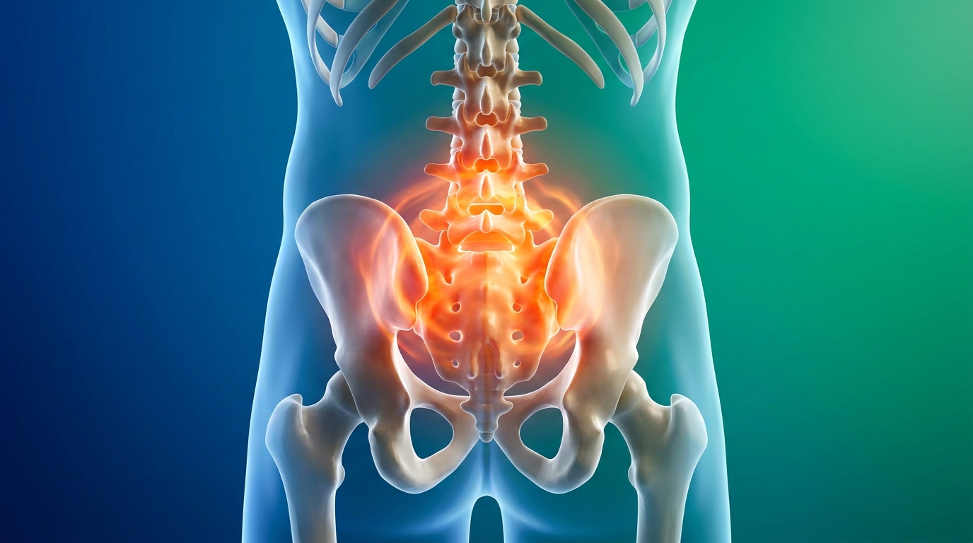
Spondyloarthritis Guide: Back Pain & Stiffness
This article is for educational purposes only and does not replace consultation with a qualified medical professional.
Waking up with severe back pain that inexplicably improves once you start moving is a hallmark sign of inflammatory joint disease, not a standard muscle strain. This distinction — between mechanical wear and immune-driven inflammation — is one of the most consequential diagnostic gaps in modern rheumatology.
Delaying evaluation for persistent morning stiffness can allow underlying inflammation to cause long-term mobility issues, effectively masking the true autoimmune nature of the condition. Uncontrolled spinal inflammation does not plateau on its own; it progresses toward structural changes that become irreversible without targeted intervention.
By the end of this guide, you will understand the clinical differences between mechanical and inflammatory back pain so you can seek appropriate rheumatological evaluation. This overview breaks down the fundamentals of spondyloarthritis, identifying distinct symptom patterns, flare timelines, and how to effectively track these metrics for your next specialist consultation.
Key Takeaways
Spondyloarthritis is a group of inflammatory diseases causing severe back pain, joint stiffness, and chronic inflammation that typically improves with physical activity.
- The 30-Minute Inflammatory Threshold: Morning stiffness lasting longer than 30 minutes strongly indicates inflammation rather than mechanical injury — a core concept this guide returns to throughout.
- Axial vs. Peripheral: Inflammation can target the spine and sacroiliac joints (axial) or outer limbs and digits (peripheral), requiring different diagnostic approaches.
- Early Evaluation: Accurate clinical charting and early MRI imaging prevent irreversible joint fusion and protect baseline function (the ability to perform everyday physical tasks without assistance).
Spondyloarthritis and Inflammatory Back Pain Fundamentals
Spondyloarthritis (SpA) encompasses a group of immune-mediated diseases characterized by chronic inflammatory back pain and severe joint stiffness. Unlike mechanical back injuries caused by heavy lifting or poor posture, inflammatory back pain stems from the immune system mistakenly attacking the body’s own spinal tissues. This fundamental difference dictates entirely different diagnostic and treatment pathways — and understanding it is the first step toward getting appropriate care.
Spondyloarthritis, a family of inflammatory rheumatic diseases, affects an estimated 1–2% of the global population, with symptom onset typically occurring between the ages of 17 and 45 (Stolwijk et al., Annals of the Rheumatic Diseases, 2016). What makes this condition particularly challenging to identify is that its earliest symptoms — dull lower back ache and morning stiffness — closely resemble far more common mechanical back problems. Many patients spend years cycling through physical therapy and general practitioners before receiving a rheumatological referral.
Inflammatory back pain typically begins before age 40 and improves with exercise, unlike mechanical back pain which worsens with activity (Sieper & Poddubnyy, The Lancet, 2017). This seemingly simple observation is clinically powerful: if a patient’s back pain reliably improves after 20–30 minutes of walking but returns after prolonged sitting or sleeping, the pain pattern points strongly toward an immune-driven origin rather than a structural or postural one.
Understanding the underlying immune mechanism is only the first step. For patients managing daily symptoms, localizing exactly where this pain manifests in the spine provides crucial information for clinical charting and specialist evaluation.
What Is Inflammatory Back Pain?
Inflammatory back pain differs from mechanical back pain in both its cause and its behavior over time. Where mechanical pain typically results from a specific physical event — a muscle tear, a herniated disc, or ligament strain — inflammatory back pain arises from the immune system producing excess inflammatory proteins (called cytokines) that attack the lining of the spinal joints and sacroiliac joints (the joints connecting the base of the spine to the pelvis).
In terms of sensation, inflammatory back pain is most often described as a deep, dull, persistent ache centered in the lower back or buttock region. It does not feel sharp or knife-like the way a muscle pull does. Patients report it as a heavy, gnawing discomfort that is worst in the early morning and gradually eases as movement stimulates blood circulation and disperses inflammatory proteins. Mechanical pain, by contrast, often intensifies with specific movements like bending or twisting, and eases with rest.
The table below contrasts the two pain types across key clinical markers:
| Feature | Mechanical Back Pain | Inflammatory Back Pain |
|---|---|---|
| Typical onset age | Any age | Under 40 years |
| Effect of rest | Improves with rest | Worsens with rest |
| Effect of exercise | May worsen | Improves with exercise |
| Morning stiffness duration | Under 30 minutes | Over 30 minutes (often hours) |
| Pain pattern | Often one-sided, specific | Diffuse, alternating sides |
| Night pain | Rare | Common (second half of night) |
| Response to NSAIDs | Partial | Often significant |
An important concept for patients to understand is non-radiographic axial spondyloarthritis (nr-axSpA) — a form of inflammatory spinal disease in which active inflammation is present and causing genuine symptoms, but X-rays appear entirely normal. This occurs because standard radiographs (X-rays) only capture established structural bone damage, not the active immune attack that precedes it. Research published in Arthritis & Rheumatology (Deodhar et al., 2020) confirmed that nr-axSpA patients experience equivalent pain burden and functional limitation to those with visible X-ray changes, yet frequently go undiagnosed for years due to “normal-looking” imaging results.
A practical patient scenario: Imagine waking at 3 AM with a dull, intense pressure across the base of the spine. Getting up and walking through the house slowly reduces the discomfort over 45 minutes. This pattern — pain forcing movement in the middle of the night, relieved by activity — is a recognized clinical indicator of inflammatory joint disease, not a standard backache.
While inflammatory pain establishes the type of condition, identifying whether the immune response targets the spine or the limbs determines the specific disease subcategory.
Axial vs. Peripheral Spondyloarthritis
Spondyloarthritis is not a single disease but a spectrum of related conditions. Clinicians classify the disease by where the primary inflammation is concentrated: in the central skeleton (axial) or in the outer joints and attachment points (peripheral).
Axial Spondyloarthritis (axSpA) — Axial, meaning “relating to the central axis,” refers to inflammation concentrated in the spine and sacroiliac (SI) joints (the two joints on either side of the lower spine where it meets the pelvis). The SI joints are almost universally the first site of inflammation in axSpA. Pain in this region typically manifests as a deep ache in the buttock area that can radiate into the thighs, and it characteristically alternates between the left and right side. Ankylosing Spondylitis (AS), a specific type of axial spondyloarthritis affecting the spine, represents the most advanced form of axSpA — it is the stage at which X-ray changes (bone erosions, fusion) become visible.
Within axSpA, clinicians differentiate between:
- Radiographic axSpA (r-axSpA / Ankylosing Spondylitis): Structural changes visible on X-ray
- Non-radiographic axSpA (nr-axSpA): Active inflammation confirmed by MRI, but no X-ray changes yet
Peripheral Spondyloarthritis — Peripheral spondyloarthritis refers to immune-driven inflammation targeting the outer limbs and the attachment points where tendons and ligaments meet bone (called entheses). Key features include:
- Dactylitis: Diffuse swelling of an entire finger or toe, giving a characteristic “sausage digit” appearance
- Enthesitis: Painful inflammation at tendon insertion points, most commonly at the heel (Achilles tendon) or the sole of the foot (plantar fascia)
- Asymmetric arthritis: Swelling and pain in large joints like the knees, ankles, or hips that does not follow a symmetrical pattern
Peripheral features can occur alongside axial disease or independently. Certain subtypes of spondyloarthritis — including psoriatic arthritis and reactive arthritis — predominantly manifest peripherally, with spinal involvement being secondary or absent altogether.
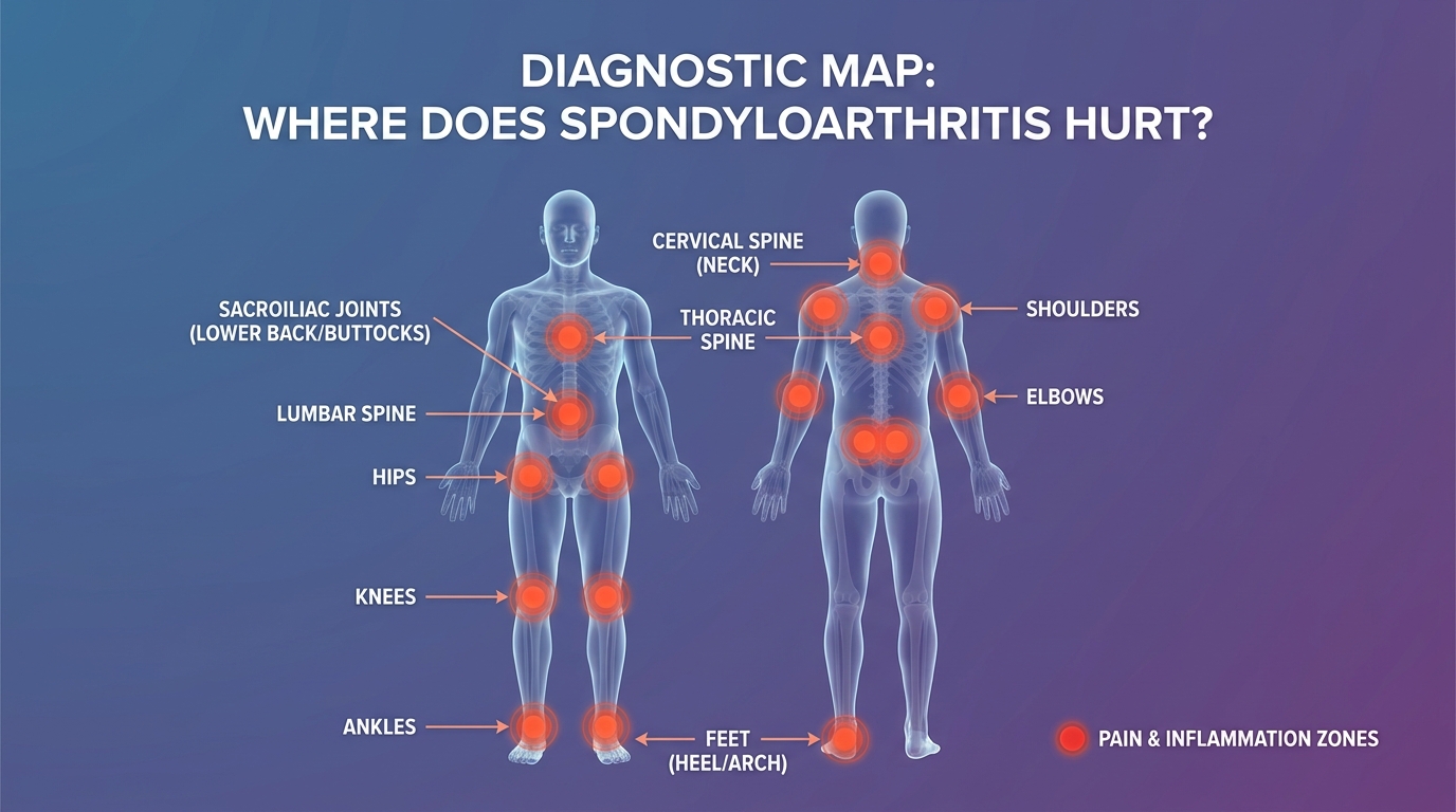
Caption: Spondyloarthritis inflammation can be concentrated in the central spine and pelvis (axial) or in the outer joints and tendon attachments (peripheral), each requiring a different diagnostic focus.
Regardless of whether the inflammation is axial or peripheral, visualizing the internal damage requires advanced imaging techniques that go beyond standard X-rays.
MRI Findings and Disease Progression
When a physician suspects inflammatory back pain or spondyloarthritis, magnetic resonance imaging (MRI) is the gold-standard diagnostic tool — particularly in early-stage disease. Understanding what an MRI can and cannot reveal helps patients frame realistic expectations before attending imaging appointments.
What an MRI will show in early Ankylosing Spondylitis:
An MRI for early ankylosing spondylitis specifically looks for active inflammatory lesions rather than structural damage. The primary findings include:
- Bone marrow edema (BME): The hallmark MRI finding in active sacroiliitis (inflammation of the sacroiliac joints). Edema appears as a bright white signal on fluid-sensitive MRI sequences (specifically STIR or T2 fat-suppressed sequences), indicating that the immune system is actively flooding the joint tissues with inflammatory fluid. The signal brightness reflects increased water content from immune-driven swelling.
- Sacroiliitis: Active inflammation of the SI joints, defined by the Assessment of SpondyloArthritis International Society (ASAS) as bone marrow edema on MRI in at least two consecutive image slices, or one lesion present on a single slice at two separate locations (ASAS, Sieper et al., Annals of the Rheumatic Diseases, 2016).
- Enthesitis lesions: Bright signal areas at tendon and ligament attachment points along the spine.
- Capsulitis and synovitis: Inflammation of the joint capsule and its fluid lining.
Why X-rays alone are insufficient: Standard X-rays detect bone density and established structural changes — erosions, sclerosis (hardening), and joint fusion. These changes take years, sometimes over a decade, to develop. For patients with nr-axSpA who are experiencing severe inflammatory pain but have no X-ray-visible damage, relying solely on radiographs will produce a “normal” result that delays diagnosis. MRI detects the active immune attack itself, not just its long-term consequences. This is precisely why rheumatology guidelines now recommend MRI of the sacroiliac joints as the primary imaging tool when inflammatory back pain is clinically suspected (van der Heijde et al., Annals of the Rheumatic Diseases, 2017).
Disease progression without intervention: If inflammation is left unmanaged, bone marrow edema can transition into structural changes. New bone formation gradually replaces damaged tissue in a process that, over years, leads to the classic “bamboo spine” appearance of advanced Ankylosing Spondylitis — where individual vertebrae fuse together, drastically reducing spinal flexibility.
With the fundamental disease mechanisms established, tracking how this internal inflammation translates into regional physical discomfort is critical for effective clinical communication with a specialist.
General Back Pain Symptoms and Characteristics
General back pain manifests differently depending on the specific region of the spine affected and the underlying cause. For patients navigating rheumatological evaluations, distinguishing between isolated lower back pain, thoracic spine pain (mid-back), and upper back and neck problems gives clinicians critical diagnostic mapping data. Lower back and sacroiliac joint pain is the presenting symptom in over 70% of axial spondyloarthritis cases, making it the most clinically significant starting point for evaluation (Rudwaleit et al., Annals of the Rheumatic Diseases, 2006).
The spine consists of three main regions, each with distinct anatomical roles and characteristic pain presentations when affected by inflammation:
- Lumbar spine (lower back): Five vertebrae (L1–L5) bearing the majority of body weight
- Thoracic spine (mid-back): Twelve vertebrae (T1–T12) attached to the rib cage
- Cervical spine (neck/upper): Seven vertebrae (C1–C7) supporting the head
Identifying which region hurts — and how that pain behaves — transforms a vague complaint of “back pain” into a precise clinical observation. Symptom location dictates diagnostic focus, influencing which imaging sequences are ordered and which specialist subspecialty is most relevant.
While the location of the pain points to the affected anatomy, the timing and intensity of the associated rigidity are what truly define an inflammatory flare.
Mapping Pain: Lumbar vs. Thoracic Spine
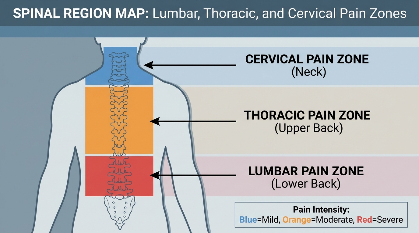
Caption: Spondyloarthritis-related inflammatory back pain most commonly begins in the lumbar region and sacroiliac joints, with thoracic and cervical involvement typically emerging in later disease stages.
Lumbar Region and Sacroiliac Joint Pain
The lumbar spine, the lower back region bearing the most body weight, is typically the first area affected in axial spondyloarthritis. Pain here originates from sacroiliitis — inflammation of the joints connecting the lumbar spine to the pelvis — and from inflammatory changes within the lumbar vertebrae themselves.
Key characteristics of lumbar inflammatory pain include:
- A heavy, dull ache centered in the lower back and buttocks, rather than a sharp or stabbing sensation
- Pain that alternates between the left and right buttock (a distinguishing feature from mechanical sciatica, which typically follows one nerve path)
- Worsening during the second half of the night, often forcing patients to stand or walk for relief
- Gradual improvement over 30–60 minutes of morning movement
Patients often describe the lumbar region as feeling “locked up” upon waking — as though the joints have stiffened into a fixed position during sleep.
Thoracic Spine Pain
The thoracic spine, the middle section of the back attached to the rib cage, becomes involved as spondyloarthritis progresses beyond the lumbar region. Inflammation here presents quite differently from lumbar pain, largely because the thoracic vertebrae are mechanically linked to the ribs.
Patients with thoracic SpA involvement often report:
- A band-like tightness wrapping around the mid-back and chest wall
- Pain that worsens with deep breathing or coughing, because the rib cage must expand during inhalation, pulling on inflamed costovertebral joints (the joints connecting ribs to the thoracic spine)
- Reduced chest expansion, measurable by a clinician using a tape measure at the nipple line during full inhalation and exhalation
- A sensation of restricted lung capacity, sometimes mistaken for a respiratory condition
Cervical Spine (Upper Back and Neck) Involvement
Upper back and neck problems in spondyloarthritis typically represent late-stage progression. Cervical involvement — inflammation of the seven vertebrae supporting the head — can result in:
- Persistent neck stiffness, especially in the morning
- Restricted rotation (difficulty turning the head to either side)
- Referred pain radiating into the shoulders or upper arms
- In rare advanced cases, atlantoaxial instability (loosening of the joint at the very top of the cervical spine), which warrants urgent specialist evaluation
Accurately describing where it hurts is only half the diagnostic equation; explaining how long it has hurt is equally vital for medical professionals.
Chronic vs. Persistent Discomfort
Understanding the language of pain is as important as understanding the biology behind it. Medical professionals use specific terminology to classify back pain by duration, and choosing the right words when describing symptoms to a clinician can significantly influence the diagnostic pathway.
Defining Chronic vs. Acute Back Pain
- Acute back pain: Pain lasting fewer than 6 weeks, typically associated with a specific injury or triggering event. Most acute episodes resolve with conservative management.
- Subacute back pain: Pain lasting 6–12 weeks, often transitioning between an acute injury and a longer-standing issue.
- Chronic back pain: Pain persisting for more than 3 months, regardless of whether a specific injury initiated it. Clinicians use the 3-month threshold because pain lasting this long is unlikely to represent a simple healing process and warrants investigation into systemic or structural causes (National Institute of Neurological Disorders and Stroke, NINDS, 2023).
The word “persistent” carries specific clinical weight. When a patient says their back pain is “persistent,” they signal that it has not resolved despite standard rest and over-the-counter pain management. This language prompts a clinician to consider chronic causes — including inflammatory conditions like spondyloarthritis.
Why Patients Underreport Pain Severity
A significant challenge in spondyloarthritis diagnosis is that patients frequently normalize chronic pain. Someone who has experienced daily lower back aching for three or four years may rate their pain as a “3 out of 10” on a standard numerical scale — because they have adapted their behavior to accommodate it. However, an objective assessment of their mobility might reveal that they can no longer bend forward beyond a limited range, cannot sit for more than 20 minutes, or have stopped all recreational physical activity.
This gap between subjective pain rating and objective functional limitation is clinically important. Patients preparing for rheumatological consultations benefit from documenting not just pain intensity, but specific functional losses: what can no longer be done comfortably and when those limitations began.
Post-Viral Musculoskeletal Inflammation
Recent research has identified post-viral triggers as a relevant differential factor in new-onset inflammatory joint pain. Post-COVID musculoskeletal syndrome — characterized by persistent joint pain, fatigue, and stiffness persisting beyond the acute infection — has been reported in a subset of patients following SARS-CoV-2 infection (Fernández-de-Las-Peñas et al., Pain, 2022). For patients whose back pain and stiffness began or noticeably worsened following a viral illness, this history is valuable clinical information to share during a specialist consultation.
When chronic baseline discomfort abruptly intensifies, patients enter a state of heightened immune activity known as a disease flare.
Stiffness, Inflammation, and Disease Flares
Back inflammation in spondyloarthritis generates profound joint stiffness that severely restricts morning mobility. Unlike minor muscle strains that loosen up immediately with gentle movement, autoimmune inflammation causes inflammatory fluid to pool within joint capsules during periods of rest. This pooling creates measurable physical swelling and resistance within the joint. The result upon waking is not just soreness, but a genuine mechanical restriction — the joint simply cannot move freely until the inflammatory fluid is dispersed through movement (Sieper et al., Nature Reviews Disease Primers, 2015).
Understanding how and why this stiffness develops — and recognizing when it crosses clinical thresholds — transforms daily symptom observations into meaningful data for a treating physician.
The 30-Minute Inflammatory Threshold
The most clinically significant self-observable marker of inflammatory joint disease is morning stiffness duration. This is the core concept behind The 30-Minute Inflammatory Threshold — the clinical benchmark separating mechanical stiffness from systemic inflammatory stiffness.
Why 30 minutes matters:
When a muscle is tight or a joint is mechanically stiff from inactivity, gentle movement typically resolves that stiffness within 10–15 minutes. This is because mechanical stiffness results from temporary fluid pooling in non-inflamed tissue, which disperses quickly with circulation.
In spondyloarthritis, the situation is fundamentally different. The joint capsules are swollen not from inactivity alone, but from active immune attack. Inflammatory cytokines — signaling proteins produced by the immune system during an attack — accumulate within and around the joint during sleep. These proteins must be physically circulated away from the joint tissues through sustained movement. This process consistently takes more than 30 minutes and often extends to 1–2 hours or longer during active disease periods.
In spondyloarthritis, debilitating morning stiffness lasting longer than 30 minutes is a primary indicator of active joint inflammation. Rheumatological assessment criteria, including the widely used Assessment of SpondyloArthritis International Society (ASAS) classification criteria, include morning stiffness duration as a formal diagnostic parameter (Sieper et al., Annals of the Rheumatic Diseases, 2009).
True joint stiffness vs. back muscle stiffness:
These two phenomena feel different and have different implications:
- Back muscle stiffness (spasms): A tight, cramping sensation in the muscular tissue alongside the spine. Resolves quickly with stretching and warmth. Does not involve the joint capsule itself.
- True joint stiffness (capsule rigidity): A deep, structural resistance at the joint itself. Feels as though the joint is physically locked. Does not resolve with stretching alone and requires sustained movement to improve.
How to track The 30-Minute Inflammatory Threshold at home:
- Note the exact time you wake up
- Begin light activity (walking, gentle movement)
- Note the exact time you can comfortably bend forward to tie your shoes without significant resistance or pain
If the gap between waking and comfortable mobility consistently exceeds 30 minutes, document this daily for one week and bring the log to a specialist appointment. This single metric carries substantial diagnostic weight.
Beyond daily morning rigidity, the disease is also characterized by broader waves of systemic inflammation known as flares.
Identifying Spondyloarthritis Flares
A disease flare, an unpredictable period of intensified immune activity, represents a significant escalation beyond the baseline symptoms of spondyloarthritis. Understanding what a flare is, what it feels like, and how long it typically lasts helps patients distinguish between their daily inflammatory baseline and an event that warrants urgent clinical attention.
What an ankylosing spondylitis flare-up feels like:
During a flare, the immune system temporarily produces a surge of inflammatory activity that overwhelms the body’s regulatory mechanisms. Patients typically report a combination of:
- Severe, localized heat: The affected joints — most commonly the sacroiliac joints and lumbar spine — feel warm or hot to the touch, indicating intense inflammation beneath the surface
- Extreme immobility: The physical restriction of movement is significantly worse than the daily baseline, sometimes making it difficult to stand, sit, or change position without assistance
- Systemic fatigue: A deep, whole-body exhaustion that is qualitatively different from ordinary tiredness — described by patients as feeling “as though the body is fighting an infection”
- Flu-like aches: Generalized muscle pain and a sense of bodily heaviness that extends beyond the spine
- Intensified upper back stiffness: During a flare, thoracic involvement can restrict chest wall expansion, making deep breathing noticeably more labored
Flare duration and timeline:
Spondyloarthritis flares are unpredictable by nature. Duration varies considerably:
- Mild flares: 2–5 days, with gradual return to baseline
- Moderate flares: 1–2 weeks of significantly elevated symptoms
- Severe flares: Several weeks of intense immune activity, potentially requiring medical intervention to manage
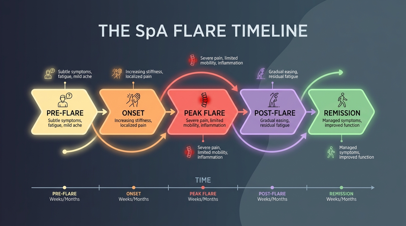
Caption: A typical spondyloarthritis flare progresses through recognizable phases — onset, peak inflammation, and gradual resolution — though the duration of each phase varies significantly between patients.
Practical flare logging strategy for doctor appointments:
Patients who document their flares provide rheumatologists with far more actionable data than those relying on memory. A simple daily log during a flare should record:
- Date and time the flare began
- Pain intensity (0–10 scale) at its worst
- Specific body regions affected
- Duration of morning stiffness on each day
- Any identified triggers (illness, physical overexertion, stress)
- Date and time symptoms returned to baseline
Because these flares are driven by systemic immune responses, the pain rarely stays confined strictly to the spine.
Related Joint and Neurological Conditions
Spondyloarthritis does not always remain confined to the central spine. As systemic inflammation persists, it can extend beyond the lumbar and thoracic vertebrae into peripheral joints and surrounding neurological structures, creating a more complex clinical presentation that requires careful differentiation from other conditions.
Peripheral joint involvement and referred pain:
Peripheral joint pain in spondyloarthritis most commonly targets the hip joints. The hips are anatomically adjacent to the sacroiliac joints and share inflammatory pathways. Hip involvement can present as deep groin pain, restricted internal rotation of the leg, or difficulty rising from a seated position. In some patients, jaw pain (temporomandibular joint, or TMJ, involvement) also occurs, reflecting the systemic reach of SpA-related inflammation.
Referred pain — discomfort that originates in one location but is felt in another — is also common. Sacroiliac joint inflammation frequently refers pain into the buttocks, thighs, and occasionally the calves, closely mimicking sciatica. This referral pattern is a common source of diagnostic confusion, leading some patients toward neurology evaluations when the true origin is rheumatological.
Differentiating SpA from other pain syndromes:
Spondyloarthritis is often confused with myofascial pain syndrome (widespread muscle and soft tissue pain without joint inflammation) and Complex Regional Pain Syndrome (CRPS, a neurological pain amplification disorder). The critical distinction lies in the nature of the inflammation: SpA involves verifiable immune-mediated joint inflammation visible on MRI and blood markers (HLA-B27, elevated CRP), while myofascial pain and CRPS involve pain amplification without structural joint damage.
A comprehensive specialist evaluation — including MRI imaging, laboratory markers, and clinical examination — is essential for accurate differentiation.
Diagnostic Coding and Clinical Charting (ICD-10)
Accurate diagnostic coding is a practical concern for patients navigating the healthcare system, not just a bureaucratic detail for clinicians. ICD-10 codes (International Classification of Diseases, 10th Revision) are the standardized alphanumeric labels assigned to diagnoses. These codes determine insurance authorization for specialist referrals, MRI scans, laboratory tests, and prescription medications.
Why coding precision matters for patients:
A code for generic “low back pain” (M54.5) is categorically different from a code for “ankylosing spondylitis” (M45) or “other spondyloarthritis” (M46). Insurance systems treat these as distinct conditions with different coverage parameters. A patient with documented inflammatory back pain who is coded only as M54.5 may find insurance denials for MRI of the sacroiliac joints or biological therapies, because those interventions are not typically indicated for simple mechanical low back pain.
How clinicians chart spinal vs. peripheral joint pain:
Accurate clinical charting distinguishes between lumbar involvement, thoracic involvement, cervical involvement, and peripheral joint involvement. Each region has distinct ICD-10 code families. Furthermore, codes differentiate between active inflammatory disease, disease in remission, and post-procedural states. Patients attending specialist appointments can request that their chart clearly reflects the specific anatomical regions affected and the inflammatory (rather than mechanical) nature of their condition.
This coding precision directly affects access to the diagnostic imaging and specialist referrals needed to confirm or rule out spondyloarthritis.
Treatments, Procedures, and Pain Management
The treatment framework for spondyloarthritis centers on two parallel goals: reducing active inflammation to prevent structural damage, and preserving or restoring baseline function (the ability to move, work, and perform daily activities). Treatment decisions are individualized based on disease subtype, severity, location of inflammation, and patient-specific factors including other health conditions and pregnancy status.
Conservative management — the first line:
Non-Steroidal Anti-Inflammatory Drugs (NSAIDs) — such as naproxen and diclofenac — are typically the first pharmacological intervention. NSAIDs reduce prostaglandin production, dampening the inflammatory cascade. Rheumatological guidelines note that a significant response to NSAIDs is itself a diagnostic indicator, as inflammatory back pain characteristically improves more substantially with NSAID therapy than mechanical pain does.
Regular, structured physical activity — particularly swimming, cycling, and spinal extension exercises — is a non-negotiable component of management. Movement disperses inflammatory cytokines, maintains joint range of motion, and counters the progressive stiffening that characterizes untreated disease.
Biological therapies for moderate-to-severe disease:
When NSAIDs provide insufficient control, biological Disease-Modifying Anti-Rheumatic Drugs (bDMARDs) — including TNF-inhibitors (such as adalimumab and etanercept) and IL-17 inhibitors (such as secukinumab) — represent a significant advancement in SpA management. These therapies target specific immune proteins driving the inflammatory cascade, offering disease modification rather than symptom suppression alone.
Surgical interventions — the final option:
Spinal fusion and joint decompression procedures are reserved for advanced structural damage that has not responded to medical management. These are not routine interventions for spondyloarthritis and carry significant consideration in the context of pre-existing bone changes from the disease itself.
All treatment decisions should be made in consultation with a board-certified rheumatologist and evaluated against individual patient safety profiles and realistic functional outcome expectations.
Finding the Right Doctor and Specialist
Navigating the medical system is critical for obtaining an accurate spondyloarthritis diagnosis. Because the symptoms of inflammatory back pain overlap with more common mechanical conditions, many patients first consult general practitioners, chiropractors, or orthopedic surgeons before reaching a rheumatologist — the specialist trained to manage autoimmune joint diseases.
Orthopedics vs. Rheumatology — who treats what:
Orthopedic surgeons specialize in structural musculoskeletal problems: fractures, disc herniations, ligament tears, and surgical correction of mechanical joint damage. Their framework is largely anatomical and procedural. Rheumatologists, by contrast, specialize in systemic immune conditions — diseases where the immune system drives joint damage. They are trained to interpret inflammatory blood markers, evaluate MRI findings for sacroiliitis, and prescribe disease-modifying biological therapies.
For a patient with suspected spondyloarthritis, the appropriate first specialist referral is almost always a rheumatologist, not an orthopedic surgeon. An orthopedic evaluation remains relevant if structural damage (such as progressive spinal fusion or hip joint deterioration) requires surgical assessment, but it does not address the underlying immune mechanism.
Preparing for a specialist consultation:
The most productive specialist appointments are built on documentation. Patients benefit from bringing:
- A written symptom diary covering at least one week (noting morning stiffness duration, pain intensity, and specific body regions affected)
- Results of any existing blood tests (especially CRP, ESR, and HLA-B27 if previously tested)
- Prior imaging (X-rays or MRI scans, including radiology reports)
- A list of all medications tried and their effectiveness
This preparation transforms a first rheumatology appointment from a general history-taking session into a focused, evidence-informed evaluation.
Limitations and Safety Considerations
Common Pitfalls in Managing Chronic Back Pain
Pitfall 1: Assuming physical therapy alone is sufficient for inflammatory disease.
Physical therapy and chiropractic care address musculoskeletal mechanics — muscle strength, joint mobility, and postural alignment. These interventions provide genuine symptomatic relief and remain a valuable component of spondyloarthritis management. However, they do not address the underlying immune mechanism driving joint inflammation. Relying exclusively on physical approaches when systemic inflammation is the root cause allows that inflammation to continue causing structural joint damage between sessions. Rheumatological medication — particularly NSAIDs and, when indicated, biological therapies — must accompany physical rehabilitation, not be replaced by it.
Pitfall 2: Treating normal X-ray results as evidence of no disease.
A normal X-ray result does not rule out spondyloarthritis. Non-radiographic axial spondyloarthritis (nr-axSpA) produces genuine, measurable inflammation on MRI long before X-ray-visible changes develop. Patients who are told “your X-ray looks fine” and discharged without further evaluation may continue to accumulate joint damage that could have been halted with early biological therapy. If symptoms align with inflammatory back pain criteria — onset under 40, improvement with exercise, morning stiffness over 30 minutes — a rheumatological referral and MRI evaluation remain appropriate regardless of X-ray findings.
Pitfall 3: Unverified regenerative medicine claims.
Some patients encountering limited success with standard therapies turn toward marketed “regenerative” treatments, including certain stem cell procedures. Patients should be aware that regenerative therapies specifically targeting spondyloarthritis remain investigational. Claims of disease reversal, complete symptom elimination, or guaranteed outcomes are not supported by peer-reviewed clinical evidence. Any such procedures should be discussed with a treating rheumatologist, and claims should be evaluated against published clinical trial data rather than marketing materials.
When to Seek Expert Rheumatological Help
Standard over-the-counter pain management and stretching are insufficient when the following patterns are present:
Red flag symptoms requiring prompt rheumatological evaluation:
- Back pain that consistently wakes you up during the second half of the night
- Morning stiffness lasting more than 30 minutes on most mornings for over 3 months
- Back pain accompanied by unexplained fatigue, weight loss, or fever
- Eye inflammation: sudden redness, pain, or light sensitivity in one eye (uveitis is a direct extra-articular feature of SpA and requires urgent ophthalmological attention)
- Swelling of an entire finger or toe (dactylitis)
- Heel pain at the Achilles tendon that is worse in the morning and not explained by footwear
Early rheumatological evaluation is not a worst-case scenario — it is the mechanism for protecting baseline mobility. Identifying inflammatory disease before structural damage develops gives patients access to therapies that can prevent the most disabling consequences of untreated spondyloarthritis.
Frequently Asked Questions
What does spondyloarthritis back pain feel like?
Spondyloarthritis back pain typically feels like a deep, dull ache localized in the lower back or buttocks that worsens with rest and improves with movement. Unlike a sharp mechanical muscle pull, this inflammatory discomfort develops gradually and often alternates between the left and right sides of the pelvis. Patients frequently experience severe morning stiffness that improves significantly after 30 to 60 minutes of physical activity or a warm shower. If back pain consistently wakes you during the second half of the night, a rheumatological evaluation is highly recommended.
How bad can spondyloarthritis get if left untreated?
If left untreated, severe spondyloarthritis can lead to permanent joint fusion in the spine, drastically reducing mobility and altering posture. Chronic, uncontrolled inflammation gradually replaces flexible spinal ligaments with rigid bone tissue in a process called ankylosis (from the Greek for “bent joint”). Research indicates that early intervention with targeted biological therapies can significantly slow or prevent structural damage in a substantial proportion of patients who maintain consistent treatment compliance (Sieper & Poddubnyy, The Lancet, 2017). Functional outcomes depend heavily on how quickly the systemic inflammation is identified and medically managed by a specialist.
What will an MRI say if you have ankylosing spondylitis?
An MRI for early ankylosing spondylitis will typically show bone marrow edema and active inflammation around the sacroiliac joints, a finding called sacroiliitis. Because standard X-rays only reveal structural bone damage that takes years to develop, fluid-sensitive MRI sequences (STIR or T2 fat-suppressed) are necessary to detect the active immune attack in early-stage disease. Radiologists look for bright white signal areas indicating increased fluid and inflammation within the pelvic bones and lower spine. Confirming these active inflammatory lesions is often the definitive step in securing an official autoimmune diagnosis before permanent joint damage occurs.
Can inflammation cause back stiffness?
Yes — systemic inflammation is the primary biological driver of severe morning back stiffness in autoimmune conditions like spondyloarthritis. During periods of rest, inflammatory cytokines (immune signaling proteins) and excess fluid accumulate within the joint capsules of the spine and sacroiliac joints. This immune response creates physical resistance that routinely takes more than 30 minutes of active movement to circulate out of the affected tissues. Distinguishing this prolonged inflammatory rigidity from brief, mechanical muscle tightness that resolves in under 15 minutes is a vital diagnostic clue for physicians evaluating chronic back pain.
Conclusion
For patients experiencing chronic lower back discomfort, recognizing spondyloarthritis early is critical for preventing permanent joint fusion. Rheumatological research demonstrates that inflammatory back pain typically begins before age 40 and uniquely improves with exercise — a pattern directly opposite to mechanical pain (Sieper & Poddubnyy, The Lancet, 2017). The most effective approach combines early MRI imaging of the sacroiliac joints, precise symptom tracking, and consultation with a board-certified rheumatologist who can interpret inflammatory blood markers and coordinate targeted therapy.
The 30-Minute Inflammatory Threshold provides a practical, self-observable benchmark: if morning stiffness consistently exceeds 30 minutes and does not resolve with gentle movement alone, this finding distinguishes immune-driven joint inflammation from ordinary mechanical strain. Tracking this single metric and distinguishing true joint capsule rigidity from muscular spasm empowers patients to advocate for the correct diagnostic tests — specifically MRI and inflammatory blood markers — rather than accepting inconclusive X-ray results as the final answer.
If you consistently wake up with severe spinal rigidity that forces you out of bed, document the exact duration of your morning stiffness for one week. Bring this clinical log — alongside any prior imaging results and blood test records — to a specialized rheumatology clinic to discuss appropriate diagnostic imaging and evidence-informed management options. Early evaluation is not alarming; it is the most direct route to protecting long-term mobility.