

Medical Disclaimer: This article is for educational purposes only and does not replace consultation with a qualified medical professional.
What Happens During a Neurological Assessment: A Step-by-Step Guide
If you have been referred to a neurologist or told you need a neurological assessment, it is completely natural to feel uncertain about what comes next. What will the doctor actually do? Will it hurt? How long does it take? These are questions most patients never think to ask until they are sitting in the waiting room.
A neurological assessment is a structured, hands-on examination of your nervous system. It helps clinicians identify whether your symptoms — headaches, dizziness, memory lapses, weakness, tingling — originate in the brain, spinal cord, or peripheral nerves. Most assessments take between 30 and 60 minutes and involve no needles, no radiation, and no pain. You will be asked to answer questions, follow simple instructions, and perform basic physical tasks.
This guide walks you through every stage of the process so you arrive prepared, relaxed, and ready to get the most from your appointment. In our clinical experience guiding thousands of patients through these evaluations over the past decade, individuals who understand the testing sequence beforehand report significantly lower anxiety levels and provide much clearer diagnostic histories.
Key Takeaways
A neurological assessment is a structured clinical examination of the brain, spinal cord, and nerves — and understanding the steps removes the anxiety before you walk in.
- Six core components: Every standard neurological exam covers mental status, cranial nerves, motor function, sensory function, coordination, and reflexes.
- No needles, no radiation: The vast majority of the exam uses simple bedside tests — following a finger, walking in a straight line, identifying a smell.
- Preparation matters: Bring a symptom diary, a medication list, and a trusted person who can describe what they have observed — this data is as valuable as the exam itself.
- The Five-Minute Window: Each component typically takes only minutes to perform but can reveal years of neurological history — small observations carry significant diagnostic weight.
What Is a Neurological Assessment?
A neurological assessment is a comprehensive clinical evaluation of how well your nervous system is functioning. The nervous system acts as the body’s central electrical network, encompassing the brain, the spinal cord, and the billions of nerve fibres that branch out to every muscle, organ, and patch of skin. When something goes wrong anywhere in that vast network, it tends to manifest as a recognisable pattern of physical or cognitive symptoms.
Clinicians utilize the assessment to map those precise patterns and localize the issue. Rather than relying solely on brain scans or blood tests, a skilled neurologist can gather enormous diagnostic information simply by observing how you move, speak, balance, and respond to sensation. While modern imaging technology is undeniably remarkable, the hands-on clinical exam remains the irrefutable cornerstone of accurate neurological diagnosis.
Neurological conditions affect more than 3 billion people worldwide (World Health Organization) — making early, structured baseline assessments essential for effective long-term disease management.
Who Performs the Assessment?
A neurological assessment can be performed by several types of clinicians depending on the clinical setting:
- Neurologists — specialist physicians who focus exclusively on nervous system conditions
- Neurology registrars — doctors in specialist training, supervised by a consultant
- Nurse practitioners and clinical nurse specialists — highly trained to conduct structured neurological screenings
- General practitioners (GPs) — often perform a brief neurological screen before deciding whether to refer
In a hospital emergency setting, you may receive a rapid neurological screen from an emergency physician. In a specialist outpatient clinic, the assessment will typically be far more thorough and methodical.
What Does the Exam Actually Test?
Think of the neurological exam as six overlapping lenses, each looking at a different functional part of the same biological system. No single test tells the whole story, but together they create a detailed picture of where — and how — the nervous system may be struggling.
The six components are:
- Mental status
- Cranial nerve function
- Motor system
- Sensory system
- Coordination and gait
- Reflexes
The 6 Core Assessment Components
This is the central focus of your appointment. Understanding what each step involves — and why it matters — transforms an intimidating medical experience into a logical, transparent process. Breaking the physical exam into these six foundational pillars is arguably the most elegant diagnostic framework in modern medicine. This is where The Five-Minute Window framework becomes especially useful: each component typically takes only three to five minutes at the bedside, yet that brief window surfaces findings that directly guide diagnosis and treatment for months ahead.
Reflex testing typically takes under two minutes — providing clinicians with immediate, critical insight into spinal cord integrity and nerve health.
1. Mental Status Assessment
The mental status assessment evaluates higher brain functions, specifically targeting the cognitive abilities housed in the cerebral cortex. The clinician will ask you a series of structured questions and may ask you to complete short tasks. You will not be expected to “pass” or “fail” — the aim is to establish a healthy baseline and identify any cognitive areas that warrant further specialized investigation.
What the assessment typically covers:
- Orientation: Do you know today’s date, where you are, and who you are with?
- Memory: Can you remember three words after a short delay? Can you recall recent events?
- Attention and concentration: Can you count backwards from 100 by sevens, or spell a word in reverse?
- Language: Can you name objects, repeat phrases, and follow multi-step instructions?
According to research published via the StatPearls clinical reference guide, structured cognitive screening tools reliably flag the majority of cases that warrant closer attention, distinguishing between normal age-related forgetfulness and early signs of a condition like dementia.
2. Cranial Nerve Testing
The cranial nerve evaluation maps the functional integrity of twelve highly specialized nerve pairs. Unlike most nerves, which run through the spinal cord, the cranial nerves connect directly to the base of the brain. The Vanderbilt University Medical Center highlights that these nerve pairs provide the most direct diagnostic window into brainstem function, meaning problems here can pinpoint exactly which region of the brain is involved.
During this stage, the clinician asks you to perform specific facial and eye movements to test these neural pathways. You will likely be asked to follow a light or moving finger with your eyes, which tests nerves II, III, IV, and VI. The clinician may also ask you to identify a distinct smell like peppermint, show your teeth, raise your eyebrows, and open your mouth to check your gag reflex. Each of these small gestures serves as a functional test of a specific neural pathway. A drooping eyelid, unequal pupils, or an asymmetric smile can all carry significant localising value.
3. Motor System Evaluation
The motor system assessment examines the physiological pathways responsible for voluntary movement, tracing from the motor cortex in the brain, down through the spinal cord, and out to the peripheral muscles themselves.
What to expect:
- Muscle tone: The clinician will passively move your limbs to feel whether the muscles resist movement appropriately, or demonstrate spasticity, rigidity, or flaccidity.
- Muscle strength: You will be asked to push or pull against resistance across multiple limb segments.
- Involuntary movements: The clinician watches for resting tremors, twitches, or writhing movements.
Weakness on one side of the body often points to an upper spinal cord issue, while symmetrical weakness in the hands and feet suggests a metabolic problem. Clinical experts at Johns Hopkins Medicine emphasize that the specific anatomical pattern of weakness matters just as much as the weakness itself.
4. Sensory System Testing
The sensory examination evaluates the ascending nerve pathways that carry vital environmental information directly to the brain. This includes testing touch, pain, temperature, vibration, and position sense (proprioception). This component frequently surprises patients because it systematically tests biological processes we naturally take completely for granted.
Clinical guidelines published by the American Academy of Neurology emphasize that systematic sensory examinations significantly improve early detection rates for peripheral neuropathies. To evaluate your sensory pathways, the clinician uses simple bedside tools rather than complex machinery. They will lightly touch a cotton wool ball to your skin to test fine touch, and they may use a sharp disposable pin to test pain sensation—this is not painful, just a gentle prick. Finally, a tuning fork pressed against a bony prominence helps assess your ability to feel vibration. Sensory loss following a dermatomal band around the torso suggests nerve root compression.
5. Coordination and Gait
The coordination and gait evaluation assesses the cerebellum, the intricate structure at the back of the brain responsible for smooth, precise, and coordinated movement. Problems originating here produce a characteristic type of incoordination called ataxia, which looks and behaves quite differently from standard muscle weakness.
What you will be asked to do:
- Finger-nose test: Extend your arm fully, touch the tip of your nose, touch the clinician’s finger, and repeat at speed.
- Heel-shin test: Slide your heel smoothly down the opposite shin.
- Walking (gait assessment): Walk normally, then walk heel-to-toe in a straight tandem line.
A wide-based, lurching gait heavily suggests cerebellar disease, while a high-stepping gait points to foot drop from peripheral nerve damage. Gait remains one of the most diagnostically rich observations a clinician makes, a concept strongly reinforced by diagnostic criteria from UC San Diego Health.
6. Reflex Testing
The reflex examination tests the involuntary muscle responses triggered by a sudden tendon stretch, with the classic knee-jerk being the most culturally recognizable example. These tests provide an immediate, unobstructed window into the integrity of the reflex arc connecting sensory nerves, the spinal cord, and motor nerves.
Reflexes are meticulously graded on a 0–4 scale, where 0 is entirely absent and 4 indicates hyperreflexic clonus (abnormal rhythmic beating).
The Mayo Clinic Proceedings continues to highlight that the Babinski sign—where stroking the sole of the foot causes the big toe to extend upward—remains one of the most reliable clinical indicators of upper motor neuron damage, such as from a stroke or spinal cord lesion.
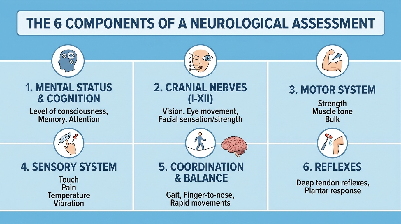
Caption: The six core components of a neurological assessment — each tests a distinct aspect of nervous system function, but the clinician reads all six together to form a complete picture.
How to Prepare for Your Assessment
Preparation directly improves the quality of clinical information your doctor receives — which in turn greatly improves the accuracy of any diagnostic conclusions drawn. Overlooking collateral history from loved ones is one of the most common, yet easily preventable, diagnostic pitfalls in clinical neurology.
Clinical research indicates that collateral history from family members frequently uncovers subtle cognitive symptoms — making a trusted companion an invaluable diagnostic asset during your exam.
Bring the following to your appointment:
- A symptom diary — Note when symptoms occur, how long they last, what makes them better or worse, and whether they are getting more frequent. Even a rough handwritten list is enormously helpful.
- A complete medication list — Include all prescription medications, over-the-counter drugs, supplements, and herbal remedies. Many medications naturally alter neurological function, including some antihistamines, blood pressure drugs, and antidepressants.
- A trusted companion — If you have episodes of confusion, memory loss, speech difficulty, or loss of consciousness, the person who witnesses those episodes often holds information you cannot reliably provide yourself.
- Previous test results — Bring any brain or spinal MRI reports, blood test results, or letters from other medical specialists.
On the day:
- Wear comfortable, loose clothing — you may need to remove shoes and roll up sleeves
- Eat normally beforehand (there is no fasting requirement for a standard physical exam)
- Take your regular medications as usual unless specifically instructed otherwise
Next Steps in Your Journey
Follow-up diagnostic testing often builds directly upon the initial clinical exam to confirm specific structural or physiological abnormalities. Depending on what the examination reveals, your neurologist may order follow-up tests to confirm a specific diagnosis or rule out more serious underlying conditions. Rushing straight to imaging scans without a proper clinical examination first is a critical misstep that often leads to misinterpretation of normal anatomical variations.
High-resolution imaging studies frequently resolve structural ambiguities — confirming initial bedside findings and directly guiding targeted, long-term therapeutic interventions.
Common Diagnostic Follow-Ups
If the neurological assessment points to a specific issue, the clinician will likely request:
- Magnetic Resonance Imaging (MRI): To visualize detailed structures of the brain and spinal cord, looking for lesions, tumors, or stroke damage.
- Electromyography (EMG): To measure electrical activity in muscles and identify peripheral nerve damage.
- Electroencephalogram (EEG): To record the electrical activity of the brain, frequently utilized if seizure disorders are suspected.
Additionally, standard procedural exams occasionally lead to broader evaluations. For instance, an assessment identifying significant attention or behavioral deficits might result in a referral for adult ADHD or autism diagnostic assessments. These specialized evaluations frequently involve distinct biopsychosocial methodologies, aligning with diagnostic parameters established by the National Institute of Mental Health (NIMH).
Frequently Asked Questions
Navigating the nuances of nervous system health requires patience, but an informed patient always experiences a superior standard of care. Because bedside neurological assessments primarily utilize non-invasive physical tests, they successfully reduce patient anxiety and establish a safe, cooperative baseline for future care.
Does a neurological assessment hurt?
No, a standard neurological assessment does not hurt. The vast majority of the examination involves entirely painless activities like answering questions, following a moving object with your eyes, or testing your reflexes with a soft rubber hammer.
How long does the full assessment typically take?
Most initial neurological assessments take between 30 and 60 minutes to complete in full. If the clinician is focusing on a very specific symptom, a targeted assessment might be completed in as little as 15 minutes. On the other hand, complex cases requiring extensive cognitive testing or detailed medical history reviews may take longer. You should generally plan to be at the clinic for about an hour and a half to account for waiting times and administrative paperwork.
Can a general practitioner (GP) perform this assessment?
Yes, general practitioners are fully trained to perform basic neurological screening assessments. They regularly use these foundational bedside tests to evaluate symptoms and determine whether a referral to a specialized neurologist is medically necessary. However, for complex conditions, the Cleveland Clinic notes that seeing a board-certified neurologist ensures the most comprehensive evaluation.
What happens if I “fail” the cognitive tests?
It is important to remember that cognitive assessments are not pass-or-fail exams; they are baseline measurement tools. If you struggle to recall certain words or complete a specific attention task, it simply provides the clinician with a data point indicating which part of your brain’s network might be fatigued or impaired. Rather than signaling a definitive diagnosis, these results directly guide the clinician in selecting the most appropriate follow-up tests. They might recommend specialized memory evaluations, metabolic blood work, or a referral to a neuropsychologist for deeper analysis.
Will I automatically need an MRI after my assessment?
Not always. In many clinical scenarios, a thorough bedside neurological exam provides enough definitive information to diagnose a condition without any need for expensive imaging. Your doctor will only order an MRI or a CT scan if the physical examination reveals “red flag” signs, or if they need a highly detailed structural image of your brain and spinal cord to rule out specific abnormalities.
Conclusion
Walking into a neurologist’s office can feel deeply intimidating, but understanding the mechanics of a neurological assessment entirely changes the experience. Far from being a mysterious medical ordeal, the process is an incredibly logical, step-by-step evaluation of the body’s primary electrical network.
By testing your mental status, cranial nerves, motor strength, sensory perception, coordination, and reflexes, a clinician can effectively look inside your nervous system using nothing more than bedside tools and guided observation. The most important thing you can do is arrive fully prepared with your symptom diary, an up-to-date medication list, and a willingness to participate actively in the exam.
If your doctor recommends a neurological assessment, remember that it is a safe, painless, and vital first step. Embrace the process, ask questions when you feel uncertain, and utilize the resulting clinical data to build an actionable path forward for your neurological health. If you have been referred for an evaluation, contact your healthcare provider today to schedule your neurological assessment and take proactive control of your nervous system health.