

Early Rheumatology Assessment: How To Protect Function
This article is for educational purposes only and does not replace consultation with a qualified medical professional. Always consult a board-certified rheumatologist for diagnosis, treatment planning, and medical advice specific to your condition.
Unexplained, persistent joint pain often leaves patients wondering if they simply slept wrong — or if something more serious is developing beneath the surface. When morning stiffness lingers beyond 30 minutes, or when symmetrical swelling appears in both wrists or knuckles, these are not signals to dismiss.
Delaying a specialist evaluation can allow silent neuroinflammatory processes — internal inflammation affecting the nervous system and joint tissues — to erode cartilage before any visible damage appears on a standard X-ray. By the time pain becomes severe, structural harm may already be irreversible.
This guide explains the critical window of opportunity for rheumatic intervention: what it is, why it matters, and exactly what functional assessments to expect during a specialist visit. You will learn how to recognize the earliest warning signs, understand the clinical tools rheumatologists use, and prepare confidently for your first consultation — because protecting your long-term joint health begins with understanding when and how to act.
Key Takeaways
An early rheumatology assessment protects joint function by identifying neuroinflammatory processes within the critical Functional Preservation Window before irreversible erosion occurs.
- The Preservation Window: Medical intervention within the first 12 weeks of symptom onset significantly improves long-term mobility outcomes according to American College of Rheumatology guidelines — acting early is the single most effective protective strategy.
- Objective Physical Assessment: Specialists use standardized tools like the Fullerton Assessment of Balance and the Fugl-Meyer Assessment to quantify joint capability accurately, removing guesswork from diagnosis.
- Proper Preparation: Bringing prior medical records and understanding the diagnostic criteria prevents clinical delays and accelerates targeted treatment planning.
The Importance of Early Assessment
Early rheumatology assessment is the critical diagnostic process used to identify autoimmune and neuroinflammatory joint conditions before permanent structural damage occurs. Acting within the first few months of symptom onset — the period clinicians call the Functional Preservation Window — allows specialists to suppress inflammation, protect cartilage integrity, and preserve long-term physical mobility, a standard validated by international rheumatology consensus.
Rheumatoid Arthritis (RA), a chronic systemic autoimmune disease, does not simply cause joint pain. It triggers a complex immune cascade that, if left unchecked, destroys the precise architecture of your joints. At the cellular level, the immune system incorrectly identifies the synovium (the thin lining of the joint) as a foreign threat. T-cells and B-cells flood the joint space, releasing pro-inflammatory cytokines like Tumor Necrosis Factor-alpha (TNF-α) and Interleukin-6 (IL-6). These chemicals cause the synovial cells to multiply rapidly, forming a thick, invasive tissue known as a pannus.
The pannus behaves almost like a localized, destructive tumor. It secretes matrix metalloproteinases (MMPs) — enzymes that aggressively dissolve the surrounding cartilage matrix and chew into the underlying subchondral bone. This underlying biological mechanism highlights why over-the-counter pain relievers are fundamentally inadequate; while they may temporarily mask discomfort, they do absolutely nothing to halt the cellular destruction orchestrated by the pannus. Understanding what drives this destruction — and why timing determines outcomes — is the foundation of all effective rheumatological care.

Caption: The Functional Preservation Window illustrates why acting within the first 12 weeks of symptom onset prevents irreversible cartilage erosion.
The Functional Preservation Window
The Functional Preservation Window represents the brief period between symptom onset and permanent joint damage where early intervention may halt disease progression. This clinical concept is arguably the most important paradigm in early rheumatology care. Clinical consensus indicates this window is typically the first 12 weeks following the appearance of consistent joint symptoms, a threshold supported by European Alliance of Associations for Rheumatology (EULAR) data. What happens inside a joint during this period dictates whether a patient maintains full function or faces a lifetime of progressive disability.
Medical intervention within the first 12 weeks of symptom onset halts synovial inflammation — preserving long-term mobility and preventing structural collapse. During these initial months, the immune system’s cellular “memory” has not yet fully locked onto the joint tissue as a permanent target. The inflammatory loop is active but vulnerable to disruption. By aggressively deploying immune-modulating therapies during this phase, clinicians can frequently induce a state of clinical remission. Once cartilage is fully degraded and bone erosion begins, however, that structural damage is strictly permanent. No currently available pharmacological therapy rebuilds lost cartilage or restores eroded bone to its original, pristine state.
This stark distinction matters enormously for patients: while joint damage is irreversible, disease progression is highly manageable if caught early. Consider two hypothetical patient trajectories. A patient who receives an early referral to a rheumatologist at week eight of symptoms — when inflammation is active but joint erosion is completely absent — may achieve deep clinical remission with standard disease-modifying therapy, retaining normal joint function for decades. Conversely, a patient who delays assessment until year two, after repeated flares have caused visible bone erosions on imaging, faces a fundamentally harder clinical challenge. Even if medications successfully halt future inflammation, the structural damage already sustained will permanently alter their joint mechanics.
Early referral to a rheumatologist is optimal precisely because it captures patients before this permanent tipping point. Standardizing care around this 12-week benchmark is the primary driver of improved functional outcomes across the medical community.
Identifying the Warning Signs
Recognizing which specific symptoms justify an urgent specialist visit is the first practical skill patients must develop. Early autoimmune joint disease rarely announces itself with dramatic, sudden incapacitation; rather, it typically presents as persistent, low-level discomfort that patients mistakenly rationalize away as aging or overexertion.
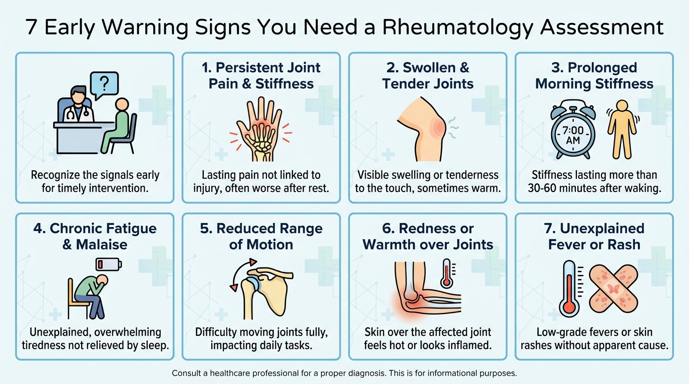
Caption: These seven warning signs represent the most clinically significant early indicators that a rheumatology assessment may be warranted.
Seven early warning signs that warrant a rheumatology assessment:
- Morning stiffness lasting more than 30 minutes — Inflammatory fluid pools in the joint capsule overnight due to a lack of muscular movement. Stiffness that takes 30 to 60 minutes to “loosen up” is a hallmark of systemic inflammation, not mechanical wear-and-tear.
- Symmetrical joint swelling — Swelling occurring in matching joints on both sides of the body (e.g., both wrists or both left and right knuckles simultaneously) strongly indicates circulating systemic autoantibodies rather than localized injury.
- Persistent fatigue disproportionate to activity — Inflammatory diseases produce severe, systemic fatigue driven by total-body immune activation, completely separate from physical exertion.
- Warmth or redness over joints — Palpable heat over a joint indicates active, acute synovitis.
- Reduced grip strength or fine motor difficulty — Struggling to turn a key, open jars, or button a shirt often points to early tenosynovitis (inflammation of the tendon sheaths) in the hands.
- Swollen joints with no recent injury — Spontaneous, unexplained swelling without a clear physical trauma requires immediate investigation.
- Symptoms persisting beyond six weeks — Inflammatory arthritis is distinguished from temporary viral arthritis or mechanical strain partly by its stubborn duration.
To confirm these physical signs, rheumatologists rely heavily on two specific blood tests that can predate physical symptoms by years. Anti-CCP (Anti-Cyclic Citrullinated Peptide antibodies) is a highly specific marker — an autoantibody produced by the immune system that mistakenly targets citrullinated proteins in the patient’s own joint tissue. A positive Anti-CCP result is considered the “smoking gun” of early RA, offering incredibly high specificity. ANA (Antinuclear Antibodies), proteins that mistakenly target the body’s own cell nuclei, are utilized to screen for a broader spectrum of systemic autoimmune conditions, including lupus and Sjogren’s syndrome.
Relying on standard X-rays alone is profoundly insufficient in early-stage disease. X-rays only detect bone erosion after it has already occurred — a failure of early intervention. Instead, advanced blood panels and musculoskeletal MRI or ultrasound imaging are utilized to detect active synovitis and autoantibody production long before any structural damage is visible on a radiograph, making them vastly superior tools for capturing the preservation window.
Core Diagnostic Criteria for RA
Rheumatologists do not diagnose Rheumatoid Arthritis based on a single blood test or a brief visual inspection. They apply a highly validated clinical scoring system developed jointly by the American College of Rheumatology (ACR) and EULAR, which officially replaced the older 1987 classification criteria.
The 2010 criteria update was a necessary paradigm shift because the older framework functionally required patients to suffer irreversible radiographic damage before earning an official diagnosis. The modern framework is specifically calibrated to catch the disease early. It scores patients across four distinct categories, with a total score of six or more points (out of ten) confirming an RA diagnosis.
ACR/EULAR 2010 RA Classification Criteria
| Category | Criteria | Points |
|---|---|---|
| Joint Involvement | 1 medium-to-large joint | 0 |
| 2–10 medium-to-large joints | 1 | |
| 1–3 small joints | 2 | |
| 4–10 small joints | 3 | |
| >10 joints (at least 1 small joint) | 5 | |
| Serology (Blood Tests) | Negative RF and negative Anti-CCP | 0 |
| Low-positive RF or low-positive Anti-CCP | 2 | |
| High-positive RF or high-positive Anti-CCP | 3 | |
| Acute-Phase Reactants | Normal CRP and normal ESR | 0 |
| Abnormal CRP or abnormal ESR | 1 | |
| Duration of Symptoms | Less than 6 weeks | 0 |
| 6 weeks or more | 1 |
RF = Rheumatoid Factor; CRP = C-Reactive Protein (an inflammation marker); ESR = Erythrocyte Sedimentation Rate (another inflammation marker).
A hypothetical scoring scenario: Consider a 45-year-old patient who presents with symmetrical swelling in both wrists and three proximal interphalangeal (finger) joints. This qualifies as “4–10 small joints” (3 points). Their lab work reveals a high-positive Anti-CCP titer (3 points) and elevated C-Reactive Protein levels (1 point). They report that the morning stiffness has persisted for eight weeks (1 point). Total Score: 8 points. Because this score exceeds the six-point threshold, a definitive RA diagnosis is confirmed.
This rigorous scoring system matters for one concrete reason: a diagnosis achieved rapidly unlocks access to disease-modifying antirheumatic drugs (DMARDs) — potent medications that suppress the underlying immune attack — before irreversible erosion advances.
Interactive Assessment Factors
Achieving a baseline diagnosis is merely the starting line of rheumatological care; it establishes a continuous, dynamic monitoring relationship between the patient and the specialist. Rheumatologists track disease activity through recurring, highly objective clinical measurements, aggressively adjusting treatment protocols as the inflammatory burden evolves.
In clinical training, specialists learn that ongoing interactive assessment factors include all of the following except subjective pain memory. Patients naturally focus intensely on how much a joint hurts today compared to last month — but subjective memory of pain is a notoriously unreliable clinical variable. Instead, rheumatologists base their ongoing assessments on concrete, measurable data that does not fluctuate based on a patient’s mood or recall. They utilize standardized disease activity indices — such as the DAS28 (Disease Activity Score in 28 joints) or the CDAI (Clinical Disease Activity Index).
These indices require the clinician to manually palpate 28 specific joints to calculate an exact Tender Joint Count (TJC) and Swollen Joint Count (SJC). These numbers are then fed into an algorithm alongside current inflammatory blood markers (CRP/ESR) and a standardized patient global assessment scale.
This strict distinction matters immensely for patients navigating long-term care. You may report feeling “better” or “worse” than your previous visit, and that qualitative context remains highly valuable. However, if your subjective pain is low but your swollen joint count and CRP levels are climbing, your rheumatologist will escalate your medication. The ongoing interactive assessment framework correctly distinguishes between disease activity (the silent, aggressive immune attack on the joint) and functional capacity (how well you manage daily tasks). Treating only one dimension inevitably leads to incomplete, inferior outcomes.
Physical and Orthopedic Evaluations
While advanced blood panels reveal the unseen neuroinflammatory markers circulating in your bloodstream, orthopedic physical assessments measure the tangible, real-world impact of that inflammation on your daily mobility. Standardized clinical tools allow rheumatologists to establish a rigid functional baseline and track objective improvements rather than relying on guesswork.
These physical tests are not intimidating or painful procedures. They are carefully engineered biomechanical activities — most of which mimic everyday movements — scored against strict, validated international benchmarks. By utilizing these tools, subtle deficits in your muscular function and joint stability are detected long before they manifest as obvious, life-altering limitations.
The Fullerton Assessment of Balance
The Fullerton Assessment of Balance is a rigorous, standardized clinical tool initially engineered to measure functional stability and identify fall risk in patients experiencing lower-body joint degradation. Within the modern rheumatology clinic, it serves a highly precise diagnostic purpose: quantifying exactly how much early joint inflammation has degraded a patient’s somatosensory feedback, balance, and lower-limb coordination.
The Fullerton Assessment of Balance utilizes a 14-item scale to accurately identify subtle mobility deficits caused by early-stage joint inflammation, according to established orthopedic guidelines. Each item in the battery evaluates a specific dimension of the body’s balance systems — integrating visual, vestibular (inner ear), and proprioceptive (joint position) data.
During the assessment, patients perform activities such as:
- Standing with feet together and eyes closed — Isolating the vestibular system and highlighting deficits in ankle proprioception caused by early synovial swelling.
- Reaching forward to retrieve an object — Testing dynamic limits of stability and the core strength necessary for safe daily function.
- Walking heel-to-toe in a straight line — Evaluating complex dynamic balance and precise lower limb motor coordination.
- Stepping over a standardized obstacle — Assessing the functional hip flexion, knee mobility, and single-leg stability required for safe navigation in uneven environments.
For a patient developing early RA in the metatarsophalangeal (toe) joints, ankles, or knees, even microscopic inflammation can measurably reduce performance across these 14 tasks — frequently before the patient themselves realizes their balance is compromised. Catching this specific deficit early allows the clinical team to integrate targeted physical therapy alongside biological medications, shielding the patient from a catastrophic fall-related injury.
Upper Extremity Fugl-Meyer Test
The Fugl-Meyer Assessment is an exhaustive, highly standardized index historically used to evaluate motor function, sensation, and joint coordination. While it originated in neurology for stroke recovery, its upper extremity component has proven exceptionally valuable for rheumatologists treating patients experiencing aggressive, early RA in the delicate joints of the hands, wrists, and shoulders.
Medical professionals and physical therapists frequently utilize standardized documentation protocols to ensure completely reproducible scoring across varying clinical settings. This rigorous standardization ensures that a patient’s hand dexterity score recorded at a local clinic is directly, mathematically comparable to a score recorded six months later at a major academic research center, yielding a flawless longitudinal record of functional change.
Early Rheumatoid Arthritis disproportionately devastates fine motor skills. The synovial sheaths surrounding the tiny joints of the fingers and wrists are typically the first tissues targeted by the autoimmune attack. This localized swelling severely reduces the precision pinch-grip and dynamic wrist extension necessary for essential tasks: turning a key, typing on a keyboard, or holding a pen. The Fugl-Meyer protocol captures these insidious losses quantitatively. Clinicians measure exact degrees of wrist extension and utilize a Jamar dynamometer to record precise grip strength variations.
Why does aggressively tracking these scores matter? The silent loss of hand dexterity is widely considered one of the most functionally devastating consequences of undertreated early RA. Patients unconsciously adapt — using two hands to lift a coffee mug, or avoiding fine motor tasks altogether — and frequently fail to register the extent of their functional loss until severe, irreversible deformities (like swan-neck or boutonniere deformities) occur. Documenting these objective scores at every visit creates an actionable record of progression, forcing intervention before those subtle adaptations become permanent physical handicaps.
Dorsiflexion and Lower Extremity Mobility
Dorsiflexion is the upward bending motion of the foot toward the shin — the critical biomechanical movement your ankle must execute when you walk, climb stairs, or rise from a seated position. Restricted dorsiflexion is universally recognized as a primary early warning sign of rheumatic disease affecting the lower extremity, and clinicians measure it using rigorous standardized techniques collectively termed dorsiflexion assessment.
Biomechanical stiffness in dorsiflexion frequently appears weeks or months before patients consciously register active ankle pain. Patients may report tripping more frequently on area rugs, feeling strangely unstable on uneven grass, or experiencing rapid fatigue when walking moderate distances. These are not signs of clumsiness; they are subtle functional changes captured through precise ankle assessment protocols that measure the exact degrees of active and passive motion available at the talocrural joint.
When ankle dorsiflexion is limited by early inflammatory effusion (fluid inside the joint capsule), the knees and hips are forced to drastically alter their movement patterns to compensate. This altered gait inevitably leads to secondary mechanical wear-and-tear in those larger joints. By utilizing foundational orthopedic physical assessment techniques, clinicians intercept this destructive chain reaction early.
Standardized lower extremity assessment techniques are applied with rigid consistency across the globe. Whether a patient is evaluated at a university hospital in North America or at a specialized, internationally accredited rheumatology center in regions like Bangkok, Thailand (such as facilities near Lad Phrao 101), the evidence-based physical assessment protocols remain identical — ensuring high-fidelity care regardless of geography.
Pediatric Rheumatology Considerations (Summary)
Rheumatic diseases are not exclusively adult conditions. Juvenile Idiopathic Arthritis (JIA) — a broad classification for autoimmune joint disease presenting before the age of 16 — is the most prevalent chronic rheumatic condition in children. Early childhood development, which encompasses the progressive acquisition of critical motor, cognitive, and social skills during the first years of life, is directly threatened when systemic rheumatic disease interrupts normal physical function.
Unlike adults who can articulate their pain verbally and describe precise functional limitations, young children often lack the vocabulary to explain their symptoms. They may simply appear withdrawn, refuse to participate in physical play, or regress in newly acquired motor skills like walking — behaviors frequently misattributed to developmental phases or behavioral issues rather than joint-driven pain.
Pediatric rheumatologists rely on a fundamentally different evaluation framework from adult orthopedics. The pediatric assessment triangle — a rapid, non-invasive visual evaluation tool examining a child’s appearance, work of breathing, and circulation to the skin — provides clinicians with an immediate impression of whether a child’s overall physiology is compromised by systemic inflammation. Identifying rheumatic disease early in childhood is exceptionally urgent, as unchecked joint inflammation during active growth phases can permanently distort bone development and impair crucial motor skill acquisition.
Navigating Rheumatology Clinics and Institutions
Identifying the pressing need for an early assessment is only half the battle — successfully securing a prompt appointment at a highly qualified institution is the next crucial step. High-tier academic medical centers inherently provide access to the latest advanced diagnostic imaging, integrated multidisciplinary clinical teams, and cutting-edge biological treatment protocols that smaller or generalist clinics simply may lack the resources to offer.
Leading Academic Medical Centers
Major universities and academic health systems drive the vast majority of global rheumatic disease research. Centers possessing dedicated, specialized rheumatology divisions — such as NYU Rheumatology (part of NYU Langone Health) or Ohio State Rheumatology (part of The Ohio State University Wexner Medical Center) — serve as national benchmarks for comprehensive autoimmune care.
The distinct advantage of being assessed at a research-driven institution extends far beyond institutional prestige. These advanced centers maintain highly active clinical trial programs, meaning eligible patients may gain access to emerging, next-generation targeted therapies years before they receive widespread commercial approval. Furthermore, multidisciplinary teams — combining board-certified rheumatologists, specialized physical therapists, and advanced musculoskeletal imaging technicians — ensure every dimension of joint function is evaluated concurrently.
For patients navigating complex diagnoses outside North America, or those aggressively seeking an authoritative second opinion, international centers offer exceptional expertise. Premium specialty clinics in Bangkok, Thailand, for example, frequently provide internationally accredited diagnostic imaging and board-certified specialist consultations within a single, highly integrated facility.
Preparing for Your Specialist Visit
Thorough preparation directly determines the clinical efficiency of your very first rheumatology consultation. A meticulously prepared patient vastly accelerates the diagnostic timeline. Conversely, an unprepared patient frequently requires multiple additional appointments simply to gather basic information that should have been compiled in advance.
Three essential items to bring to your first rheumatology appointment:
- Comprehensive laboratory results — Supply any recent blood work from your primary care physician, explicitly including full blood counts, inflammatory markers (CRP, ESR), and any previously run autoantibody panels (Anti-CCP, ANA).
- Diagnostic imaging reports — Bring physical discs or digital access codes for any recent X-rays, MRI scans, or musculoskeletal ultrasound reports of the affected joints.
- A detailed written symptom timeline — Accurately document when symptoms first appeared, explicitly note which specific joints are affected, track the daily pattern of morning stiffness, and record how the pain varies across the day.
Managing expectations during the initial visit is equally vital. The primary focus of a first consultation is comprehensive data gathering and safety monitoring, not necessarily finalizing an immediate diagnosis or handing over a prescription. Specialists must establish a rigorous objective baseline before initiating powerful immune-modulating medications.
Clinical Literature and Textbooks (Summary)
The rigorous physical assessment techniques and complex diagnostic criteria you will experience in the rheumatology clinic did not emerge arbitrarily. They are painstakingly standardized through decades of peer-reviewed medical literature — including double-blind clinical trials, extensive case studies, and systematic reviews published in authoritative journals — that collectively define modern evidence-based practice.
Medical professionals continually review published clinical case studies specifically to refine their ability to detect the absolute earliest, most subtle biological signs of joint erosion. Standard X-rays only become positive for erosion after structural damage has already accumulated. Published clinical case literature, by contrast, exhaustively documents the pre-erosive imaging patterns, highly atypical autoantibody profiles, and ambiguous early presentations that experienced clinicians must recognize.
For patients who wish to develop a highly informed, working knowledge of their own condition, foundational rheumatology textbooks provide comprehensive, transparent coverage of diagnostic criteria, pharmacological treatment algorithms, and functional assessment protocols. Understanding the academic framework guiding your specialist helps you ask far more precise questions and engage productively in shared clinical decision-making.
Professional Rheumatology Conferences (Summary)
The concept of the Functional Preservation Window is not a static, historical definition — it is a highly dynamic clinical construct that global experts continuously refine as vast amounts of new evidence emerge. The primary venue for this rapid refinement is the international medical conference circuit, where leading rheumatologists, immunologists, and clinical researchers present breaking trial data, fiercely debate emerging protocols, and establish the updated guidelines that filter down into local clinical practice.
The two dominant global organizations driving this rigorous process are the American College of Rheumatology (ACR) and the European Alliance of Associations for Rheumatology (EULAR). The ACR’s annual meeting serves as the primary stage where major updates to diagnostic criteria and biological treatment recommendations are first presented before formal publication.
For patients, the practical implication of this global academic ecosystem is highly reassuring: current clinical guidelines are significantly more accurate and evidence-based than historical protocols, precisely because this conference ecosystem ensures the continuous, rigorous updating of medical standards worldwide.
Safety, Limitations & Alternatives
Common Pitfalls in Early Diagnosis
Several heavily documented clinical pitfalls routinely delay early rheumatological diagnosis, and each carries a severe, irreversible functional cost for the patient.
Pitfall 1 — Dismissing inflammatory symptoms as normal aging. Persistent joint stiffness and spontaneous swelling are frequently, and incorrectly, attributed to “getting older” or mechanical overexertion. This rationalization is particularly dangerous for patients under 40, who mistakenly assume they are not at risk for systemic autoimmune disease. Persistent, symmetrical joint symptoms in any adult warrant immediate medical investigation, not passive observation.
Pitfall 2 — Over-reliance on standard X-rays. Standard radiographs are effective for detecting established, permanent bone erosion but possess notoriously poor sensitivity for early-stage synovial inflammation. A “normal” X-ray result absolutely should not be interpreted as a clean bill of joint health. Blood panels (measuring Anti-CCP and ANA) and MRI imaging provide actionable data months or years before an X-ray shows damage.
Pitfall 3 — Expecting regenerative therapies to replace disease-modifying drugs. Regenerative medical approaches, including platelet-rich plasma (PRP) therapy and stem cell interventions, do not cure, reverse, or halt rheumatoid arthritis. They may offer supportive, localized functional benefits in highly selected patients — for example, reducing localized tendon pain — but they cannot halt a systemic autoimmune attack. Patients pursuing regenerative approaches must do so strictly under the ongoing supervision of a board-certified rheumatologist.
When to Seek Immediate Expert Help
Two specific clinical scenarios require an immediate, urgent specialist evaluation — not a routinely scheduled outpatient referral.
Scenario 1: Sudden, severe joint swelling accompanied by fever. This specific symptom combination strongly indicates either septic arthritis (a catastrophic joint infection) or a rapidly escalating autoimmune flare. Septic arthritis can permanently destroy a joint’s architecture within days if left untreated. Do not wait for a standard appointment; proceed directly to an emergency department.
Scenario 2: Rapid, unexplained loss of motor function. If you experience a sudden, accelerating inability to grip objects, fully extend your fingers, walk normally, or perform routine fine-motor tasks over a period of days or weeks, this signals highly aggressive disease progression or nerve compression. Rapid medical intervention during a severe flare may preserve physical capabilities that watchful waiting will permanently destroy.
Frequently Asked Questions
Why is early diagnosis of rheumatoid arthritis important?
Early diagnosis of rheumatoid arthritis is essential because it prevents irreversible cartilage and bone destruction. Within the first few months of symptom onset, neuroinflammatory processes begin degrading joint structures at the synovial membrane. Initiating targeted medical intervention during this functional preservation window halts structural damage. Clinical data shows that patients treated within 12 weeks maintain significantly higher long-term mobility. Delays in assessment directly correlate with a much higher risk of permanent physical disability.
What is the functional assessment of early rheumatoid arthritis?
The functional assessment of early rheumatoid arthritis evaluates a patient’s objective physical capability, balance, and motor skills rather than just subjective pain levels. Rheumatologists utilize standardized clinical tools, such as the Fugl-Meyer assessment or Fullerton balance test, to precisely quantify dexterity and dynamic stability. These metrics establish an objective baseline for aggressively tracking disease progression. Documenting functional limitations early allows specialists to tailor physical therapy and medical interventions accurately to the specific deficit. Results naturally vary based on the specific joints affected by early synovial inflammation.
Can arthritis be cured if detected early?
Autoimmune arthritis cannot be entirely cured, but early detection frequently leads to deep clinical remission. When rheumatologists intervene during the earliest stages of disease, they utilize potent medications to suppress the immune system and halt the neuroinflammatory attack on the joints. This proactive suppression prevents the progressive physical erosion that permanently cripples untreated patients. Patients who successfully achieve early remission frequently maintain fully normal joint function and a high quality of life for decades. Sustained remission strictly requires ongoing clinical monitoring and rigid adherence to prescribed treatment.
Is it good to diagnose arthritis early?
Yes, diagnosing arthritis early is universally considered the single most critical factor in successful long-term disease management. Early identification allows specialists to immediately deploy disease-modifying therapies before the joint cartilage is permanently dissolved by inflammatory enzymes. Protecting the structural integrity of the joint aggressively prevents the future need for highly invasive orthopedic joint-replacement surgeries. Extensive ACR data demonstrates that early diagnostic strategies drastically reduce long-term patient disability rates. Patients should consult a board-certified specialist immediately upon noticing persistent, unexplained joint swelling or severe morning stiffness.
Conclusion
For patients experiencing persistent joint pain, a prompt rheumatology assessment protects long-term mobility by aggressively halting irreversible cartilage erosion before it accumulates. Medical intervention within the first 12 weeks of consistent symptoms preserves structural joint integrity, a timeline consistently validated by international rheumatology guidelines. The most effective protective approach combines the early identification of warning signs, comprehensive autoantibody blood panels, advanced physical assessments, and evidence-led medical care from a board-certified specialist.
The Functional Preservation Window represents a profoundly narrow, highly critical timeframe. Acting decisively within this period is the literal difference between managing a highly controllable condition and living with permanent, irreversible joint destruction. The deep fear of losing independence that drives patients to seek medical information is entirely justified — and entirely actionable, provided the correct diagnostic steps are taken before structural harm becomes permanent.
Do not wait for localized joint pain to evolve into debilitating physical dysfunction. Gather your recent laboratory results and musculoskeletal imaging reports, meticulously document your daily symptom timeline with specific details regarding morning stiffness, and schedule a consultation with a board-certified rheumatologist to secure your joint health.