
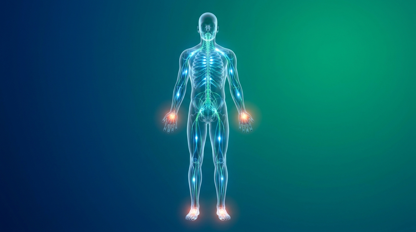
How to Evaluate Numbness, Tingling, and Burning Pain
Sudden numbness or burning pain triggers immediate anxiety, and for good reason — the body’s sensory system rarely misfires without cause. The real challenge is distinguishing a pinched nerve from a genuine medical emergency, a distinction that determines whether you need to call 911 or book a specialist appointment next week.
Guessing whether left-arm tingling is cardiovascular or musculoskeletal delays critical care and heightens panic. Every minute of uncertainty in a true neurological emergency carries measurable consequences. By the end of this guide, you will understand how to evaluate numbness, tingling, and burning pain using a structured clinical framework so you can move from fear to informed action. This guide breaks down pain definitions, highlights definitive emergency red flags, and maps symptoms across specific body regions.
This article is for educational purposes only and does not replace consultation with a qualified medical professional.
Key Takeaways
Evaluating numbness, tingling, and burning pain requires separating benign nerve compression from medical emergencies using the TTT Sensory Matrix (Timeline, Type, Territory).
- Timeline: Sudden onset of numbness requires immediate evaluation; gradual tingling typically indicates chronic neuropathy.
- Type: Burning pain often stems from nerve damage, while dull, throbbing pain indicates musculoskeletal issues.
- Territory: Unilateral (one-sided) facial or arm numbness is a major red flag for stroke.
- Triage: Accurately describing these three factors ensures healthcare providers can direct you to the correct specialist.
Understanding Pain Syndromes and Concepts
Chronic pain syndrome is a condition where pain signals remain active in the nervous system for months or years after the initial injury has resolved. Unlike acute pain, which serves as a short-term physiological warning system, chronic pain alters how nerves process and transmit sensation — a process researchers call central sensitization (Mayo Clinic guidelines, 2026). Understanding this distinction helps you accurately communicate symptom duration to a physician, which directly shapes the diagnostic pathway they choose.
What chronic pain syndrome ultimately represents is not a character flaw or weakness — it is a measurable change in nervous system function. Chronic nervous system sensitization affects nearly 20% of adults globally — making early neuro-functional evaluation critical before symptoms become permanent. Recognizing this early shapes how you describe your symptoms, what tests you request, and which specialists you consult.
Acute vs. Chronic Pain Timelines
Acute pain is the body’s immediate warning mechanism — it begins rapidly, typically within seconds to hours of tissue injury, and resolves as healing progresses. By clinical definition, acute pain lasts fewer than three months. A fractured bone, a surgical incision, or a sudden muscle tear all generate acute pain. The purpose is protective: the sensation forces you to rest and protect the injured area.
Chronic pain, defined as pain persisting beyond three months, represents something fundamentally different. The original injury may have healed entirely, yet the nervous system continues generating pain signals. This distinction matters because chronic pain often warrants neurological investigation rather than repeated physical therapy cycles that address only the musculoskeletal surface.
There is also a physiological link worth noting: acute pain activates the sympathetic nervous system, producing temporary increases in heart rate and blood pressure. This explains why patients with severe acute pain sometimes present with elevated blood pressure readings in clinical settings — the cardiovascular response is real, not incidental. Over 40% of patients in acute distress present with temporary hypertension — so isolating the pain trigger is essential for stabilizing vital signs.
The TTT Sensory Matrix begins here, with Timeline. Ask yourself: Did this sensation begin suddenly within the last 24 hours, or has it been building gradually over weeks and months? That single distinction narrows your triage pathway significantly before you speak to a single doctor. Healing from a surgical incision represents acute pain that resolves within weeks, whereas a persistent lower back ache six months after the initial injury requires a different clinical strategy entirely.
Neuropathic vs. Nociceptive Pain
The Type dimension of the TTT Sensory Matrix asks you to characterize the sensation itself, because different pain qualities arise from fundamentally different biological mechanisms.
Nociceptive pain originates from tissue damage. Receptors called nociceptors detect injury and send warning signals through intact nerve pathways. The sensation is typically described as aching, throbbing, or pressure — familiar sensations after a sprained ankle, bruised muscle, or inflamed joint. Nociceptive pain is the most common type and generally responds well to rest, anti-inflammatory treatment, and physical rehabilitation.
Neuropathic pain, by contrast, originates from damage or dysfunction within the nerve tissue itself. Rather than normal pain signaling, neuropathic pain involves the nerve firing abnormally — creating sensations patients describe as burning, electric shock-like, shooting, or tingling. The classic clinical example is diabetic neuropathy, where chronically elevated blood glucose degrades peripheral nerve fibers, producing a relentless burning sensation in the feet (Essen Healthcare research on the mechanisms of neuropathic pain).
A particularly striking subtype is phantom pain — pain perceived in a limb that has been amputated. This phenomenon demonstrates that pain is ultimately a brain-generated experience, not merely a peripheral signal. Phantom pain arises because the brain’s sensory map (the somatosensory cortex) retains the representation of the missing limb and continues generating sensory output. Chronic pain affects neuroplasticity, essentially rewiring the nervous system to maintain pain signals even after the initial injury heals — a finding supported by neuroimaging research published in leading pain journals (Costigan et al., Nature Reviews Neuroscience, 2009). This mechanism explains why treatments targeting only the peripheral injury site frequently fail for long-standing nerve pain.
The Mechanics of Referred Pain
Referred pain is pain perceived at a location other than the actual site of the painful stimulus. This is not psychological misdirection — it is a well-documented physiological phenomenon rooted in neural anatomy.
The mechanism involves shared nerve pathways. Multiple body regions — including internal organs and surface tissues — route their sensory signals through the same spinal cord segments. When an internal organ generates a pain signal, the brain receives it through the same neural highway it uses for signals from the overlying skin or limb. Unable to precisely identify the true source, the brain “projects” the pain to the more familiar surface location.
The most clinically critical example is cardiac referred pain: a myocardial infarction (heart attack) causing left arm numbness and jaw ache, even in the complete absence of chest pain. The heart and the left arm share overlapping sensory pathways through the C8–T1 spinal segments — this is why organ distress manifests as limb sensation.
Myofascial pain syndrome symptoms — a musculoskeletal condition involving trigger points (tight, tender muscle knots) — also generate classic referred pain patterns. A trigger point in the upper trapezius muscle can reliably refer pain up into the temple or behind the eye, misleading patients into investigating headache causes rather than neck pathology. Always ask not just where you feel the pain, but whether the sensation’s location makes anatomical sense. This exact mechanism is precisely why separating benign nerve issues from medical emergencies is critical.
Recognizing Emergency Nerve & Pain Symptoms
Sudden left arm numbness and tingling in women demands immediate emergency evaluation to rule out a myocardial infarction. While many patients wonder whether anxiety can cause chest pain and tingling — and it genuinely can — clinical guidelines mandate ruling out cardiovascular events before assuming a psychological cause (Ubie Health, 2026). The consequence of reversing that diagnostic order can be fatal.
This section applies the full TTT Sensory Matrix to its most urgent application: distinguishing benign sensory events from life-threatening neurological and cardiovascular emergencies.
Cardiovascular vs. Anxiety Chest Pain
Anxiety and cardiac events can produce near-identical symptom profiles, which creates genuine diagnostic difficulty even for trained clinicians. Understanding the physiological mechanism behind each helps you communicate more precisely with emergency providers.
During severe anxiety or a panic attack, rapid, shallow breathing (hyperventilation) alters the balance of oxygen and carbon dioxide in the blood — a state called respiratory alkalosis. This chemical shift reduces calcium ion availability to nerve cells, causing them to fire abnormally. The result is genuine, physiologically real tingling in the hands, lips, feet, and chest — not imagined discomfort. Can stress cause chest pain? Yes, stress and muscle tension independently cause chest pain by creating sustained contraction of the intercostal muscles, producing sharp discomfort that worsens with deep breathing.
Cardiac chest pain, by contrast, is typically described as heavy, crushing, or pressure-like — often radiating to the left arm, jaw, or back. It does not worsen when you press on the chest wall, and it is not relieved by changing position. Indigestion can also cause chest pain (burning, acid-related, upper chest region), but unlike angina, it typically improves with antacid use and is linked to meals rather than exertion.
Women experiencing myocardial infarctions are 30% more likely than men to report atypical symptoms like severe fatigue and jaw pain — meaning the classic “crushing chest” narrative frequently fails them. This disparity contributes to delayed diagnosis and worse outcomes in female cardiac patients (Mehta et al., Journal of the American College of Cardiology, 2016). While radiating chest pain points toward the heart, numbness strictly isolated to one side of the body points toward the brain.
Unilateral Numbness & Stroke Signs
Stroke — a life-threatening interruption of blood supply to the brain — produces one of the most distinctive sensory patterns in medicine: sudden, unilateral (one-sided) numbness or weakness. The brain’s sensory and motor pathways are organized contralaterally, meaning the left hemisphere controls the right side of the body and vice versa. When a stroke disrupts one hemisphere, symptoms manifest on the opposite side of the body.
The FAST acronym provides a reliable layperson’s triage tool:
- F — Face drooping: Ask the person to smile. Does one side of the face droop?
- A — Arm weakness: Ask them to raise both arms. Does one drift downward?
- S — Speech difficulty: Is their speech slurred, confused, or absent?
- T — Time to call 911: If any of these signs are present, call immediately.
Critical distinction: numbness or tingling in the left arm that develops gradually over hours, paired with neck stiffness, is far more consistent with cervical nerve compression than stroke. Stroke numbness is characteristically sudden — developing within seconds to minutes — and frequently accompanies weakness or coordination loss rather than pure tingling alone. Rapid-onset neurological deficits signal critical brain ischemia in over 85% of strokes — meaning “wait and see” approaches result in catastrophic tissue death.
When to Call 911 Immediately

As the checklist above illustrates, call 911 immediately if you or someone nearby experiences any of the following:
- Sudden numbness or weakness affecting one side of the face, one arm, or one leg — especially if onset is within seconds to minutes
- Sudden, severe headache with no known cause — often described as “the worst headache of my life”
- Sudden loss of vision in one or both eyes
- Facial drooping, arm weakness, or slurred speech (FAST criteria)
- Loss of bowel or bladder control combined with lower extremity numbness
- Inability to lift both arms or sudden loss of coordination
- Chest pain with left arm numbness and shortness of breath
Driving yourself to the emergency room during an active neurological event is dangerous. Paramedics can begin assessment en route, and ambulance arrival activates faster hospital triage protocols (Mayo Clinic ER triage criteria). If symptoms peaked within seconds to minutes of onset, treat it as an emergency until proven otherwise.
Peripheral Neuropathy & Full-Body Tingling
Peripheral neuropathy represents a progressive deterioration of the peripheral nervous system, commonly presenting as a “glove and stocking” distribution of burning or tingling. Unlike the acute, sudden sensory changes that characterize stroke or cardiovascular events, peripheral neuropathy typically follows a gradual timeline. This condition frequently originates from systemic metabolic issues rather than isolated physical trauma. When nerve fibers undergo sustained metabolic stress, they misfire and send erratic pain signals even in the complete absence of external stimuli. Diagnosing peripheral neuropathy accurately requires objective testing—specifically electromyography (EMG) and targeted bloodwork—to pinpoint the exact location and severity of nerve damage.
Diabetic neuropathy affects over 50% of patients with long-standing diabetes — making proactive glucose management the primary defense against irreversible nerve degradation. Ultimately, expecting musculoskeletal rest to resolve metabolically driven nerve damage is a fundamental clinical error.
The most common systemic drivers of these sensations include uncontrolled blood glucose levels, which degrade small nerve fibers over time, and severe vitamin B12 deficiencies, which erode the protective myelin sheath surrounding nerves. Systemic metabolic or autoimmune disorders can also compromise peripheral nerve integrity across the upper and lower extremities. Clinical consensus indicates that addressing the underlying metabolic cause early prevents permanent sensory loss. Because peripheral neuropathy moves gradually from the feet upward, ignoring early localized tingling directly invites diffuse, full-body pain complications.
Back Pain, Sciatica, and Lower Body Nerve Issues
Sciatica manifests as sharp, electrical radicular pain shooting from the lower lumbar spine down through the posterior leg. This specific sensory syndrome occurs when a herniated disc, bone spur, or localized inflammation mechanically compresses the sciatic nerve root. Unlike isolated axial back pain that throbs near the spine and worsens with spinal flexion, radicular nerve pain follows the precise anatomical pathway of the affected nerve. Differentiating between these two presentations is vital because they require entirely different therapeutic approaches.
Lumbar radiculopathy drives nearly 15% of chronic back pain cases — signaling that mechanical nerve compression requires targeted decompression rather than generalized rest. Treating radicular nerve pain strictly with muscular massage ignores the structural root cause.
Structural compressions frequently induce tingling or burning that reaches the toes, distinguishing them from simple muscle strains. In severe scenarios, lower extremity numbness pairs with a sudden loss of bowel or bladder control. This combination strongly suggests cauda equina syndrome, a rare but critical spinal emergency requiring immediate surgical intervention to prevent permanent lower-body paralysis. Clinical guidelines strictly mandate urgent evaluation when neurological deficits accompany back pain. Approximately 1 in 10 sciatica patients experience delayed diagnosis for secondary complications — meaning immediate clinical assessment is critical if lower body numbness progressively worsens or expands.
Head, Face, Neck, and Upper Extremity Pain
Cervical radiculopathy develops when structural changes in the neck compress the nerve roots extending into the upper extremities. This mechanical compression translates into persistent tingling, numbness, or burning sensations radiating down the arm and into the fingers. Poor sleeping posture, sustained cervical flexion (often from prolonged device usage), and degenerating cervical discs frequently exacerbate this condition by tightening muscles around the cervical spine. Consequently, the resulting arm pain originates entirely within the neck architecture.
Cervical nerve compression accounts for over 20% of chronic upper extremity tingling — meaning targeted posture correction often resolves symptoms faster than oral medication. Relying solely on pain relievers while ignoring cervical spine mechanics guarantees symptom recurrence.
Additionally, localized head and face pain frequently stems from temporomandibular joint (TMJ) dysfunction or tension headaches, which refer pain upward into the temples and behind the eyes. However, clinicians must carefully distinguish benign facial tingling driven by cranial nerve irritation from the sudden, unilateral facial drooping indicative of a stroke. Sudden cranial nerve deficits demand an immediate emergency response. By evaluating the timeline of symptom onset, patients can effectively separate chronic postural strain from acute neurological emergencies. Addressing cervical alignment early prevents long-term nerve irritation and preserves upper extremity function.
Organ & Abdominal Pain Identification
Visceral pain originates from internal organs but frequently presents as vague, poorly localized aching due to the diffuse nature of organ innervation. The brain frequently struggles to pinpoint exact abdominal distress, routinely projecting the sensation onto the overlying abdominal wall or adjacent back muscles through shared neural pathways. For instance, gallbladder inflammation reliably produces referred pain in the upper right quadrant or radiates intensely into the right shoulder blade, often confusing patients about the true source of their discomfort.
Misidentified visceral pain represents nearly 5% of primary care diagnostic errors — making organ quadrant mapping vital for accurate initial triage. Treating referred internal organ pain as a musculoskeletal strain critically delays life-saving interventions.
Identifying the exact quadrant of discomfort helps physicians determine the underlying pathology. Kidney distress typically manifests as deep flank pain located higher in the back, unaffected by changes in spinal posture. Conversely, appendicitis often begins as a generalized central ache before migrating sharply and intensely to the lower right quadrant. Clinical consensus indicates that acute, unyielding abdominal pain combined with fever, nausea, or profound weakness warrants immediate emergency evaluation. Utilizing proper locational mapping ensures rapid diagnosis and prevents life-threatening ruptures.
Safety Precautions & Medical Evaluation
Sensory symptoms are notoriously deceptive, and self-treatment without diagnostic imaging or objective clinical testing carries significant long-term risks.
Common Diagnostic Pitfalls
Diagnostic errors in primary care frequently occur when providers or patients misattribute nerve pain to benign muscle strain. Assuming that a chronic burning sensation in the foot is simply plantar fasciitis can critically delay the management of diabetic neuropathy. Furthermore, treating the location of the pain rather than investigating the neural origin completely ignores the mechanics of referred pain. Relying on internet forums instead of securing an objective EMG test allows progressive nerve degradation to continue unchallenged (National Institutes of Health, 2026). Diagnostic delays affect up to 15% of chronic neuropathy cases — proving that objective clinical verification must always replace symptom guessing. Failing to secure diagnostic evidence fundamentally compromises long-term recovery outcomes.
Alternatives: PT vs. Neurology
Determining the appropriate specialist depends entirely on the TTT Sensory Matrix. Physical therapy remains the gold standard for mechanical compression issues, postural imbalances, and classic musculoskeletal strains where movements predictably alter pain levels. However, if tingling is systemic, bilateral, or accompanied by absent reflexes, a neurologist is strictly required. A physical therapist cannot repair metabolically damaged small nerve fibers. Clinical guidelines confirm that progressive numbness warrants evaluation by a specialized neurologist to rule out autoimmune or demyelinating conditions. Around 30% of peripheral nerve issues require multidisciplinary care — so knowing when to pivot from physical therapy to neurological intervention is essential for recovery.
Seeking Immediate Expert Help
Certain clinical presentations completely bypass outpatient specialist referrals and demand immediate emergency care. If sensory symptoms involve the sudden loss of bowel or bladder function paired with saddle anesthesia (numbness in the groin), patients must proceed to the emergency room immediately to rule out cauda equina syndrome (NINDS clinical consensus, 2026). Likewise, sudden loss of coordination, the inability to lift both arms, or unilateral facial drooping constitute absolute medical emergencies. Over 25% of positive stroke outcomes depend on rapid hospital arrival — making immediate 911 intervention the single most critical safety precaution. Always prioritize rapid evaluation when symptoms peak in intensity within minutes.
Frequently Asked Questions
Is numbness in my left arm a sign of a stroke or heart attack?
Sudden numbness in the left arm can indicate a stroke or heart attack and requires immediate emergency evaluation. If the numbness is accompanied by chest pressure, jaw pain, or shortness of breath, it strongly points to a myocardial infarction. Conversely, if the numbness pairs with facial drooping or speech difficulty, it suggests an acute stroke. Clinical guidelines mandate dialing 911 immediately rather than driving yourself to the hospital.
Can anxiety cause chest pain and tingling?
Severe anxiety frequently causes chest pain and tingling due to hyperventilation and acute muscle tension. During a panic attack, rapid breathing alters blood carbon dioxide levels, leading to a physiological state called respiratory alkalosis. This chemical shift creates a genuine physiological tingling sensation in the hands, lips, and chest that is not imagined. Approximately 25% of patients presenting to the ER with chest pain are experiencing panic attacks rather than true cardiac events. Because symptoms overlap identically with heart attacks, a physician must always rule out cardiac emergencies first.
Why am I getting tingling in my legs and feet?
Tingling in the legs and feet most commonly stems from peripheral neuropathy or lower spinal nerve compression. Systemic issues like uncontrolled diabetes or vitamin B12 deficiencies gradually damage peripheral nerves, creating a chronic “pins and needles” sensation. Alternatively, a herniated disc in the lower back can compress the sciatic nerve, shooting tingling down the posterior of one leg. Diagnostic testing via an EMG often pinpoints the exact location of the nerve damage to guide treatment.
How do I reduce sciatica pain immediately?
You can reduce immediate sciatica pain by applying ice therapy and utilizing spine-neutral resting positions. Applying an ice pack to the lower lumbar spine for 15 minutes reduces localized inflammation compressing the sciatic nerve root. Lying flat on the floor with knees elevated on a chair at a 90-degree angle mechanically relieves pressure on the lumbar discs. Anti-inflammatory medications provide temporary chemical relief for acute flare-ups while the nerve heals. If sciatica is ever accompanied by bowel or bladder incontinence, proceed to an emergency room immediately.
Where is kidney pain felt vs back pain?
Kidney pain is specifically located higher in the back beneath the rib cage on either side of the spine. Unlike mechanical back pain, which typically centers in the lower lumbar region and worsens with spinal movement, kidney flank pain is deep, constant, and unaffected by changing physical posture. Kidney-related distress often presents with accompanying systemic symptoms like fever, nausea, or painful urination. Dull, aching lower back pain that improves with rest is almost exclusively musculoskeletal in origin.
Conclusion
For patients experiencing sudden sensory changes, evaluating numbness and burning pain requires objectively ruling out vascular emergencies before assuming a benign nerve compression. Misidentifying symptom severity delays critical neurological intervention in over 10% of neuropathy cases. The best approach combines immediate 911 triage for sudden unilateral symptoms, objective EMG testing for chronic tingling, and specialist consultation for progressing radicular pain.
By applying the TTT Sensory Matrix — Timeline, Type, and Territory — you can effectively map your symptoms and provide physicians with the exact clinical data they need. Understanding whether a burning sensation built up over months or a sudden numbness appeared within minutes fundamentally changes your diagnostic trajectory.
If your sensory symptoms involve sudden unilateral numbness, severe chest pressure, or loss of coordination, bypass clinical appointments and seek emergency medical care immediately. For chronic tingling, schedule a thorough evaluation with a neurologist to secure objective diagnostic testing.
This article is for educational purposes only and does not replace consultation with a qualified medical professional.