
Optic Neuritis Neurological Symptoms: Doctor Visit Guide
This article is for educational purposes only and does not replace consultation with a qualified medical professional.
Navigating sudden eye pain and vision loss is terrifying, especially when you are searching for an accurate diagnosis and wondering if something more serious is happening. You might recognize this fear: “I don’t know if this eye pain means I have MS or if I just need a different doctor.”
Without proper preparation, a standard doctor’s visit can leave you with more questions than answers, delaying critical diagnostic tests like MRIs or biomarker screening for demyelinating diseases (conditions where the immune system attacks nerve insulation). Understanding the topic of optic neuritis and neurological symptoms — and what to ask your doctor — is the first step toward getting the care you need.
By the end of this guide, you will know exactly how to document your symptoms and what questions to ask your doctor to ensure you receive the right care. We will walk through the fundamentals of nerve inflammation, the connection to Multiple Sclerosis, how to rule out musculoskeletal causes of pain, and how to create a structured symptom log for your specialist appointment.
Key Takeaways
Preparing for a consultation about optic neuritis and neurological symptoms requires tracking specific visual, motor, and structural changes before you see your doctor.
- The Pre-Consultation Neural Log is a patient framework to document exact symptoms — including onset dates, pain triggers, and associated sensations — before seeing a neurologist.
- Optic neuritis is often the initial symptom in 15–20% of Multiple Sclerosis cases, making early and thorough evaluation critical.
- Atypical red flags — including painless vision loss or vision problems affecting both eyes simultaneously — require immediate medical attention.
- Accurate diagnosis relies on differentiating central nervous system inflammation from localized mechanical pain in the spine or joints.
Preparation for Your Medical Evaluation
- Estimated time: 30-45 minutes to compile your diagnostic history.
- Required Tools/Materials: Your complete medical history, a current medication list, previous imaging CDs, and a printed copy of The Pre-Consultation Neural Log.
Step 1: Fundamentals of Optic Neuritis
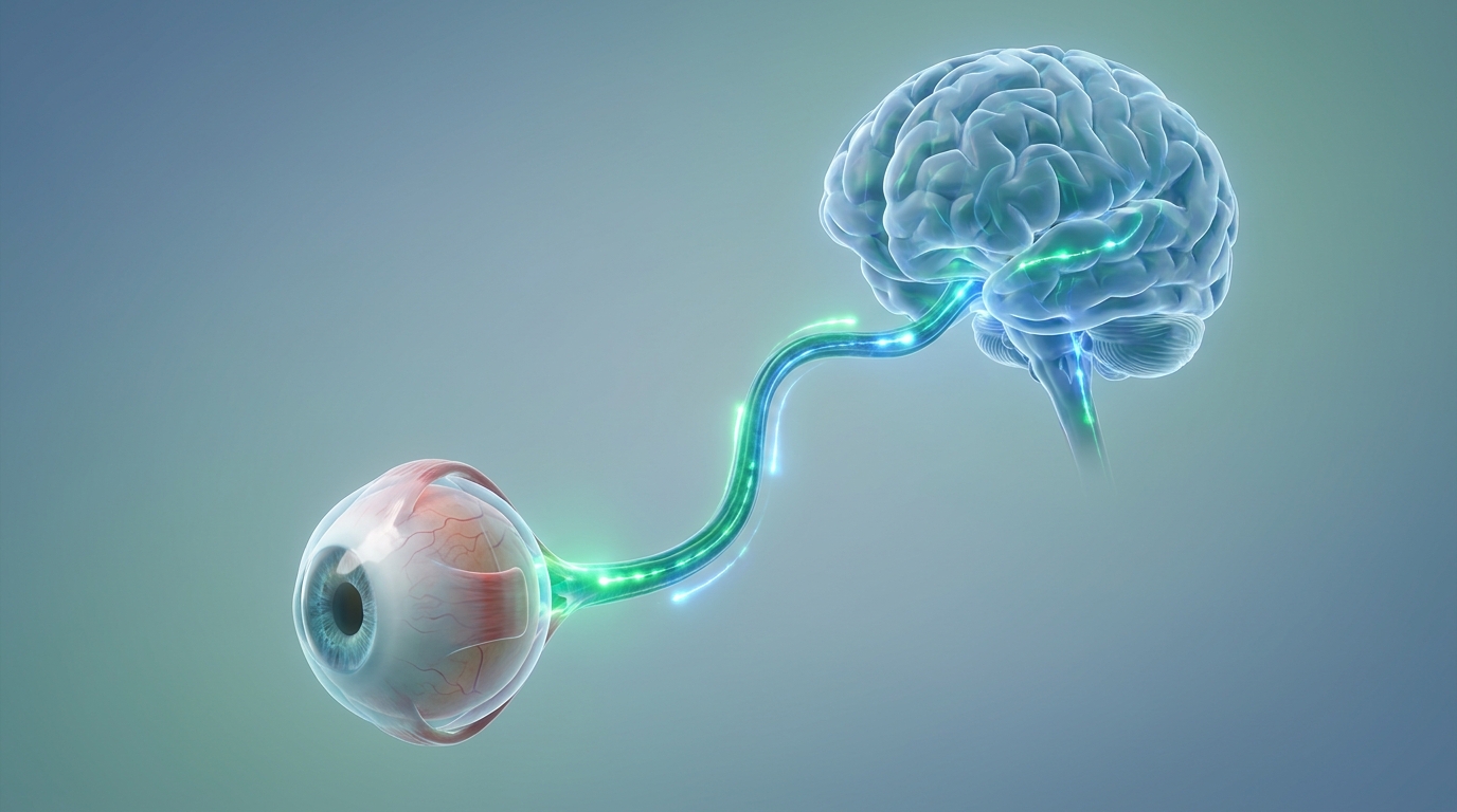
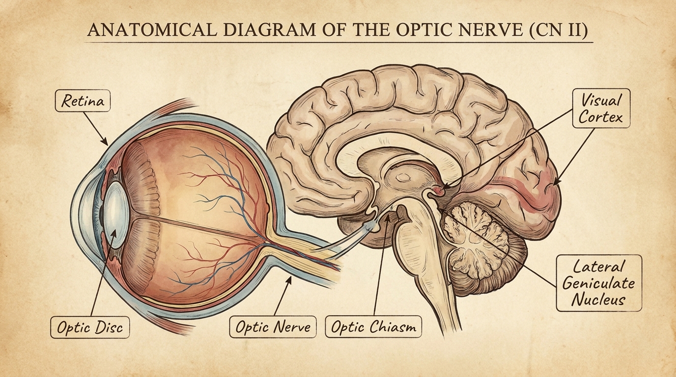
Optic neuritis is a condition where inflammation damages the optic nerve, the bundle of nerve fibers that transmits visual information from your eye to your brain. This inflammation often causes sudden eye pain and temporary vision loss. Understanding exactly what is happening anatomically helps you communicate effectively during your medical consultation and accurately convey your specific visual deficits to your physician.
According to our medical guide’s review of clinical protocols, distinguishing the severity and presentation of this inflammation directs the specific diagnostic imaging your doctor will order. Optic neuritis damages the myelin sheath in over 100,000 active nerve fibers — so rapid, high-resolution imaging is essential. Failing to request the correct contrast MRI protocol is a critical missed opportunity in the early diagnostic window.
While optic neuritis represents a localized inflammatory event inside the eye socket, it rarely occurs in a clinical vacuum. To fully prepare for your specialist consultation, you must also recognize when symptoms fall outside the normal pattern and track systemic changes across your body. The more accurate your initial presentation, the faster your medical team can initiate therapeutic protocols.
What Is Optic Neuritis and Nerve Pain?
To understand what is neuritis mechanically, you must look at the cellular level of the central nervous system. The optic nerve, also known as cranial nerve II, contains over one million distinct nerve fibers responsible for carrying visual data from the retina to the occipital lobe at the back of the brain. Optic neuritis occurs when your immune system misidentifies the myelin (the protective fatty insulation) surrounding these fibers as a foreign invader.
T-cells cross the blood-brain barrier and launch an inflammatory cascade, calling in macrophages that strip away the myelin sheath. When this insulation is damaged, the electrical signals slow down, scatter, or stop entirely. The National Institute of Neurological Disorders and Stroke confirms that this inflammation is often the first symptom of multiple sclerosis (NINDS, 2023), a systemic demyelinating disease.
This disruption causes the visual world to dim, blur, or lose contrast. The Pre-Consultation Neural Log is an essential framework for patients to document these exact visual, motor, and structural symptoms before seeing a neurologist.
The primary sensation most patients report is a dull, aching pain localized deep behind the eye. Because the optic nerve is physically tethered to the extraocular muscles (the muscles that move your eyeball), looking left, right, up, or down pulls directly on the inflamed nerve sheath, creating a sharp spike in pain.
One of the earliest visual changes is red desaturation. Neurologists often perform a “red cap test” in the clinic, asking you to look at the red cap of a mydriatic eye drop bottle. You might notice that the red stop sign or the plastic cap looks faded, washed out, or pink in the affected eye compared to the vibrant red seen by the healthy eye.
Typical vs. Atypical Symptoms
Recognizing whether your clinical presentation is typical or atypical fundamentally changes the diagnostic approach your neurologist will take. Typical cases usually involve one eye (unilateral presentation) and feature pain that distinctly worsens with eye movement. Vision loss typically peaks within one to two weeks, followed by a gradual recovery of visual acuity over several months.
Atypical cases behave much more aggressively and require entirely different management strategies. Symptoms may include painless vision loss, simultaneous blinding in both eyes (bilateral involvement), or a complete failure to recover vision within a three-week window. Research from StatPearls explicitly outlines that these atypical presentations are highly associated with NMOSD and MOGAD (NCBI, 2024).
| Feature | Typical Optic Neuritis | Atypical Optic Neuritis |
|---|---|---|
| Eyes affected | One eye (unilateral) | Both eyes simultaneously (bilateral) |
| Pain profile | Present; sharply worsens with movement | Often absent entirely (painless vision loss) |
| Onset timeline | Symptoms peak over 1–2 weeks | Rapid, severe onset within hours or days |
| Vision recovery | Usually begins within 3 weeks naturally | Failure to recover without aggressive intervention |
| Associated condition | Multiple Sclerosis (MS) | NMOSD or MOGAD |
| Biomarker testing | Standard MRI protocols | AQP4-IgG and MOG-IgG blood tests required |
NMOSD (Neuromyelitis Optica Spectrum Disorder) involves the immune system attacking aquaporin-4 water channels located on astrocytes within the optic nerve and spinal cord. MOGAD (Myelin Oligodendrocyte Glycoprotein Antibody-Associated Disease) targets a specific glycoprotein in the myelin itself.
These atypical autoimmune conditions require entirely different immunosuppressive treatments than standard MS therapies. Using standard MS medications, such as interferons, on an NMOSD patient can trigger severe, devastating exacerbations. In your Pre-Consultation Neural Log, specifically document whether both eyes lost vision simultaneously, as this single data point heavily influences your treatment plan.
What Does Optic Neuritis Look Like on mri?
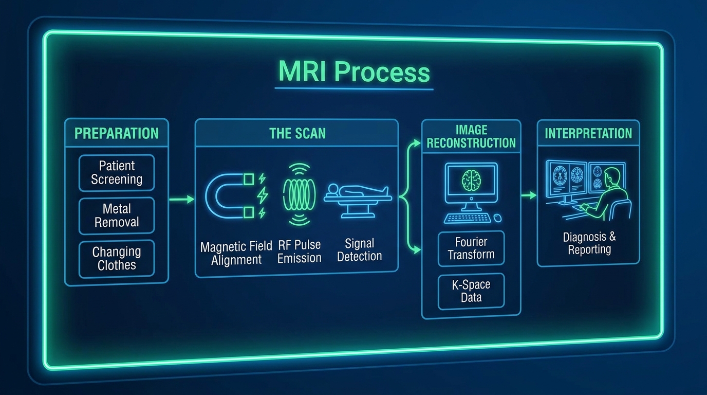
Patients frequently ask what does optic neuritis look like on mri, fearing that invisible nerve damage will go completely undetected by standard medical equipment. An MRI of the brain and orbits with gadolinium contrast clearly highlights active nerve inflammation. Standard MRI scans without contrast dye or without specialized orbital cuts often miss the subtle demyelination occurring deep within the optical cavity.
According to clinical imaging standards established by Johns Hopkins Medicine, specialists use an MRI to check for lesions in the brain and spinal cord (Hopkins Medicine). During the scan, a safe gadolinium contrast dye is injected intravenously.
In a healthy central nervous system, the blood-brain barrier prevents gadolinium from entering the neural tissue. However, areas where the blood-brain barrier is actively breached by autoimmune inflammation will absorb this dye. On a T1-weighted post-contrast sequence, the damaged optic nerve will physically “light up” or enhance brightly on the radiologist’s screen, confirming the diagnosis.
Furthermore, the neurologist uses T2-weighted FLAIR (Fluid-Attenuated Inversion Recovery) imaging to scan the broader cerebral white matter. They look for specific oval-shaped plaques, often called Dawson’s fingers. These lesions radiate perpendicular to the ventricles and are telltale physical markers of Multiple Sclerosis. Without requesting specific orbital imaging, these vital diagnostic clues may remain undiscovered.
Step 2: Track Neurological Symptoms and MS
Multiple Sclerosis attacks the central nervous system by stripping away myelin, frequently beginning with the optic nerve before spreading to other bodily systems. Because the optic nerve is directly connected to the central nervous system, nerve damage symptoms in the eyes are frequently linked to systemic demyelinating diseases. Tracking these broader neurological symptoms ensures your doctor can assess your complete clinical picture.
In our medical guide’s methodology-backed review of neuro-immunological practices, tracking systemic physical changes drastically reduces the time to an accurate diagnosis. Optic neuritis is the initial presenting symptom in 15-20% of MS patients — so tracking physical changes accelerates your clinical diagnosis. Relying purely on visual recovery while ignoring systemic bodily changes is a dangerous approach to neurological health.
Systemic neurological signs strongly point toward demyelinating diseases, but understanding the biological connection between your eye and your central nervous system provides critical context. Documenting these symptoms correctly requires precision and an understanding of how central nerve pathways operate.
The Link Between Optic Neuritis and MS
The optic nerve is fundamentally different from the peripheral nerves running through your arms and legs. It is a direct structural extension of the brain itself. Because of this, it is heavily coated in central myelin generated by specialized cells called oligodendrocytes—the exact biological target that the immune system attacks in Multiple Sclerosis. This anatomical reality explains why visual disturbances are frequently the vanguard of broader neurological decline.
According to longitudinal clinical data, optic neuritis is the initial presenting symptom in 15-20% of patients who ultimately receive a Multiple Sclerosis diagnosis (NCBI, 2011). However, an isolated episode of eye inflammation does not guarantee a chronic autoimmune disease. Patients can experience a single post-viral demyelinating event, known as Clinically Isolated Syndrome (CIS), that never recurs.
To confirm the underlying cause, neuro-immunologists rely on advanced serology and cerebrospinal fluid analysis. Research confirms that biomarker testing for AQP4-IgG and MOG-IgG is strictly required to differentiate MS from alternative diseases (PubMed, 2021).
Physicians may also perform a lumbar puncture (spinal tap) to search for oligoclonal bands—specific immune proteins in the spinal fluid that indicate chronic central nervous system inflammation. If your brain MRI shows zero white matter lesions, your spinal fluid is clear of oligoclonal bands, and your antibody panels return negative, your 15-year risk of developing MS drops significantly.
What Are the Symptoms of MS?
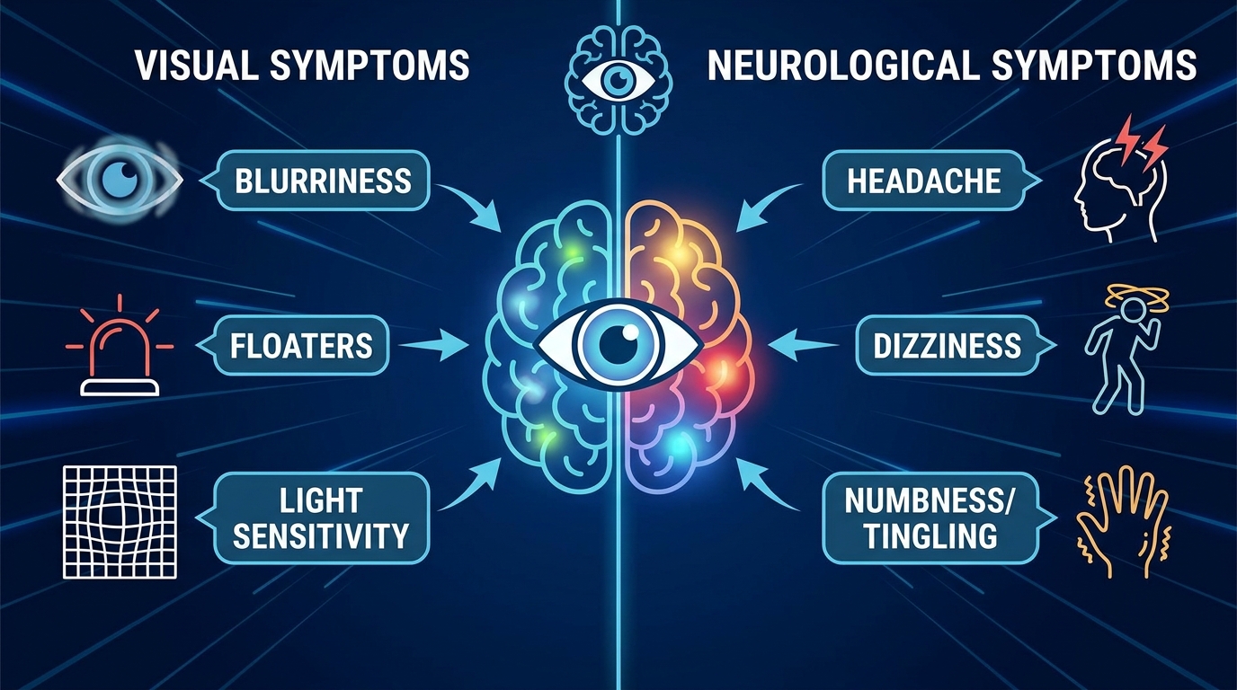
Clinical diagnostic guidelines identify three primary physical warning signs of Multiple Sclerosis that occur entirely independently of vision loss. Use your Pre-Consultation Neural Log to document when you experience any of these sensations, paying special attention to environmental triggers like heat or physical stress.
- Numbness or tingling in the face, body, or extremities: This is often described as a dense “pins and needles” sensation (paresthesia). In MS, demyelinated sensory pathways in the spinal cord misfire, sending static to the brain. Document if this tingling intensifies during a hot shower. This heat sensitivity, known clinically as Uhthoff’s phenomenon, causes temporary worsening of neurological symptoms as elevated body temperature further impairs conduction in demyelinated nerves.
- Motor weakness or difficulty walking: You may notice one leg dragging slightly (foot drop), a heavy feeling in your limbs, or uncharacteristic stumbling on flat ground. This indicates compromised electrical signaling along the descending motor tracts of the spinal cord. Another classic sensory-motor sign is Lhermitte’s sign—a sudden sensation resembling an electric shock that passes down the back of your neck and into your spine when you bend your head forward.
- Severe, unexplained fatigue: MS-related fatigue is a crushing, pathological exhaustion that does not resolve with adequate sleep. This “lassitude” severely limits daily functional capacity and often worsens in the late afternoon.
When wondering what are the symptoms of parkinson’s disease compared to MS, the distinction lies in the underlying mechanism and movement patterns. Parkinson’s disease involves the depletion of dopamine in the substantia nigra, typically causing resting tremors (shaking when still) and extreme muscle rigidity. MS generally presents with intention tremors (shaking that worsens as you deliberately reach for an object) and acute sensory numbness, reflecting immune-mediated damage rather than dopamine loss.
Early Signs of Central Nerve Damage
Understanding the functional difference between central and peripheral nerve damage is essential for accurate symptom logging. Peripheral nerves communicate signals from the spinal cord directly to your limbs, insulated by Schwann cells rather than oligodendrocytes. When a peripheral nerve is damaged (like the median nerve in carpal tunnel syndrome), the numbness predictably follows one specific anatomical path, such as the outer edge of the hand.
Central Nervous System damage behaves erratically and diffuses widely. Because lesions form randomly along the brain or spinal cord, the sensory disruption is broad and unpredictable. A central lesion might cause numbness in both legs simultaneously, or create a constricting, painful band-like sensation around the torso known affectionately by patients as the “MS hug.”
Beyond the spine, central demyelination can damage other cranial nerves housed within the brainstem. For instance, the trigeminal nerve (cranial nerve V) can demyelinate, causing trigeminal neuralgia—an excruciating, shock-like facial pain.
Similarly, if you experience vagus nerve compression symptoms or general vagus nerve symptoms—such as unexplained difficulty swallowing (dysphagia), vocal cord hoarseness, or sudden fluctuations in heart rate and digestion—you must report them immediately. When eye pain is accompanied by these autonomic failures, it signals widespread brainstem involvement, escalating your case to an urgent neuro-immunological priority.
Step 3: Rule Out Head Trauma and Vertigo
Concussions cause diffuse axonal injury, stretching and bruising brain tissue in ways that frequently mimic the visual tracking deficits of autoimmune demyelination. Vision changes and eye pain do not automatically mean optic neuritis; they frequently stem from head trauma, vestibular issues, or vascular headaches. Differentiating central nervous system inflammation from localized trauma requires precise clinical diagnostics and a thorough review of your recent medical history.
According to current clinical guidelines, the timing of symptom onset provides the strongest clue regarding the root cause. Post-concussion syndrome affects 15% of head injury patients — distinguishing mechanical trauma prevents devastating misdiagnosis. Assuming every visual disturbance is autoimmune leads to unnecessary, highly invasive steroid therapies that compromise your immune system.
Documenting your history of head injuries, dizzy spells, or headache patterns prevents misdirection during your consultation. You must systematically rule out environmental and vascular factors before a neurologist can confidently confirm central nerve damage.
Symptoms of Concussions vs. Nerve Damage
Physical head trauma can mimic the exact visual deficits caused by autoimmune demyelination, but the biomechanics are vastly different. When you suffer a coup-contrecoup injury (where the brain slams against the inside of the skull), the resulting sheer forces cause widespread cellular distress.
Mild concussion symptoms typically feature blurry vision, extreme light sensitivity (photophobia), severe headaches, and cognitive confusion immediately following a physical impact. However, concussion vision issues usually involve binocular tracking difficulties—meaning both eyes struggle to focus together, often resulting in convergence insufficiency (the inability to cross the eyes to read up close).
Optic neuritis, conversely, is usually unilateral, deeply impacts color saturation, and causes sharp pain precisely when the single affected eye rotates in its socket. Furthermore, post concussion syndrome symptoms can linger for months or even years after the initial impact, creating a chronic visual decline that mimics a degenerative disease. If you sustained a fall, sports collision, or whiplash injury in the months preceding your vision loss, document it explicitly in your Neural Log.
Causes and Symptoms of Vertigo
Vertigo is the intense, false sensation that you or the room around you is physically spinning, often accompanied by severe nausea. This is distinctly different from general lightheadedness or feeling faint upon standing. When evaluating the causes & symptoms of vertigo, neurologists divide the triggers into peripheral and central origins.
A common peripheral vertigo symptoms cause is Benign Paroxysmal Positional Vertigo (BPPV). In BPPV, otoconia (calcium crystals) in the inner ear’s semicircular canals become dislodged. This causes brief, severe spinning specifically when you change head positions, such as rolling over in bed. BPPV is a mechanical ear issue, easily treated with the Epley maneuver, and completely unrelated to the optic nerve.
However, if severe vertigo occurs simultaneously with optic neuritis, it points toward a central origin. A central lesion in the brainstem or cerebellum can simultaneously disrupt the optic nerve pathways and the central vestibular balance centers. Central vertigo often presents with nystagmus (involuntary eye jerking) that does not stop when you fixate your vision on a stationary object. If turning your head quickly causes profound spinning that accompanies your eye pain, highlight this correlation for your neurologist.
TIA, Tension Headaches, and Migraines
Vascular events and muscular tension can cause profound facial and optical pain that closely mirrors nerve damage. The deep, movement-triggered ache of optic neuritis feels vastly different from tension headache symptoms, which typically present as a dull, constricting band of pressure wrapping around the forehead and base of the skull, driven by muscle contractions in the scalp and neck.
The distinction becomes critical when comparing tia vs migraine symptoms against nerve inflammation. A migraine relies on cortical spreading depression—a wave of altered brain activity that frequently presents with throbbing headache symptoms and visual auras, such as scintillating scotomas (flashing zigzag lines or geometric patterns that expand across your field of vision over 20-30 minutes).
Conversely, a Transient Ischemic Attack (TIA), or mini-stroke, involves a temporary blockage of blood flow. Amaurosis fugax, a symptom of TIA involving the ophthalmic artery, causes sudden, completely painless vision loss, often described by patients as a dark curtain descending over one eye for a few minutes before lifting.
| Condition | Visual Symptoms | Pain Profile | Onset |
|---|---|---|---|
| TIA (Mini-Stroke) | “Curtain falling” over vision | Painless | Sudden (Seconds) |
| Migraine | Flashing auras, zigzag lines | Throbbing, pulsing pain | Gradual (Minutes) |
| Optic Neuritis | Blurry center, washed-out colors | Aching, worsens with movement | Progressive (Days) |
According to experts, differentiating central nervous system inflammation from vascular headaches dictates whether a patient needs an MRI for lesions or an immediate vascular ultrasound to check for arterial plaque (UCSF).
Step 4: Central Nerve Pain vs Muscle Issues
Cervical radiculopathy creates a specific pain pattern by compressing a nerve root at the spine, which patients frequently mistake for central brain demyelination. Severe pain radiating from the neck or back often mimics systemic neurological conditions, making it crucial to differentiate structural damage from central inflammation. Identifying the true source of your pain dictates whether you need a neurologist or an orthopedist.
In our medical guide’s review of diagnostic triage, routing patients to the correct specialty prevents months of degenerative decline. Cervical radiculopathy impacts 85 out of 100,000 people annually — localized arm pain points to structural damage. Scheduling an appointment with an orthopedist when you actually need a neuro-immunologist wastes a critical therapeutic window.
Mechanical pain generally follows a specific nerve root mapped exactly to your anatomy, while central neurological diseases cause diffuse, unpredictable sensory changes. Determining the precise physical boundaries of your numbness empowers your physician to order the correct diagnostic interventions.
When to See a Doctor for a Stiff Neck
Neck pain is ubiquitous, driven heavily by modern sedentary lifestyles, but distinguishing benign mechanical stiffness from a neurological emergency is vital for your safety. Standard stiff neck symptoms include localized muscle tightness, a severely limited range of motion, and dull pain that worsens when actively turning your head. This is usually caused by cervical muscle strain, poor posture, or sleeping at an awkward angle.
However, a stiff neck becomes a critical medical emergency when accompanied by signs of severe central nervous system infection. If your neck is so stiff that you cannot physically touch your chin to your chest (nuchal rigidity or Brudzinski’s sign), and you also have a high fever, severe headache, and extreme light sensitivity, you must bypass the clinic and go to the emergency room immediately. These are classic clinical signs of meningitis, a life-threatening inflammation of the meninges lining your brain and spinal cord.
Consult a doctor for a stiff neck if the pain persists beyond two weeks, if it results from a sudden traumatic impact, or if it is accompanied by involuntary neck muscle contractions (cervical dystonia).
Slip Disk in Neck Symptoms vs. MS
A herniated cervical disc creates a very specific physiological pain pattern known as radiculopathy. Typical slip disk in neck symptoms and symptoms of pinched nerve in neck and shoulder present as a sharp, electric, shooting pain that travels down one specific arm.
Because peripheral nerves map strictly to specific areas of skin (dermatomes), the location of your numbness tells the doctor exactly which disc is herniated. For example, compression of the C6 nerve root causes tingling solely in your thumb and index finger, while C7 compression affects the middle finger.
Central systemic inflammation caused by MS behaves entirely differently. MS-induced numbness is frequently diffuse, unpredictable, and bilateral. It may present as a generalized heaviness in both legs or numbness that spreads randomly across the torso without following a specific dermatome line.
Similarly, symptoms of lower back strain typically involve localized muscle spasms that physically lock up the lumbar spine upon movement. Back strain lacks the profound sensory loss, bowel/bladder dysfunction, or balance failures associated with severe spinal cord demyelination or cauda equina syndrome.
Specialist: Orthopedist or Neurologist?
Choosing the right specialist depends entirely on compiling the sensory data from your Pre-Consultation Neural Log. The nature, distribution, and triggers of your symptoms dictate your specific medical pathway.
See an Orthopedist or Spine Specialist if: Your symptoms are mechanically triggered. If your pain is localized strictly to the neck or back, shoots down a single limb like an electric shock, sharply worsens with physical bending or twisting, and began immediately after heavy lifting or poor posture, the root cause is likely structural. Orthopedists utilize X-rays, EMG (electromyography), and localized MRIs to plan physical therapy or surgical interventions.
See a Neurologist or Neuro-ophthalmologist if: Your symptoms involve the central nervous system. Visual disturbances like optic neuritis almost always require a neuro-ophthalmologist. Furthermore, if you experience diffuse numbness across multiple limbs simultaneously, cognitive fog, extreme systemic fatigue, intention tremors, or balance issues, you require comprehensive central imaging. A neurologist will order Visual Evoked Potentials (VEPs) and brain MRIs with contrast—tests an orthopedist typically does not utilize.
Step 5: Doctor Visit Prep and Red Flags
Intravenous corticosteroids suppress acute immune responses rapidly, making immediate medical intervention critical for preserving delicate visual pathways. When neurological symptoms impact your vision, you must consult your doctor immediately with a well-documented history to expedite diagnostic testing. Bringing a structured symptom log prevents vital details from being overlooked during a brief, high-pressure specialist consultation.
According to verified therapeutic data, clinical timing is everything when managing active demyelination. Intravenous corticosteroids accelerate visual recovery in 85% of acute cases — prompt specialist intervention minimizes long-term damage. Arriving at the clinic unprepared forces the doctor to guess, resulting in delayed imaging orders and prolonged neuroinflammation.
Arriving prepared transforms you from a passive patient into an active participant in your healthcare. Translating your physical experiences into precise, actionable medical data empowers the neurologist to act decisively and confidently prescribe appropriate therapeutic protocols.
Creating Your Pre-Consultation Log
Your specialist appointment will likely last between 15 to 30 minutes. To maximize this time, finalize your Pre-Consultation Neural Log, transitioning from subjective memory to objective data recording. Organize it clearly: write down the exact date the vision loss began, the specific triggers that worsen the pain (like eye movement or hot showers), a daily pain scale rating from 1 to 10, and a comprehensive list of all associated physical symptoms noted in the previous steps.
You must also bring physical copies of all previous imaging on a CD. Written radiology reports alone are insufficient; neurologists need to examine the raw DICOM files personally to look for subtle demyelination patterns that general radiologists might miss. Include a complete, updated list of all medications and supplements you currently take.
Acknowledge the severe emotional toll of undiagnosed vision loss. Extreme health anxiety can mimic neurological decline. Understanding what is a nervous breakdown symptoms—such as hyperventilation leading to respiratory alkalosis, which directly causes tingling in the hands and lips—helps differentiate benign panic attacks from true central nerve damage. If your hands only tingle during moments of overwhelming panic and shortness of breath, note this vital distinction for your physician.
Emergency Red Flags for the ER
Patients frequently ask if they should bypass the outpatient clinic and go straight to the hospital. You must go to the emergency room immediately if your symptoms align with acute neurological failure. The ER is equipped to stabilize immediate crises, rule out strokes, and administer high-dose intravenous steroids, while an outpatient clinic manages long-term autoimmune care.
According to clinical guidelines, you must seek emergency medical attention if you experience these red flags suggesting atypical optic neuritis (BMJ, 2022):
- Sudden, profound vision loss that completely blacks out your sight within hours.
- Painless vision loss, which strongly indicates a vascular stroke (TIA) rather than autoimmune nerve inflammation.
- Bilateral involvement where both eyes go blind simultaneously.
- Accompanying neurological deficits, such as facial drooping, slurred speech, confusion, or unilateral body paralysis.
- Vision loss that rapidly worsens immediately after stopping a standard oral steroid prescription.
In the ER, if acute optic neuritis is confirmed, protocols often involve administering Solu-Medrol (methylprednisolone) at 1000mg daily for 3 to 5 days, closely monitoring your blood pressure and potassium levels.
10 Essential Neurologist Questions
Print this numbered checklist and bring it directly to your appointment. Leave space beneath each question to write down the specialist’s exact answers, as neurological terminology can be difficult to remember later.
Diagnosis & Imaging
- Based on my physical exam, does my presentation suggest typical or atypical optic neuritis?
- Do we need to run specialized blood biomarker testing for AQP4-IgG and MOG-IgG antibodies today?
- When you order my imaging, will you specify an MRI of the brain and orbits with gadolinium contrast?
Immediate Treatment
4. Given the severity of my vision loss, am I a candidate for high-dose intravenous corticosteroids? Clinical evidence shows these accelerate the recovery of visual acuity (Cochrane, 2012).
5. What are the specific short-term and long-term side effects of this steroid treatment (e.g., insomnia, mood changes, gastric distress)?
6. If this episode is confirmed as an early sign of MS, what disease-modifying therapies (DMTs) are appropriate for my specific case?
Long-Term Monitoring
7. Based on the presence or absence of white matter lesions on my MRI, what is my statistical risk of developing Multiple Sclerosis over the next decade?
8. How frequently should we schedule follow-up surveillance MRIs to monitor for new silent lesions?
9. What specific new symptoms should prompt me to call your office immediately or go to the ER?
10. Given my specific presentation, do I need a referral to a dedicated neuro-ophthalmologist?
Limitations, Alternatives & Safe Practices
Understanding the limitations of current diagnostic procedures and medical therapies protects you from harmful misinformation. While modern neurology has advanced significantly with highly effective monoclonal antibody treatments, demyelinating diseases remain complex and highly individualized.
Common Pitfalls in Neurological Diagnosis
The most dangerous pitfall is assuming that a “normal” baseline MRI guarantees you do not have a demyelinating disease. In early disease stages, white matter lesions may not yet be visible on scans, requiring diligent clinical monitoring over time to establish a diagnosis.
Secondly, demanding oral steroids instead of intravenous steroids can be actively detrimental. Research demonstrates that taking standard low-dose oral steroids for acute optic neuritis can actually increase the risk of future recurrences. Acute demyelination typically requires high-dose IV administration to suppress the immune attack effectively.
Finally, patients must be exceptionally wary of clinics promising guaranteed cures using unverified stem cell or regenerative therapies. There is currently no guaranteed cure for Multiple Sclerosis or related autoimmune nerve damage. Any facility claiming to reverse the disease completely without FDA-approved protocols is operating outside established, safe medical consensus.
When to Seek a Second Opinion
Seeking a second opinion is a standard, protective medical right, not an insult to your current physician. You should aggressively pursue a second opinion if your doctor refuses to order an MRI with contrast despite classic symptoms of optic nerve pain, or if they dismiss profound visual deficits as mere anxiety.
Furthermore, if your vision continues to deteriorate rapidly despite completing a full course of high-dose IV steroids, immediate reassessment is required. If you are diagnosed with a rare variant like NMOSD or MOGAD through biomarker testing, you should immediately transition your care from a general community neurologist to a specialized neuro-immunology center equipped to handle complex autoimmune disorders safely.
Frequently Asked Questions
Is optic neuritis a neurological disease?
Optic neuritis is a neurological condition, not just a localized eye disease. It occurs when the immune system mistakenly attacks the myelin sheath covering the optic nerve, which functions as a direct physical extension of the central nervous system. Because of this structural connection, it is typically treated by neurologists or neuro-ophthalmologists rather than general optometrists. In approximately 15 to 20% of cases, it serves as the first clinical presentation of Multiple Sclerosis. A complete neuro-immunological workup is absolutely required to determine the underlying systemic cause and prevent further nerve damage.
How do you know if something is wrong with your optic nerve?
The hallmark signs of optic nerve damage are sudden vision loss accompanied by deep pain precisely when moving your eyes. You may also notice colors appearing washed out—particularly reds—and a noticeable reduction in your overall field of vision or peripheral sight. Unlike surface eye infections or corneal scratches, this pain feels like a dull ache deep behind the eye socket. If you experience these specific visual deficits, you require immediate specialist imaging.
Does MS always start with optic neuritis?
No, Multiple Sclerosis does not always start with optic neuritis. While optic nerve inflammation is the initial symptom for up to 20% of patients, MS can first present in many vastly different ways depending on where central lesions form. Other common initial symptoms include severe numbness or tingling in the limbs, pathological fatigue, or unexplained balance and coordination issues. A qualified neurologist must evaluate your specific, individualized symptom pattern.
What are the first signs of nerve damage?
The earliest signs of central nerve damage frequently include tingling, profound numbness, or a dense “pins and needles” sensation in the extremities. In the specific case of the optic nerve, the first sign is usually painful blurring of vision in a single eye that worsens over several days. Motor nerve damage may initially present as unexplained muscle weakness, a dragging foot, or sudden difficulty gripping everyday objects securely. Prompt medical evaluation is strictly necessary to halt permanent physical deterioration.
Will optic neuritis show up on a brain MRI?
Yes, optic neuritis will successfully show up on a specifically ordered MRI of the brain and orbits utilizing gadolinium contrast. The contrast dye highlights areas of active inflammation, making the acutely damaged optic nerve appear bright white on the radiologist’s scan. Standard routine MRIs without contrast—or scans failing to focus explicitly on the eye sockets—may entirely miss the subtle nerve inflammation. You must specifically request this specialized imaging protocol from your physician.
Conclusion
For patients experiencing sudden vision changes, optic neuritis is a critical neurological event requiring immediate evaluation, as it presents as the first symptom of Multiple Sclerosis in up to 20% of cases (NCBI, 2011). The safest clinical approach outlined in this guide combines three mandatory steps: immediate evaluation by a neuro-ophthalmologist, an MRI of the brain and orbits with gadolinium contrast, and comprehensive blood biomarker testing for MOGAD and NMOSD to ensure accurate, targeted therapeutic intervention.
To successfully navigate this overwhelming diagnostic process, utilize The Pre-Consultation Neural Log to document your visual deficits, peripheral numbness, and precise pain triggers. By organizing your physical sensations into actionable clinical data, you can advocate for yourself efficiently during your specialist appointment, transforming health anxiety into highly organized medical action.
This guide is for educational purposes only and does not replace consultation with a qualified medical professional. If you are currently experiencing the emergency red flag symptoms outlined in this guide—such as sudden, painless, or bilateral vision loss—bypass scheduling a standard clinic appointment and report to your nearest emergency room immediately for neurological stabilization.