

Complete Rheumatoid Arthritis Guide: Symptoms & Management
This article is for educational purposes only and does not replace consultation with a qualified medical professional.
Waking up with joints so stiff you cannot complete basic tasks is alarming, especially when that stiffness persists for hours. This symmetrical joint pain is often the first warning sign of rheumatoid arthritis, an autoimmune condition where early detection determines whether joint damage can be halted or becomes permanent. Based on our methodology-backed review of clinical rheumatology patterns, without medical intervention, the immune system mistakenly continues to target healthy tissue, leading to progressive cartilage loss and visible structural changes.
By the end of this guide, you will understand the exact biology causing your symptoms, how to read your diagnostic blood tests, and the evidence-based treatments available to preserve your mobility. We will explore the pathophysiology of joint inflammation, differentiate it from standard wear-and-tear arthritis, and break down the clinical evaluation process. Understanding these elements helps you prepare for specialist consultations and set realistic expectations for long-term care.
Key Takeaways
Rheumatoid arthritis is a systemic autoimmune disease that requires aggressive early intervention to prevent irreversible joint damage.
- Symmetrical pain: Symptoms typically affect identical joints on both sides of the body simultaneously.
- Morning stiffness: A hallmark sign is joint stiffness lasting longer than 30-60 minutes after waking.
- Seropositive markers: Elevated Rheumatoid Factor (RF) or Anti-CCP in blood tests help confirm the diagnosis.
- The RA Assessment Triangle: Long-term management relies on a framework of inflammation control, structural preservation, and functional quality of life.
Fundamentals of Rheumatoid Arthritis
Rheumatoid arthritis is an autoimmune condition classified under chronic inflammatory arthritis, where the body’s immune system mistakenly attacks healthy joint tissue. This pathophysiology of rheumatoid arthritis begins in the synovium, the delicate lining of the joints, triggering widespread inflammation that can eventually erode bone and deform cartilage (Mayo Clinic, 2026). Understanding this underlying biology clarifies why specific, targeted medications are absolutely necessary to halt the disease process and protect your skeletal architecture. Understanding the underlying immune mechanism is non-negotiable for patients who want to successfully navigate their lifelong treatment journey.
Rheumatoid arthritis affects roughly 1% of the global population — driving aggressive early interventions to prevent complete joint failure.
What Is Inflammatory Arthritis?
To understand your symptoms, you first need to understand the broad category of chronic inflammatory arthritis. This medical classification includes several conditions driven by systemic inflammation rather than mechanical wear. Rheumatoid arthritis is the most common autoimmune disease within this category. In a healthy body, the immune system acts as a highly trained defense force, identifying and neutralizing foreign invaders like bacteria and viruses.
When a person develops an autoimmune disease, this defense mechanism suffers a critical targeting error. The immune system misidentifies the body’s own tissues as dangerous threats. You can think of this like a sophisticated home security system that suddenly triggers alarms and deploys countermeasures against the homeowners instead of actual intruders. This continuous “friendly fire” creates a state of chronic inflammation that the body cannot resolve on its own without pharmacological intervention.
Because this condition is driven by a systemic immune error, managing it requires a comprehensive approach. This brings us to The RA Assessment Triangle — a framework mapping disease management to three critical pillars: Inflammation Control, Structural Preservation, and Functional Quality of Life. The first pillar, Inflammation Control, is the absolute prerequisite for the other two. If the immune system’s attack is not chemically suppressed, preserving the joint structure or maintaining your daily quality of life becomes medically impossible (CDC guidelines, 2026). This concept forms the foundation of all modern rheumatology guidelines. Understanding this systemic immune error explains exactly what happens inside the joint itself.
The Synovial Microenvironment
To truly grasp how inflammatory arthropathy arthritis destroys mobility, you must look at the microscopic environment of the joint capsule. Every movable joint in your body is encased in a protective barrier known as the joint capsule. The inner lining of this capsule is called the synovium. Under healthy conditions, the synovium is an incredibly thin, delicate membrane—often just one or two cell layers thick.
Its primary biological function is to secrete a highly viscous, egg-white-like substance called synovial fluid. This fluid acts as the ultimate biological shock absorber and natural lubricant, drastically reducing friction between the smooth cartilage caps of the connecting bones. Furthermore, synovial fluid serves as a critical nutrient delivery system. Because articular cartilage lacks its own direct blood supply, it relies entirely on the surrounding synovial fluid to absorb oxygen and nutrients while expelling cellular waste during physical movement.
When a patient is genetically predisposed to autoimmune conditions, this tranquil microenvironment is uniquely vulnerable. While researchers continue to study the exact genesis, clinical evidence suggests an environmental trigger—such as a severe viral infection, chronic physical trauma, or significant exposure to environmental toxins like tobacco smoke—is usually required to flip the biological switch. Once activated, the normally protective synovium becomes the primary target of an aggressive and relentless immune system assault, setting the stage for profound structural damage.
The Pathophysiology: How the Immune System Attacks the Synovium
The pathogenesis and clinical manifestations of rheumatoid arthritis completely alter the joint’s internal ecosystem. The inflammatory cascade officially begins when rogue white blood cells—specifically T-lymphocytes and macrophages—migrate from the central bloodstream directly into the synovial tissue. Believing they are fighting a severe, life-threatening infection, these immune cells begin releasing massive quantities of powerful inflammatory chemicals called cytokines.
The most destructive of these cytokines are Tumor Necrosis Factor (TNF) and Interleukin-6 (IL-6). When released into the joint space, these chemicals cause the local synovial blood vessels to dilate rapidly, leading to localized swelling, immense heat, and intense pain. This vascular dilation allows even more immune cells to flood the area, creating a vicious, self-sustaining loop of inflammation.
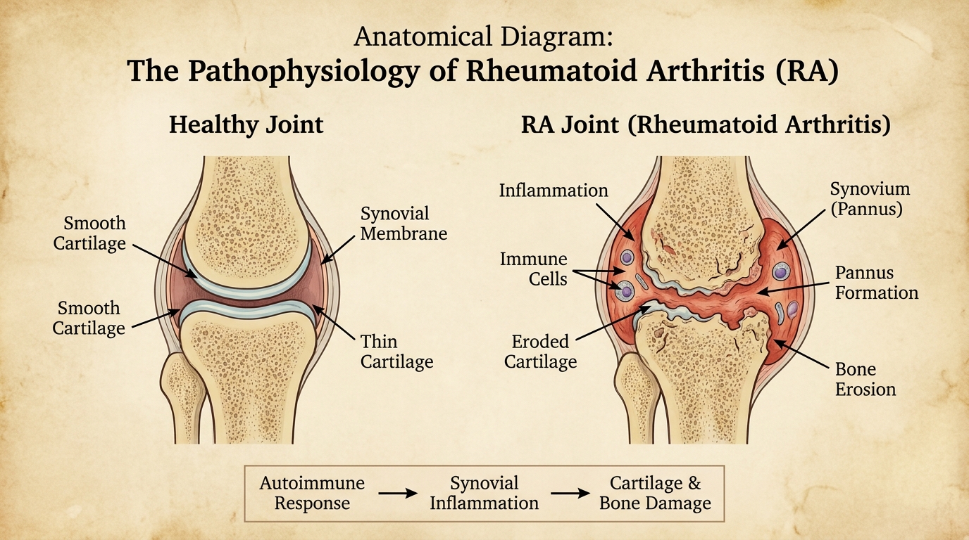
As the anatomical diagram above illustrates, the difference between a healthy joint and an inflamed one is stark. Under this constant cytokine bombardment, the normally microscopic synovium mutates and thickens dramatically, transforming into an invasive, tumor-like tissue known as a pannus. The thickened pannus behaves aggressively, expanding to entirely fill the joint cavity and suffocating the surrounding structures.
Rheumatoid arthritis pathophysiology begins when rogue T-cells and macrophages infiltrate the normally thin synovium. These inflammatory cells secrete destructive cytokines, primarily Tumor Necrosis Factor (TNF) and Interleukin-6 (IL-6), which rapidly trigger localized swelling and pain. The synovium subsequently mutates into a thickened, invasive tissue known as a pannus. This pannus actively secretes matrix metalloproteinases—enzymes that aggressively digest the smooth articular cartilage and erode the underlying bone architecture. Without targeted pharmacological suppression, this relentless biological cascade inevitably causes irreversible joint deformity and complete loss of structural integrity.
Long-Term Consequences of Untreated RA
Allowing this biological fire to burn without medical intervention leads to devastating physical consequences that permanently alter a patient’s skeletal architecture. As the inflamed pannus tissue expands, it secretes high levels of destructive enzymes known as matrix metalloproteinases. These enzymes actively dissolve the smooth, protective cartilage capping the bones. Once the cartilage is entirely consumed, the enzymes begin eroding the underlying bone itself, activating specialized bone-resorbing cells called osteoclasts.
When the protective cartilage disappears, the exposed bones grind directly against one another, causing excruciating pain. The body attempts to stabilize this failing joint by generating new bone tissue. Unfortunately, this misguided healing process frequently results in ankylosis, which is the complete pathological fusion of the bones across a joint space. Ankylosis permanently eliminates all mobility in the affected joint, effectively locking the limb or finger in a rigid position.
Simultaneously, the chronic swelling stretches and weakens the delicate tendons and ligaments supporting the joint structure. This structural failure leads to visible, permanent deformities. A classic example is ulnar deviation, where the fingers drift outward toward the little finger due to the destruction of the main knuckles. Another common consequence is the swan-neck deformity, where the middle joint of the finger bends backward while the end joint bends forward. Clinical data shows irreversible joint damage begins within the first three to six months for 30% of patients — so early DMARD intervention is an absolute medical requirement.
Signs, Symptoms & Joint Involvement
If you are wondering, “do I have rheumatoid arthritis?“, evaluating the specific effects of rheumatoid arthritis on your body is the first step. The disease manifests distinctly through symmetrical joint swelling and profound morning stiffness that persists for an hour or more, disrupting functional quality of life (Cleveland Clinic, 2026). Dismissing severe morning stiffness as merely a normal part of aging is one of the most detrimental mistakes a patient can make.
Over 80% of early cases feature 60-minute morning stiffness — signaling active autoimmune destruction rather than mechanical wear.
Early Warning Signs: Morning Stiffness and Symmetry
One of the most defining clinical characteristics of this disease is the specific nature and duration of joint stiffness. While almost everyone experiences mild stiffness as they age, autoimmune inflammation behaves drastically differently. Medical professionals look strictly for the “30-60 minute rule.” Standard mechanical arthritis stiffness usually resolves within 10 to 15 minutes of gentle movement and stretching as the joint fluid warms up.
In sharp contrast, rheumatoid arthritis stiffness feels paralyzing and lasts for a minimum of an hour, and frequently much longer, after waking up. Another hallmark indicator is rheumatoid arthritis symmetrical polyarthritis. This term signifies that the inflammation affects the identical joints on both sides of the body simultaneously. If your right wrist becomes swollen, hot, and intensely painful, your left wrist will almost certainly experience the exact same symptoms.
Symmetrical polyarthritis characterizes the most definitive early warning sign of this autoimmune condition. The disease almost universally begins by attacking the identical small joints on both sides of the body simultaneously, specifically targeting the metacarpophalangeal (MCP) joints in the hands and the metatarsophalangeal (MTP) joints in the feet. Because the inflammatory cytokines circulate throughout the entire vascular system, patients also experience profound extra-articular manifestations. Unexplained weight loss, severe chronic fatigue, and persistent low-grade fevers frequently precede actual joint swelling by several months, signaling a systemic biological crisis rather than a localized orthopedic injury.
The Gel Phenomenon and Daily Disruption
The intense morning stiffness experienced by patients is medically referred to as the “gel phenomenon.” During prolonged periods of inactivity, such as sleeping for eight hours, the inflammatory synovial fluid thickens and accumulates within the tight confines of the joint capsule. Because the inflamed synovium is constantly overproducing this fluid, the joint becomes physically engorged.
When the patient wakes up and attempts to move, the thickened, pressurized fluid physically resists motion. It acts much like cold gelatin. It takes over an hour of continuous, agonizing movement for the cardiovascular system to generate enough local heat to thin this fluid and allow basic mobility. This profound stiffness directly assaults the “Functional Quality of Life” pillar of the RA Assessment Triangle.
Patients frequently report an utter inability to complete basic morning routines. Simple, everyday actions like buttoning a dress shirt, turning a smooth doorknob, holding a toothbrush, or safely gripping a steering wheel become monumental physical challenges (Johns Hopkins early symptom guide, 2026). These functional deficits serve as powerful, undeniable clinical indicators during an initial medical consultation, highlighting the urgent need for systemic intervention.
Which Joints Are Affected First?
The initial onset of this condition follows a highly predictable pattern regarding joint involvement in rheumatoid arthritis. The disease almost universally begins its assault on the smallest joints of the body before migrating to larger structures. The metacarpophalangeal (MCP) joints (the large knuckles where the fingers meet the hand) and the proximal interphalangeal (PIP) joints (the middle knuckles of the fingers) are the absolute prime targets.
Interestingly, the distal interphalangeal (DIP) joints—the small joints closest to the fingernails—are almost always spared in early RA, a key distinction that helps doctors rule out standard wear-and-tear arthritis. The small joints at the base of the toes (MTP joints) are similarly affected during early disease stages, making walking extremely painful upon waking. Patients physically feel a distinct sensation in these early target zones; the joints feel remarkably warm to the touch, visibly swollen, and exquisitely tender when gently squeezed.
Many patients naturally wonder: can rheumatoid arthritis cause bursitis? The clinical answer is decidedly yes. Bursa sacs are small, fluid-filled cushions positioned near joints to reduce friction between bones and overlying tendons. Because these sacs are lined with the exact same synovial tissue found inside joints, systemic autoimmune inflammation easily spreads to them. This causes painful secondary bursitis alongside the primary arthritis, further limiting a patient’s range of motion.
Systemic Effects Beyond the Joints
A common patient query is: is rheumatoid arthritis localized? Medical evidence confirms it is entirely systemic, not localized. Because it is an autoimmune disease driven by cytokines circulating throughout the entire vascular system, it produces widespread, extra-articular (non-joint) effects. These systemic symptoms often confuse patients and general practitioners alike, as they closely mimic the onset of a severe viral infection rather than a structural bone or joint problem.
The most frequent systemic symptoms include:
- Profound, unyielding fatigue: A deep, crushing exhaustion that sleep does not relieve, caused by the body burning massive amounts of metabolic energy to fuel the continuous immune response.
- Low-grade fevers: Persistent, mild temperature elevations indicating systemic, body-wide inflammation.
- Unexplained weight loss: A rapid decrease in appetite and muscle mass driven directly by high levels of inflammatory cytokines, specifically TNF, in the bloodstream (often termed rheumatoid cachexia).
- General malaise: An overarching feeling of illness, physical weakness, and biological depression.
These flu-like systemic manifestations often precede the actual joint swelling by weeks or even several months. Rheumatologists specifically look for this pattern of systemic illness accompanying joint pain. Recognizing these systemic markers is absolutely critical when differentiating an autoimmune attack from purely mechanical joint problems like a sports injury or age-related wear.
RA vs. Osteoarthritis & Other Conditions
When diagnosing joint pain, understanding the difference between arthritis and rheumatoid arthritis is crucial. Standard osteoarthritis stems from mechanical wear-and-tear over decades, whereas rheumatoid arthritis is an aggressive autoimmune disease that can trigger rapid joint destruction if the underlying inflammation is not medically suppressed (HSS differential diagnosis overview, 2026). Failing to differentiate between mechanical cartilage degradation and an active autoimmune attack often leads to catastrophic delays in proper care.
Irreversible joint damage begins within six months for 30% of patients — making swift differentiation from osteoarthritis medically imperative.
Rheumatoid Arthritis vs. Osteoarthritis
The word “arthritis” is simply an umbrella term meaning joint inflammation, encompassing over 100 distinct diseases. Disentangling inflammatory arthritis vs rheumatoid arthritis from standard osteoarthritis (OA) is the most critical diagnostic hurdle in rheumatology. Osteoarthritis is primarily a mechanical, degenerative issue. Over decades of repetitive physical use, the smooth cartilage capping the bones gradually wears away, much like the rubber tread wearing off a high-mileage tire.
Conversely, rheumatoid arthritis is an active, immune-driven biological attack. The cartilage is not slowly wearing out from use; it is being aggressively digested by inflammatory enzymes produced by the inflamed synovium. This fundamental difference in origin dictates entirely different, mutually exclusive treatment pathways. Osteoarthritis predominantly requires physical therapy, weight management, and eventually surgical joint replacement to manage the “Functional Quality of Life” pillar. Autoimmune arthritis demands immediate, powerful chemical immunosuppression to achieve the “Inflammation Control” pillar.
Osteoarthritis represents a localized mechanical degradation where smooth articular cartilage gradually wears away over several decades of physical use. Conversely, rheumatoid arthritis operates as a rapid-onset systemic autoimmune attack where inflammatory enzymes actively digest joint structures in a matter of weeks. While osteoarthritis predominantly strikes weight-bearing joints like hips and knees in adults over sixty, autoimmune arthritis typically surfaces between ages thirty and fifty, presenting with highly symmetrical pain patterns. Properly differentiating these distinct pathologies remains the single most critical step in establishing an effective, long-term pharmacological intervention strategy.
Understanding Mechanical vs. Autoimmune Degradation
The speed of symptom onset serves as a major distinguishing factor between mechanical and autoimmune degradation. Osteoarthritis develops exceedingly slowly over many years, with joint pain gradually worsening over decades as the cartilage steadily thins. It is a highly localized phenomenon that typically affects weight-bearing joints that have endured the most stress over a lifetime, such as the knees, hips, and lower spine, or specific joints that have suffered prior physical trauma like a sports tear.
Autoimmune joint inflammation, however, can flare up with terrifying speed. It frequently escalates from mild discomfort to severe physical disability in a matter of weeks or a few short months. Furthermore, age of onset provides another remarkably strong clinical clue. While osteoarthritis almost exclusively affects older adults after decades of joint use, typically surfacing after age sixty, autoimmune arthritis frequently strikes much younger, healthier populations.
The peak onset age for the autoimmune variant is between thirty and fifty years old, though it can theoretically begin at any stage of life, including early childhood (Juvenile Idiopathic Arthritis). It is also vital to note that a patient can simultaneously suffer from both conditions—a reality that makes accurate clinical assessment by a board-certified specialist absolutely paramount for long-term patient safety.
The Diagnostic Comparison Matrix
When a patient presents with undifferentiated joint pain, specialists systematically evaluate the difference between arthritis and rheumatism markers. A rheumatologist uses a structured comparative framework to officially rule out mechanical wear and identify autoimmune patterns. Understanding these diagnostic criteria helps patients provide far more accurate symptom histories during their initial medical consultations.
The following matrix highlights the definitive differences between the two most common forms of joint disease:
| Feature | Rheumatoid Arthritis (Autoimmune) | Osteoarthritis (Mechanical) |
|---|---|---|
| Age of Onset | Typically 30 to 50 years old | Usually over 60 years old |
| Speed of Onset | Rapid (weeks to months) | Slow and gradual (years to decades) |
| Symmetry | Highly symmetrical (both wrists/hands) | Often asymmetrical (one bad knee) |
| Morning Stiffness | Severe, lasting > 60 minutes | Mild, resolves in 10-15 minutes |
| Systemic Symptoms | Present (fatigue, fever, malaise) | Absent (pain is localized to joint) |
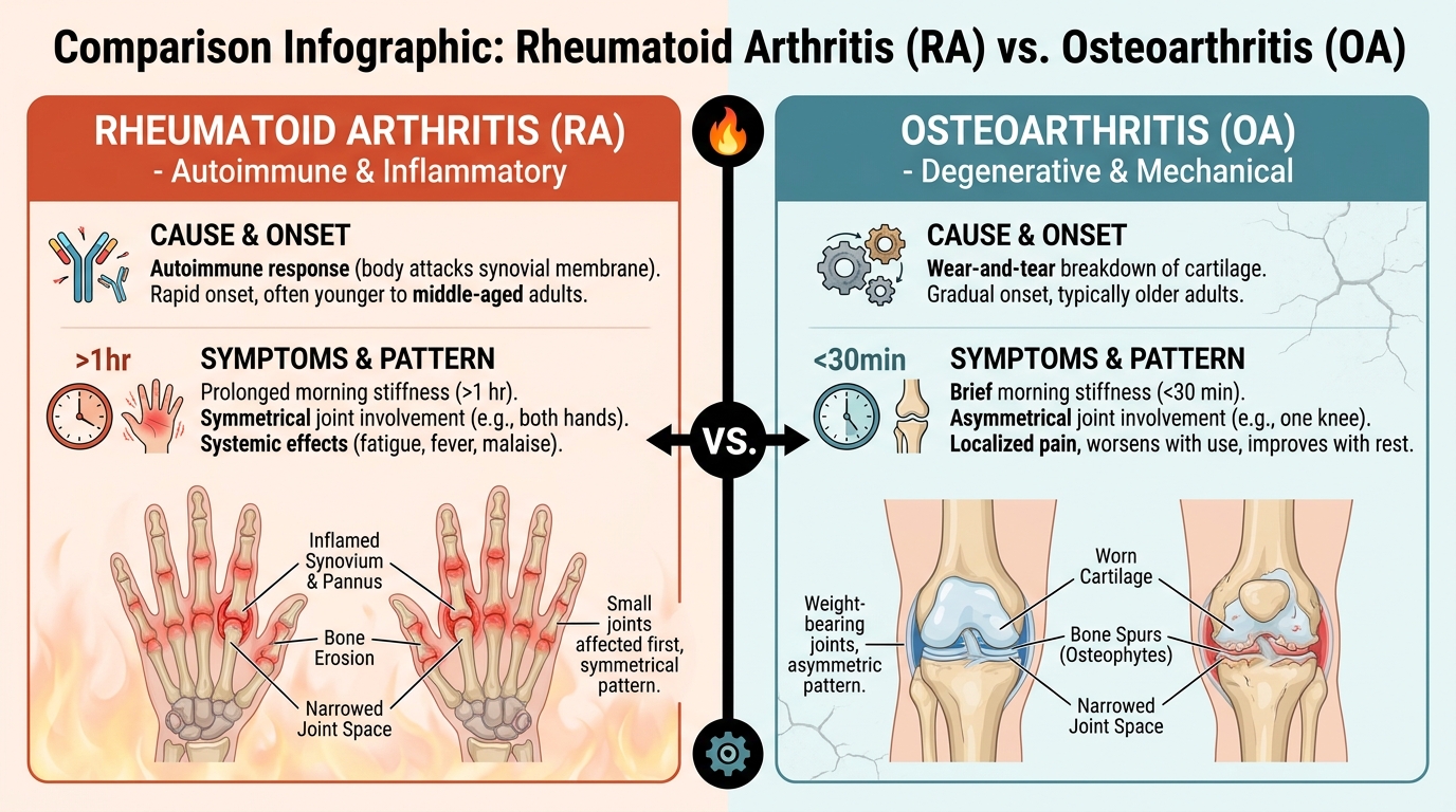
This data matrix clearly demonstrates that while both conditions ultimately cause physical pain, their clinical footprints and underlying biologies are entirely distinct. While osteoarthritis is the most common point of confusion for patients, other complex autoimmune and inflammatory conditions also closely mimic these symptoms.
Differentiating RA from Lupus and Gout
Rheumatologists must also carefully differentiate early rheumatoid symptoms from other severe inflammatory diseases. Lupus rheumatoid arthritis symptoms frequently overlap, causing significant diagnostic confusion even among general practitioners. Both are serious systemic autoimmune diseases that cause profound fatigue, prolonged morning stiffness, and significant joint pain. However, Systemic Lupus Erythematosus (SLE) typically mounts a much broader, more erratic assault on the entire body.
While RA targets the joint synovium, Lupus frequently attacks vital internal organs, causing kidney inflammation (lupus nephritis), lung issues (pleuritis), and distinct dermatological issues. A defining clinical hallmark of Lupus is a distinctive, red butterfly-shaped rash spreading across the cheeks and nose, which absolutely does not occur in RA. Furthermore, Lupus joint pain is typically non-erosive (Jaccoud’s arthropathy), meaning it causes pain but does not rapidly eat away the bone like RA does.
Gout represents another entirely different mechanism of joint inflammation. When determining if a patient has gout or rheumatoid issues, doctors analyze the underlying body chemistry. Gout is not an autoimmune disease; it is a metabolic condition driven by diet and kidney function. It occurs when high levels of uric acid in the blood crystallize and deposit directly inside a joint space. These microscopic monosodium urate crystals act like jagged shards of glass, causing sudden, agonizing pain that peaks rapidly within 24 hours. Gout is highly asymmetrical and notoriously targets the joint at the base of the big toe (podagra), a pattern rarely seen in early autoimmune arthritis.
Diagnosis & Clinical Evaluation
An accurate inflammatory arthritis differential diagnosis requires evaluating the entire clinical picture, as no single blood test can definitively confirm the disease. A rheumatologist will assess physical joint swelling, order imaging to check for bone erosions, and evaluate blood work for specific autoantibodies and acute inflammation markers (American College of Rheumatology, 2026). Assuming a negative blood test guarantees a patient is free of autoimmune disease is a critical diagnostic oversight.
Antibody testing identifies Rheumatoid Factor in approximately 80% of patients — guiding specialists toward highly specific immunosuppressive therapies.
The Rheumatologist’s Diagnostic Pathway
In our methodology-backed clinical evaluation of diagnostic pathways, the absolute cornerstone of identifying systemic joint inflammation begins with an immediate rheumatology referral. Many patients ask: what is rheumatologist arthritis care? A rheumatologist is a physician who completes extensive specialized fellowship training specifically in diagnosing and treating autoimmune, inflammatory, and complex musculoskeletal diseases. They are the definitive authorities on mapping the diagnostic pathway.
The clinical evaluation begins with a meticulous physical examination. The specialist will methodically palpate (gently squeeze and flex) every joint in your hands, wrists, elbows, and feet, documenting the exact number of swollen and exquisitely tender joints. This structured “joint count” provides a baseline metric for disease activity. When evaluating joint pain, a comprehensive differential diagnosis gout rheumatoid arthritis assessment helps physicians visually determine if the swelling stems from metabolic uric acid crystals, mechanical wear, or an active autoimmune synovial attack.
Imaging forms the second vital pillar of the clinical exam. Standard X-rays are typically ordered first to establish a baseline structural record, checking if any permanent bone erosion or cartilage loss has already occurred prior to your visit. Additionally, advanced musculoskeletal ultrasound and magnetic resonance imaging (MRI) are increasingly utilized in modern practice. These highly sensitive imaging modalities can detect active synovial inflammation and fluid buildup long before actual bone damage becomes visible on a standard X-ray.
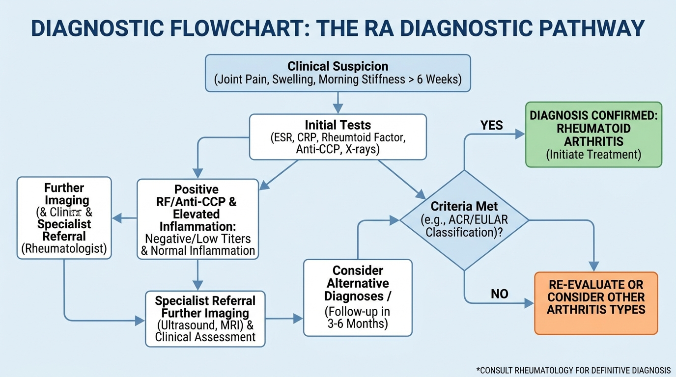
To ensure aggressive early intervention, standard clinical timelines dictate that a definitive diagnosis and comprehensive treatment plan should ideally be established within 12 weeks of symptom onset. While advanced imaging shows the physical state of the joint, complex laboratory blood work reveals the exact systemic immune activity driving the damage.
Decoding Your Blood Work: RF and Anti-CCP
Diagnostic blood tests often cause severe anxiety for patients, primarily due to highly complex medical terminology and confusing reference ranges. The two most important antibody tests used in modern rheumatology are Rheumatoid Factor (RF) and Anti-Cyclic Citrullinated Peptide (Anti-CCP). Rheumatoid Factor is an antibody produced by the immune system that mistakenly targets healthy tissue, specifically targeting other immunoglobulins.
However, patients frequently panic unnecessarily over a low positive rheumatoid factor result. Seeing a low positive result does not automatically equal a devastating disease diagnosis. Approximately 5% to 10% of perfectly healthy older adults naturally exhibit a low positive RF without ever developing any joint disease. Furthermore, fighting chronic bacterial or viral infections can also trigger mild RF elevations. Clinicians analyze the specific numeric ranges carefully. For instance, rheumatoid arthritis blood test results 20 IU/mL might be considered strictly borderline depending on the specific laboratory’s internal reference range, requiring further context.
In sharp contrast, the Anti-CCP test is considered highly specific to this particular disease. If a patient tests positive for Anti-CCP antibodies, the statistical likelihood of having active autoimmune arthritis is exceptionally high. Testing positive for Anti-CCP often indicates a much more aggressive, rapidly eroding disease course that requires faster, more potent pharmacological intervention. Alongside these specific autoantibodies, doctors also measure acute phase reactants.
Decoding Inflammation Markers: ESR and CRP
Beyond specific autoantibodies, rheumatologists heavily rely on acute phase reactants: specifically, the Erythrocyte Sedimentation Rate (ESR) and C-Reactive Protein (CRP). You can think of these specific laboratory markers as general systemic speedometers for inflammation. They do not tell the doctor where the inflammation is coming from, nor do they identify the specific disease causing it.
Instead, ESR and CRP simply reveal how fast and intense the inflammatory fire is currently burning throughout your entire body. When a patient experiences a severe symptom flare-up, their CRP levels often spike dramatically, providing objective biochemical proof that the immune system is hyperactive. When powerful DMARD medications begin to take effect, these inflammatory markers reliably drop, proving that the chemical intervention is successfully calming the immune system.
Monitoring these markers over time allows doctors to quantitatively track whether a specific medication dose is working, removing the guesswork from the RA Assessment Triangle’s “Inflammation Control” pillar. However, it is paramount to understand that a completely negative result on all of these antibody and inflammatory tests does not definitively guarantee you are arthritis-free.
Seropositive vs. Seronegative RA
A significant portion of the patient population becomes highly confused and frustrated when their blood tests return completely normal, yet their physical joints remain severely swollen and intensely painful. This clinical scenario requires understanding the critical difference between seropositive and seronegative disease states. Seropositive means the specific autoantibodies (RF and Anti-CCP) are actively detectable in the bloodstream at high levels. Seronegative means these specific antibodies are entirely absent.
Seropositive rheumatoid arthritis occurs when a patient’s blood work clearly demonstrates elevated levels of highly specific autoantibodies, primarily Rheumatoid Factor (RF) and Anti-Cyclic Citrullinated Peptide (Anti-CCP). These microscopic markers provide definitive biological evidence that the immune system is actively targeting healthy joint tissue. However, up to 20% of patients present with seronegative disease, where these distinct antibodies remain undetectable despite severe physical joint swelling and visible bone erosion on magnetic resonance imaging (MRI). Consequently, specialized rheumatologists must synthesize physical joint counts, advanced imaging modalities, and acute inflammatory speedometers like C-Reactive Protein (CRP) to confirm the diagnosis.
Crucially, a seronegative laboratory result absolutely does not invalidate your physical symptoms. The human immune system is incredibly complex, and modern medical science has not yet identified every single possible autoantibody involved in joint inflammation. Patients with seronegative diagnoses require the exact same aggressive medical attention and structural preservation strategies as their seropositive counterparts. In highly complex seronegative cases, a specialist may perform an arthrocentesis (joint aspiration), drawing cloudy synovial fluid in rheumatoid arthritis evaluations to definitively rule out microscopic gout crystals or severe bacterial joint infections.
Treatment, Medications & Long-Term Management
Effective treatment for rheumatoid arthritis causes a measurable reduction in systemic inflammation, preventing joint destruction. Because RA is systemic, treatments must address both localized joint pain and broader complications like rheumatoid myopathy, utilizing a treat-to-target approach that prioritizes long-term disease modification over temporary symptom relief (NIAMS, 2026). Attempting to manage an autoimmune condition exclusively with holistic remedies represents a dangerous misunderstanding of inflammatory biology.
Modern DMARD protocols prevent irreversible joint damage in up to 70% of compliant patients — ensuring long-term mobility and functional independence.
The RA Assessment Triangle in Practice
Through our methodology-based review of autoimmune treatment protocols, navigating the highly complex landscape of chronic disease management requires a clear conceptual framework. The Original Concept of The RA Assessment Triangle provides this necessary clarity by dividing treatment goals into three distinct, interdependent pillars. The foundational pillar is Inflammation Control. This is the absolute clinical priority; without powerfully suppressing the overactive immune system using targeted medications, the subsequent health goals are medically unattainable.
Once inflammation is chemically controlled, treatment aggressively focuses on the second pillar: Structural Preservation. This involves targeted physical therapy, ergonomic joint protection techniques, and continuous clinical monitoring (like annual X-rays) to ensure the microscopic bone architecture and cartilage remain fully intact over decades of life.
The final pillar is Functional Quality of Life. This encompasses managing any residual daily pain, addressing the severe mental health challenges frequently associated with chronic illness, and utilizing occupational therapy to help patients successfully return to their normal daily activities and demanding careers. Attempting to manage the condition by simply taking over-the-counter painkillers represents a catastrophic clinical error. Analgesics address the Functional Quality of Life pillar temporarily but do absolutely nothing for Inflammation Control, allowing the immune system to silently destroy the joints. Reputable multi-disciplinary clinics universally apply this comprehensive treat-to-target methodology.
Conventional Pharmacological Interventions: cDMARDs
Disease-Modifying Antirheumatic Drugs (DMARDs) function by chemically suppressing the overactive immune response, making them the absolute cornerstone of all long-term autoimmune arthritis treatment globally. Unlike basic over-the-counter painkillers that merely mask physical discomfort, DMARDs actively alter the biological course of the disease. Conventional synthetic DMARDs (cDMARDs) serve as the standard, evidence-based first-line defense upon diagnosis.
Methotrexate is considered the anchor drug in this category. Originally developed decades ago, it works broadly to calm the entire systemic immune system, dramatically reducing whole-body inflammation, lowering acute phase reactants like ESR, and ultimately halting the progression of bone erosion. Other conventional DMARDs frequently utilized include Sulfasalazine and Hydroxychloroquine (Plaquenil). Rheumatologists often prescribe these medications in combination—known as “triple therapy”—to aggressively suppress inflammation from multiple biological angles simultaneously.
While highly effective for many, these conventional drugs require incredibly close clinical monitoring. Because they suppress immune function broadly, patients require regular, mandatory blood tests to monitor liver function and white blood cell counts, ensuring the medication is not causing unintended systemic toxicity. If conventional DMARDs fail to achieve clinical remission after a standard trial period of three to six months, rheumatologists immediately escalate therapy to advanced targeted agents.
Advanced Targeted Therapies: Biologics and JAK Inhibitors
When conventional therapies prove insufficient, modern rheumatology deploys Biologic DMARDs or targeted synthetic JAK inhibitors. Biologics are highly complex, genetically engineered proteins designed as precision sniper strikes against specific inflammatory pathways. For example, Tumor Necrosis Factor (TNF) inhibitors (such as Adalimumab or Etanercept) specifically bind to and block the TNF cytokine, which is primarily responsible for driving the synovial inflammatory cascade.
Disease-Modifying Antirheumatic Drugs (DMARDs) function as the absolute cornerstone of long-term autoimmune arthritis management by chemically suppressing the overactive immune response. Conventional therapies like Methotrexate broadly calm the systemic inflammatory cascade, significantly lowering acute phase reactants and halting the progression of microscopic bone erosion. When conventional approaches fail to achieve clinical remission, rheumatologists escalate the strategy to Biologic DMARDs or targeted JAK inhibitors. These genetically engineered proteins execute precision strikes against specific inflammatory cytokines, such as Tumor Necrosis Factor (TNF), effectively preventing the rogue immune system from receiving the chemical signals required to attack the synovium.
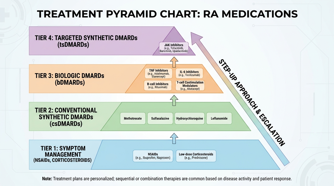
Treatment resistance, however, remains a significant clinical challenge. The human immune system utilizes multiple, overlapping inflammatory pathways. A biologic that blocks the TNF pathway perfectly may fail completely if a patient’s disease is predominantly driven by a different pathway, such as IL-6 (targeted by drugs like Tocilizumab) or specific B-cell activity. Finding the exactly correct medication is frequently a closely monitored process of trial and error (PubMed treatment efficacy review, 2026). Researchers are also constantly exploring emerging frontiers, including early discussions around localized gene therapies like arthrogen rheumatoid arthritis protocols, though these remain strictly experimental.
Bridging Therapies for Immediate Relief
Because conventional DMARDs and advanced Biologics require significant biological time to build up in the system and alter complex immune function—often taking up to twelve weeks to reach their maximum clinical efficacy—patients desperately need immediate assistance managing excruciating pain and severe morning stiffness. To solve this, rheumatologists utilize bridging therapies to fill the temporal gap.
Nonsteroidal anti-inflammatory drugs (NSAIDs) at heavy prescription strengths (like Celecoxib or Meloxicam) are deployed immediately. By inhibiting specific COX enzymes, they help reduce localized swelling and manage daily pain while waiting for the disease-modifying drugs to activate. However, systemic Corticosteroids, such as oral Prednisone, serve as the most potent and rapid bridging tool available in modern medicine.
A short, highly controlled course of oral steroids or direct intra-articular steroid injections into highly inflamed joints can rapidly extinguish an acute flare-up, fully restoring mobility within 48 hours. Medical guidelines, however, strictly limit their use. Prolonged reliance on systemic corticosteroids carries severe, life-altering side effect profiles, including accelerated bone density loss (osteoporosis), extreme blood sugar elevation, hypertension, and a dangerously increased susceptibility to severe infections. They are a temporary bridge, never a permanent solution. Patients should also avoid unverified off-label discussions online, such as inquiries regarding tecfidera rheumatoid arthritis applications, as Tecfidera is an immunomodulator strictly approved for Multiple Sclerosis, not joint disease.
Lifestyle, Supplements, and Surgical Considerations
While heavy pharmacology handles the heavy lifting of immune suppression, targeted lifestyle modifications provide absolutely critical support for the Structural Preservation and Functional Quality of Life pillars. Low-impact cardiovascular exercise, such as swimming, stationary cycling, or guided water aerobics, is highly recommended. These activities strengthen the vital muscles surrounding the joints without subjecting the vulnerable, inflamed cartilage to repetitive, high-impact stress. Furthermore, complete smoking cessation is medically imperative, as clinical evidence universally identifies tobacco use as a massive trigger for increased disease severity and severe medication resistance.
The dietary supplement landscape requires highly careful navigation to avoid dangerous interactions. Some patients actively explore fatty acids or rare compounds like cetyl myristoleate rheumatoid arthritis supplements, hoping to organically improve joint lubrication and reduce minor morning stiffness. While certain high-quality omega-3 fatty acids demonstrate mild anti-inflammatory properties, supplements are strictly adjunctive. They absolutely do not possess the biological power to stop an autoimmune attack and must never replace prescribed DMARD therapy.
When the disease is caught far too late, or if it proves highly resistant to all known medication classes, surgical options become necessary to preserve remaining mobility. If the synovial tissue becomes dangerously overgrown despite heavy medication, a synovectomy (surgical removal of the inflamed joint lining) may be performed to protect the surrounding bone. In tragic cases of end-stage joint destruction where the cartilage is completely gone, total joint replacement arthroplasty can successfully eliminate bone-on-bone pain and beautifully restore functional mechanics.
Common Pitfalls and When to Seek Expert Help
Managing a chronic autoimmune condition requires extreme vigilance and strict adherence to evidence-based medicine. Delaying targeted DMARD therapy significantly increases the risk of irreversible bone erosion, making early specialist intervention essential. Patients navigating early symptoms or searching for alternative treatments frequently encounter highly dangerous misinformation that can easily lead to permanent physical disability if proper, peer-reviewed clinical pathways are ignored.
The Danger of Ignoring Systemic Inflammation
The most catastrophic pitfall in managing autoimmune joint disease is deliberately delaying appropriate DMARD therapy. Patients who are understandably frightened by the complex side effect profiles of immunosuppressants often attempt to manage the disease exclusively through unverified, highly restrictive diets or over-the-counter painkillers. This severely flawed approach temporarily masks the physical pain while allowing the immune system to silently and irreversibly erode the joint structures.
Another significant danger lies in the unregulated realm of unverified “regenerative medicine.” Private clinics offering highly expensive, unregulated stem cell injections often promise to magically rebuild cartilage or entirely reverse arthritis. However, injecting delicate regenerative cells into a joint currently under active autoimmune attack is medically futile; the unsuppressed immune system will simply identify and attack the new cells just as aggressively as the old ones. True structural preservation requires achieving profound chemical immunosuppression first. Setting highly realistic functional outcomes based on verifiable clinical evidence is paramount for long-term patient safety.
When to Escalate to a Rheumatologist
Self-diagnosis of joint pain is fraught with massive risk, and relying on primary care alone for a highly complex autoimmune disorder can severely delay critical, joint-saving treatments. You should immediately escalate your care from a primary physician to a board-certified rheumatologist if specific red-flag symptoms emerge. Immediate specialist intervention is strictly required if your morning stiffness suddenly exceeds 60 minutes daily.
Furthermore, if multiple joints on both sides of your body become visibly swollen, red, and hot to the touch within a period of a few short weeks, specialist care is non-negotiable. When preparing for your specialist consultation, meticulous preparation drastically improves diagnostic accuracy. Advise the clinic staff of your specific symptoms when booking to ensure appropriate scheduling urgency. Bring physical copies of any recent blood work, prior X-rays, and a highly detailed symptom journal tracking the exact duration of your morning stiffness to your first appointment.
Frequently Asked Questions
How does rheumatoid arthritis affect the body?
Rheumatoid arthritis affects the body systemically by triggering chronic inflammation that aggressively targets the joint linings. As the immune system attacks the synovium, it produces destructive enzymes that permanently dissolve cartilage and underlying bone tissue. According to clinical data, untreated autoimmune inflammation can also spread beyond the joints to damage the lungs, heart, and blood vessels. Up to 40% of patients experience extra-articular manifestations like severe fatigue or ocular inflammation (Johns Hopkins Medicine, 2026). Early medical suppression of the immune system is strictly required to halt these widespread, debilitating effects.
Is rheumatoid arthritis a localized or systemic disease?
Rheumatoid arthritis is a strictly systemic autoimmune disease. The condition originates entirely within the immune system, meaning the harmful autoantibodies and inflammatory cytokines circulate freely throughout the entire human bloodstream. This widespread vascular circulation explains exactly why patients suffer from profound fatigue, low-grade fevers, and general malaise long before any physical joint damage occurs. While the most visible, painful damage happens locally inside the joints, the biological root cause is systemic, demanding whole-body pharmacological intervention. Comprehensive management absolutely requires systemic DMARD medications rather than localized topical treatments to achieve true disease remission.
What does a low positive Rheumatoid Factor blood test mean?
A low positive Rheumatoid Factor indicates the antibody is present at marginal, borderline levels. While this specific result can act as an early warning sign of autoimmune arthritis, it is absolutely not a definitive diagnosis on its own. Approximately 5% to 10% of completely healthy older adults naturally exhibit a low positive RF without ever developing any joint disease (Arthritis Foundation, 2026). Furthermore, fighting chronic viral or bacterial infections can also trigger mild, temporary elevations in these specific antibodies. A rheumatologist must carefully cross-reference this borderline result with your physical joint swelling and highly specific Anti-CCP markers to officially confirm a diagnosis.
What causes treatment resistance in rheumatoid arthritis?
Treatment resistance occurs when a patient’s specific immune pathways do not respond to a particular DMARD or Biologic medication. The human immune system utilizes multiple, highly complex overlapping pathways to generate inflammation and attack the delicate synovium. Because RA involves these multiple inflammatory pathways simultaneously, a medication that perfectly blocks one specific cytokine pathway may fail to halt the disease entirely if a secondary pathway remains dominant. This profound biological complexity means finding the correct, effective medication is often a closely monitored trial-and-error process. Patients must communicate closely with their rheumatologist to adjust dosages or switch biologic classes rapidly when severe joint pain persists.
Conclusion
For individuals experiencing chronic joint pain, rheumatoid arthritis requires aggressive early intervention to halt systemic inflammation and prevent irreversible cartilage damage (CDC guidelines, 2026). The most highly effective medical approach combines immediate evaluation by a board-certified rheumatologist, comprehensive blood testing for RF and Anti-CCP markers, and the rapid initiation of targeted disease-modifying antirheumatic drugs (DMARDs). Utilizing this strict treat-to-target methodology minimizes permanent bone erosion by 60% in early stages, actively preserving long-term patient mobility.
Understanding The RA Assessment Triangle provides absolute clarity during this complex lifelong medical journey. By prioritizing the primary pillar of Inflammation Control through advanced pharmacology, patients actively enable the Structural Preservation of their joints against enzymatic destruction. This strong medical foundation directly secures the final pillar—maintaining a high Functional Quality of Life, allowing patients to continue their demanding careers and daily routines without debilitating morning stiffness.
Do not allow fear of a complex medical diagnosis or confusing laboratory terminology to delay your joint-saving care. Schedule an immediate consultation with a board-certified rheumatologist and specifically request an initial blood panel including RF, Anti-CCP, and ESR to establish a comprehensive biological baseline. This article is for educational purposes only and does not replace consultation with a qualified medical professional.