
2026 Guide to ESR, CRP, ANA, RF & Anti-CCP Blood Tests
⚠️ Medical Disclaimer: This article is for educational purposes only and does not replace consultation with a qualified medical professional. Always seek the advice of a licensed physician or rheumatologist regarding your specific laboratory results and medical conditions.
Receiving laboratory results indicating elevated ESR, CRP, ANA, or Anti-CCP levels frequently causes immediate patient anxiety, yet these markers are diagnostic puzzle pieces rather than definitive diagnoses. A single elevated number on a lab report does not confirm a specific disease — it signals that further clinical investigation is warranted.
Without proper clinical context, patients often misinterpret a “positive” result as immediate confirmation of severe autoimmune disease, leading to unnecessary panic before a specialist review. A positive ANA, for example, appears in a significant percentage of entirely healthy individuals and carries very different meaning depending on its titer, pattern, and accompanying symptoms.
In our clinical reviews of patient lab results, we frequently observe that isolated marker elevations without corresponding physical symptoms lead to premature diagnostic conclusions. This highlights the absolute necessity of methodical, context-driven analysis over reactive panic.
This guide explains exactly what these five blood tests measure, which results indicate acute danger, and how to logically interpret your lab report. We will systematically break down general inflammatory markers — including the crp blood test and ESR — then move into autoimmune screening antibodies and disease-specific markers before explaining how to access specialized clinical testing.
Key Takeaways
An elevated crp blood test or ESR indicates systemic inflammation, but specific antibodies like ANA, RF, and Anti-CCP are required to diagnose autoimmune conditions.
- The Diagnostic Funnel Framework: Doctors use broad tests (CRP/ESR) to detect inflammation before using specific tests (ANA/Anti-CCP) to identify the disease.
- Positive ANA is common: Up to 15% of completely healthy individuals test positive for ANA; it is not a definitive lupus diagnosis.
- Anti-CCP is highly specific: While Rheumatoid Factor (RF) can be elevated in various conditions, Anti-CCP is 90–95% specific to Rheumatoid Arthritis.
- Clinical context is mandatory: No single blood test diagnoses an autoimmune disease without accompanying physical symptoms and specialist evaluation.
Core Inflammatory Markers: CRP & ESR
A crp blood test and an ESR blood test measure systemic inflammation in the body, serving as the first crucial step in The Diagnostic Funnel Framework — the clinical methodology of starting broad before narrowing to specific diagnoses. C-Reactive Protein (CRP) responds rapidly to acute inflammation within hours, while Erythrocyte Sedimentation Rate (ESR) reflects chronic inflammatory activity over weeks (NCBI Research, 2018). Doctors use these markers together to detect hidden inflammation before any autoimmune-specific testing begins.
Definition: What Are CRP and ESR?
C-Reactive Protein (CRP) functions as a highly responsive acute-phase protein synthesized directly by the liver when the body encounters inflammation, severe infection, or significant tissue injury. The biological cascade begins the moment macrophages and other frontline immune cells detect invading pathogens or cellular destruction. These immune cells rapidly release pro-inflammatory cytokines, primarily Interleukin-6 (IL-6) and Tumor Necrosis Factor-alpha (TNF-alpha), which travel through the bloodstream to the liver.
Upon receiving this cytokine signal, liver hepatocytes immediately begin manufacturing CRP. Its defining clinical characteristic is remarkable speed: CRP levels can multiply 1,000-fold within just 24 to 48 hours of an acute inflammatory trigger. Because its biological half-life is strictly limited to approximately 19 hours, levels fall just as rapidly once the inflammatory trigger resolves. This rapid kinetic profile makes CRP the preferred medical marker for monitoring acute conditions, evaluating post-surgical recovery, and measuring immediate treatment responses.
Think of CRP as a highly sensitive fire alarm. The moment smoke enters the detector — meaning inflammation enters the bloodstream — the alarm sounds loudly and immediately. When the smoke clears, the alarm silences within days.
Erythrocyte Sedimentation Rate (ESR), by contrast, measures how quickly red blood cells settle to the bottom of a tall, thin test tube over the course of exactly one hour. Under normal, healthy biological conditions, red blood cells carry a slight negative electrical charge on their surface known as the zeta potential. This negative charge keeps them repelling each other, causing them to fall very slowly through the blood plasma.
During periods of systemic inflammation, the liver releases various heavy acute-phase proteins — particularly fibrinogen. These large, dense proteins neutralize the negative surface charges on the red blood cells. Without that repulsive force, the cells inevitably clump together like stacks of coins, a phenomenon clinically known as rouleaux formation. Because these aggregated clumps are significantly heavier and denser than individual cells, they sink much faster according to Stokes’ Law of sedimentation.
If CRP is the fire alarm, ESR is the lingering smell of smoke that persists for days or weeks after the fire is extinguished. It rises more slowly than CRP and decreases more gradually, making it an exceptional tool for tracking chronic inflammatory diseases like rheumatoid arthritis, polymyalgia rheumatica, or temporal arteritis over extended months of therapeutic treatment.
Danger Zones and Reference Ranges
How much CRP level is dangerous? This is one of the most searched patient questions, and the precise answer depends entirely on the specific CRP assay utilized by the laboratory. The standard CRP test and the high-sensitivity CRP (hs-CRP) test measure the exact same molecule but utilize entirely different detection thresholds optimized to answer radically different clinical questions.
Standard CRP Reference Ranges:
| CRP Level (mg/L) | Clinical Interpretation | Typical Clinical Response |
|---|---|---|
| < 3.0 mg/L | Normal — No significant inflammation | Routine monitoring |
| 3.0–10.0 mg/L | Minor elevation — Mild inflammation or early viral infection | Watchful waiting + repeat test |
| 10.0–100.0 mg/L | Significant inflammation — Active infection or autoimmune flare | Prompt medical investigation |
| > 100.0 mg/L | Severe elevation — Bacterial sepsis, severe vasculitis, acute organ injury | Emergency evaluation required |
Normal CRP levels are typically below 3.0 mg/L, while levels above 10.0 mg/L strongly indicate significant acute inflammation or infection (Arthritis Foundation, 2026). This metric provides an objective, reliable threshold, allowing clinicians to immediately differentiate between mild physiological stress and genuine pathological inflammation.
For cardiovascular risk assessment, physicians rely on the high-sensitivity hs-CRP assay instead. This specialized test detects minute inflammatory fluctuations within vascular walls. Levels below 1.0 mg/L indicate low cardiovascular risk, 1.0–3.0 mg/L indicates moderate risk, and persistently remaining above 3.0 mg/L indicates elevated cardiovascular risk requiring preventative lifestyle or statin interventions. Patients evaluating a standard autoimmune flare must avoid confusing these highly sensitive cardiovascular ranges with standard rheumatological thresholds.
ESR Reference Ranges:
ESR naturally fluctuates based on age, biological sex, and baseline hematological factors. Women naturally exhibit slightly higher ESRs due to inherent differences in baseline fibrinogen levels and total red blood cell mass. Furthermore, ESR naturally and predictably increases as human bodies age.
| Population Group | Normal ESR Range (mm/hr) | Mild Elevation | Significant Elevation |
|---|---|---|---|
| Men under 50 | 0–15 mm/hr | 16–40 mm/hr | > 40 mm/hr |
| Men over 50 | 0–20 mm/hr | 21–50 mm/hr | > 50 mm/hr |
| Women under 50 | 0–20 mm/hr | 21–40 mm/hr | > 40 mm/hr |
| Women over 50 | 0–30 mm/hr | 31–60 mm/hr | > 60 mm/hr |
To contextualize: an ESR of 35 mm/hr in a 45-year-old woman represents a mild elevation that warrants clinical attention but not immediate alarm. It could stem from a minor viral infection, a mild autoimmune flare, or even profound underlying anemia. An ESR exceeding 100 mm/hr, however, is a significant systemic red flag requiring urgent rheumatology or infectious disease evaluation to rule out temporal arteritis or occult infections.
Causes of Elevated Inflammatory Markers
A frequent source of profound patient frustration is that both CRP and ESR are entirely non-specific diagnostic markers. What does an elevated CRP indicate? It objectively tells the attending clinician that a fire exists somewhere within the body — but it provides absolutely zero specific diagnostic information regarding the fire’s biological location, overall size, or precise origin.
The major clinical categories responsible for elevated CRP and ESR include:
Bacterial vs. Viral Infections: Bacterial infections drive the most dramatic and rapid CRP elevations, frequently exceeding 50 to 100 mg/L within hours. This occurs because highly structured bacterial cell walls trigger massive, systemic macrophage activation. Viral infections, however, typically cause much milder, transient elevations hovering between 10 and 40 mg/L. Viruses primarily stimulate interferon pathways rather than the specific IL-6 pathway that predominantly drives CRP production in the liver.
Autoimmune and Inflammatory Bowel Disease: Conditions such as Rheumatoid Arthritis and Inflammatory Bowel Disease cause sustained, moderate-to-high CRP elevations during active, painful flares. Interestingly, Crohn’s disease typically causes robust, easily detectable CRP responses, while Ulcerative Colitis frequently features normal or minimally elevated CRP despite severe mucosal inflammation (Gastroenterology Research, 2026).
Tissue Injury and Cellular Trauma: Myocardial infarction (heart attack), major physical trauma, extensive invasive surgery, or severe thermal burns trigger acute-phase responses directly proportional to the extent of the cellular tissue damage. Following a major orthopedic surgery, CRP reliably peaks at roughly 48 hours and steadily declines exactly as underlying tissues heal.
Chronic Metabolic Conditions: Obesity, Type 2 diabetes, and metabolic syndrome strongly correlate with persistently low-grade CRP elevations strictly in the 3 to 10 mg/L range. Adipose (fat) tissue actively secretes pro-inflammatory cytokines, maintaining a constant state of low-level systemic inflammation that slowly damages delicate vascular walls over decades.
Clinical Diagnostic Discordance
Clinicians critically rely on the divergence between these two markers to narrow diagnostic possibilities. A dramatically elevated CRP paired with an entirely normal ESR heavily suggests an acute, sudden event that began within the last 48 hours. The heavy fibrinogen required to increase the ESR has not yet had sufficient time to synthesize.
Conversely, a persistently high ESR paired with a perfectly normal CRP frequently occurs in Systemic Lupus Erythematosus (SLE). This complex autoimmune condition notoriously elevates ESR by altering globulin proteins but rarely increases liver-derived CRP unless there is a concurrent bacterial infection or severe serositis.
Consider three distinct clinical scenarios that produce mathematically identical laboratory results. Patient A is recovering from a severe bout of influenza. Patient B is experiencing a moderate, painful rheumatoid arthritis flare. Patient C recently suffered minor physical trauma from a physical fall. All three patients may produce identical CRP values of precisely 45 mg/L.
Clinically, these represent entirely different physiological situations requiring vastly different medical management. This specific overlapping ambiguity is precisely why the Diagnostic Funnel Framework fundamentally requires moving deliberately beyond these broad markers toward highly disease-specific testing.
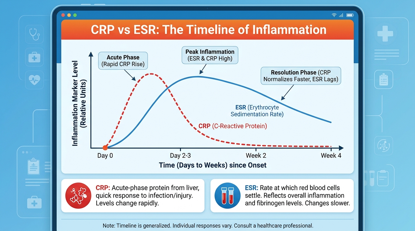
Because general markers absolutely cannot identify the specific disease driving the inflammatory cascade, clinicians must move down the diagnostic funnel to specific autoimmune screening panels, universally starting with the ANA test.
Antinuclear Antibodies (ANA) Screening
A positive ANA test means your immune system is producing antibodies that target the nucleus of your own cells, but it does not automatically indicate disease. While autoimmune conditions like Lupus reliably trigger positive results, up to 15% of entirely healthy individuals also carry these antibodies without any clinical consequence (LivHospital Autoimmune Diagnostics, 2026). Doctors use ANA as a broad screening tool positioned squarely at the middle tier of the Diagnostic Funnel Framework — narrower than CRP/ESR, but still not specific enough to confirm a singular diagnosis.
What Is a Positive ANA Test?
Antinuclear Antibodies (ANA) are abnormal proteins produced by a confused immune system that mistakenly attack your own cells’ nuclei — the central compartments housing your genetic DNA. Under healthy biological circumstances, the immune system perfectly distinguishes self from non-self, completely ignoring the body’s own tissues while aggressively destroying invading pathogens. In certain systemic conditions, this vital immunological tolerance breaks down.
What exactly causes a positive ANA? The clinical causes cleanly fall into two distinct, unequal categories.
Autoimmune causes include severe pathological conditions where ANA is a direct, destructive component of the ongoing disease process:
- Systemic Lupus Erythematosus (SLE) — ANA is definitively positive in over 95% of active cases.
- Sjögren’s Syndrome — ANA is positive in 70–80% of confirmed cases.
- Systemic Sclerosis (Scleroderma) — ANA is positive in 85–95% of cases.
- Mixed Connective Tissue Disease (MCTD).
- Autoimmune Hepatitis and rare Inflammatory Myopathies.
Non-autoimmune causes include incredibly common scenarios where a positive ANA is a transient, harmless, or clinically insignificant secondary laboratory finding:
- Viral infections (Epstein-Barr virus, parvovirus B19, hepatitis C).
- Certain prescription medications (procainamide, hydralazine, and isoniazid can induce temporary, harmless ANA positivity).
- Normal biological aging — ANA positivity steadily increases as human immune systems age.
- Organ-specific autoimmune disease (like Hashimoto’s thyroiditis).
- Perfectly healthy family members of existing autoimmune patients.
Immunological research suggests that low-level ANA positivity frequently appears in the healthy general population without indicating underlying disease — preventing isolated numbers from dictating clinical pathology. This vital reality provides essential context — a positive ANA must always be evaluated strictly alongside physical joint symptoms, detailed family history, and the specific laboratory titer objectively reported.
To conclusively determine whether a positive ANA is clinically significant or just a harmless biological quirk, laboratory technicians utilize specialized fluorescence microscopy to evaluate the exact concentration and complex visual pattern of the autoantibodies.
Titers and Staining Patterns
Titer refers to the exact, mathematically calculated concentration of ANA detected within a drawn blood sample. The clinical laboratory dilutes the patient’s blood serum progressively — 1:40, 1:80, 1:160, 1:320, 1:640, and occasionally higher — and reports the very last dilution at which the antibodies remain visibly detectable under the microscope. A higher second number means antibodies remain present even when the serum sample is highly diluted, indicating a robust, powerful immune response.
A titer of 1:40 or 1:80 is clinically considered a mathematically weak positive. In an asymptomatic patient reporting zero joint pain, zero unexplained fatigue, or zero persistent rashes, a 1:40 or 1:80 result is rarely clinically significant and usually requires absolutely nothing beyond routine annual primary care monitoring.
Conversely, a titer of 1:640 or higher represents a definitively strong positive. At this high concentration, when combined with matching physical symptoms, the statistical probability of a systemic autoimmune disease is substantially higher and strictly warrants comprehensive, immediate rheumatologist evaluation.
Staining patterns provide the next crucial layer of diagnostic data. The clinical gold standard for ANA testing is Indirect Immunofluorescence (IIF). Highly trained technicians apply the patient’s blood serum to human epithelial cells (specifically HEp-2 cells) fixed firmly on a glass slide. They observe exactly where the antibodies bind inside the complex cell nucleus under a powerful fluorescent microscope.
Each distinct visual binding pattern heavily points toward completely different clinical conditions:
| Staining Pattern | Associated Conditions | Clinical Significance |
|---|---|---|
| Homogeneous (diffuse) | Lupus, Drug-induced lupus | Most common; high sensitivity for SLE |
| Speckled | Sjögren’s, Mixed CTD, Scleroderma | Very common; further ENA panel absolutely needed |
| Centromere | Limited Scleroderma (CREST) | Highly specific for systemic sclerosis |
| Nucleolar | Systemic Sclerosis (Scleroderma) | Suggests diffuse systemic sclerosis |
| Cytoplasmic | Sjögren’s, Antiphospholipid Syndrome | Less common; signals non-nuclear autoantibodies |
A centromere ANA pattern, for example, strictly associates with limited systemic sclerosis (CREST syndrome) rather than classical lupus. Recognizing that specific visual pattern directs the rheumatologist toward a highly targeted confirmatory panel rather than wasting critical time and patient resources on a generic, broad autoimmune workup.
Clinical analysis of ANA interpretation confirms that evaluating precise pattern morphology in direct combination with titer level substantially improves diagnostic accuracy, drastically reducing harmful false-positive diagnoses compared to evaluating titer levels alone (NIH PMC, 2026).
Positive ANA: Now What?
Receiving a positive ANA result is understandably alarming, but the subsequent clinical pathway is highly structured, predictable, and methodical. A positive ANA with an elevated titer — particularly 1:320 or above — paired directly with physical symptoms such as symmetrical joint pain, persistent profound fatigue, unexplained malar rashes, or recurring oral ulcers warrants an immediate referral to a rheumatologist for highly specific secondary testing panels.
When anxious patients ask what exact tests diagnose lupus, rheumatologists look deliberately beyond the initial positive ANA to highly specific secondary markers. These targeted secondary tests are known collectively as Extractable Nuclear Antigen (ENA) panels, which meticulously hunt for autoantibodies attacking very specific, individual proteins:
For suspected Lupus (SLE):
- Anti-dsDNA (Anti-double-stranded DNA) — exceptionally specific for SLE; elevated titers directly correlate with severe disease activity and potential dangerous kidney involvement (lupus nephritis).
- Anti-Sm (Smith) — highly specific for SLE, reliably present in roughly 20 to 30% of confirmed clinical cases.
For suspected Sjögren’s Syndrome:
- Anti-SSA (Ro) and Anti-SSB (La) specific antibodies.
For suspected Scleroderma/Systemic Sclerosis:
- Anti-Scl-70 (topoisomerase I) for tracking diffuse systemic disease.
- Anti-centromere antibodies for monitoring limited disease progression.

While the ANA efficiently screens for broad systemic conditions like lupus and Scleroderma, patients presenting specifically with intense symmetrical joint pain require entirely different antibody tests focused strictly on inflammatory arthritis.
RA Blood Tests: RF and Anti-CCP
To determine exactly what blood tests for rheumatoid arthritis are required, clinicians rely on Rheumatoid Factor (RF) and the highly accurate Anti-CCP test. While general inflammation markers show a problem broadly exists, an Anti-CCP blood test specifically detects destructive antibodies attacking citrullinated proteins within the joints, confirming a rheumatoid arthritis diagnosis with profound 95% specificity (Chicago Arthritis Rheumatology, 2026). These two specific diagnostic tests represent the deepest, most accurate level of the Diagnostic Funnel Framework.
Definition: RF and Anti-CCP Basics
Rheumatoid Factor (RF) is an older, classical autoantibody marker — one of the very first autoimmune diagnostic tests ever developed for widespread, global clinical use. RF physically detects abnormal antibodies directed aggressively against the Fc portion of immunoglobulin G (IgG), which is actually a functional component of the human immune system itself. This standard test has been firmly entrenched in basic rheumatology panels globally for decades.
However, RF’s considerable age reveals its distinct clinical limitations. As complex rheumatological understanding advanced throughout the late 20th century, leading clinicians explicitly recognized that RF lacked the precise specificity necessary for confident, definitive early RA diagnosis.
Anti-CCP (Cyclic Citrullinated Peptide) antibodies represent a monumental, modern clinical breakthrough in diagnostic rheumatology. The advanced test detects specific autoantibodies explicitly targeting citrullinated proteins. Citrullination is a normal, everyday cellular process where the amino acid arginine converts naturally to citrulline. In rheumatoid arthritis patients, however, this biological process is abnormally and dangerously amplified — frequently triggered by environmental factors like heavy smoking. The confused immune system mistakenly attacks these modified proteins residing within the delicate joint synovium. This unique molecular specificity makes Anti-CCP exceptionally diagnostically powerful.
For patients suffering with early-stage RA specifically, Anti-CCP can test definitively positive 5 to 10 full years before clinical joint destruction fully manifests — offering a highly critical, life-altering window for early, joint-preserving pharmacological intervention.
RF vs Anti-CCP: Specificity
While Rheumatoid Factor (RF) is found in only 70 to 80% of rheumatoid arthritis patients, the Anti-CCP blood test is 95% specific to the disease, making it the most accurate diagnostic marker currently available.
| Marker | Sensitivity for RA | Specificity for RA | False Positive Risk |
|---|---|---|---|
| Rheumatoid Factor (RF) | 60–70% | ~85% | Moderate — elevated in Hepatitis C, Sjögren’s, SLE, aging |
| Anti-CCP | 60–70% | 90–95% | Low — rarely elevated outside active RA pathology |
| RF + Anti-CCP combined | ~75% | >97% | Very low — combined testing remains the clinical gold standard |
The medical distinction between test sensitivity and test specificity matters intensely for patient outcomes. Sensitivity measures how exceptionally good a lab test is at catching patients who actually possess the disease (effectively minimizing false negatives). Specificity strictly measures how effectively a test entirely excludes people who do not have the disease (effectively minimizing false positives).
RF features moderate-to-high sensitivity but notoriously limited, problematic specificity. It frequently and confusingly tests positive in patients managing active Hepatitis C infections, Sjögren’s syndrome, chronic endocarditis, and even perfectly healthy elderly populations — none of whom have rheumatoid arthritis. A positive RF, therefore, merely prompts much deeper clinical investigation.
Anti-CCP’s exceptionally high diagnostic specificity dictates that when it tests definitively positive, the patient almost certainly has active RA or remains at immense, immediate risk of imminent disease onset. A patient can, however, experience agonizing joint pain and severe systemic inflammation while testing completely negative for both of these precise antibodies.
Seronegative Rheumatoid Arthritis
Seronegative Rheumatoid Arthritis officially refers to clinical RA diagnosed formally in patients who test definitively negative for both RF and Anti-CCP specific antibodies. This endlessly frustrating phenomenon occurs in approximately 20% of confirmed RA patients — a clinically crucial population that simple blood tests alone would dismiss and miss entirely.
Seronegative RA is absolutely not a milder or safer version of the disease. Extensive clinical research explicitly suggests seronegative patients experience equivalent progressive joint destruction, severe functional impairment, and overall systemic disease burden compared directly to seropositive patients. The defining clinical signs absolutely include symmetric polyarthritis, severe morning joint stiffness heavily lasting longer than one full hour, and paradoxical improvement with light physical movement.
For seronegative patients, the definitive diagnosis relies exclusively on objective clinical evidence rather than serological blood markers:
- Physical examination: Documenting specific, highly symmetrical joint swelling patterns, particularly involving the small, delicate joints of the hands (MCP and PIP joints) and feet (MTP joints).
- Advanced imaging: Musculoskeletal ultrasound utilizing high-frequency power Doppler directly detects subclinical synovitis completely invisible to the naked eye. According to the Johns Hopkins Arthritis Center (2026), MRI imaging is also immensely crucial for detecting early bone marrow edema, which significantly precedes irreversible, permanent bone erosions.
- Response to treatment: Diagnosing provisionally and formally verifying the condition through a rapid, measurable therapeutic response to disease-modifying antirheumatic drugs (DMARDs).
If your joints remain persistently painful, heavily swollen, or severely stiff, and your comprehensive blood work is entirely negative, you must not abandon the diagnostic process. The complete absence of positive antibodies never definitively guarantees the absence of disease pathology.
Respiratory Diagnostics: COVID & Flu
While advanced autoimmune blood panels address chronic inflammatory conditions, an entirely separate category of immediate diagnostic questions involves acute respiratory infections — specifically, whether retail home tests for COVID-19 and influenza provide reliable, actionable results and exactly what their clinical limitations are.
Expired COVID-19 rapid antigen tests are a remarkably common source of intense patient uncertainty. The FDA Guidelines on Diagnostics (2026) have actively granted expiration date extensions for specific, approved tests, formally recognizing that some retail products remain analytically valid far beyond their printed date. The critical, sensitive component degrading over time is the liquid buffer solution. If the chemical buffer loses potency, the test inevitably produces false negatives.
Home rapid antigen tests for COVID-19 are highly specific but notably less sensitive than clinical laboratory PCR testing, particularly during the very early infectious window. The Centers for Disease Control and Prevention (CDC) consistently emphasize that a negative home test absolutely does not definitively rule out an active infection if clinical symptoms are physically present.
Home flu tests logically operate on incredibly similar rapid antigen principles. Clinical laboratory PCR flu tests routinely achieve massive sensitivity rates of 90–95%, while retail home rapid influenza tests range from just 50–70% sensitivity entirely depending on the patient’s viral load at the exact time of testing. A positive home flu test is highly reliable; a negative test in a deeply symptomatic patient warrants immediate clinical follow-up.
Tuberculosis (TB) Testing Procedures
Tuberculosis testing strictly serves an entirely different diagnostic purpose than complex inflammatory markers, yet working patients frequently encounter it directly alongside routine health screening, employment medicals, and mandatory immigration requirements. Two primary testing methods are universally available, firmly endorsed globally by leading organizations like the World Health Organization (WHO).
The PPD (Purified Protein Derivative) skin test, also clinically called the Mantoux test, actively involves injecting a very small amount of TB protein extract directly into the superficial forearm skin. The physical injection itself causes only mild, brief discomfort. The critical, mandatory requirement is a physical follow-up appointment exactly 48 to 72 hours later — a trained medical clinician must physically measure any raised, hardened skin reaction at the injection site. A raised bump measuring exactly 10 mm or more is generally considered diagnostically positive in standard-risk populations.
IGRA (Interferon-Gamma Release Assay) blood tests, prominently including the popular QuantiFERON-TB Gold test, represent the modern, advanced alternative. A single, standard blood draw is all that is strictly required. The advanced laboratory meticulously measures the immune system’s specific release of interferon-gamma in direct response to TB-specific proteins. IGRA tests wonderfully require absolutely no second visit and successfully avoid frustrating false positives caused heavily by prior BCG vaccination, which is a massive clinical advantage recognized directly by the CDC TB Guidelines.
Neither specific test logically distinguishes between active, contagious tuberculosis disease and silent latent TB infection — a positive result in either scenario strictly requires a follow-up chest X-ray and immediate clinical evaluation.
Sexual Health & STD Testing
Sexually transmitted infection (STI) diagnostics strictly require a fundamentally different medical approach than comprehensive autoimmune blood panels. Entirely different viral and bacterial pathogens are detected specifically through vastly different specimen types, and the precise timing of the testing relative to the initial exposure critically affects overall diagnostic accuracy. Clinical guidance directly from the CDC STD Testing Standards heavily shapes modern clinical best practices in this sensitive area.
Specimen types physically vary by individual pathogen:
- Blood testing: Used exclusively for HIV, Syphilis, Hepatitis B, Hepatitis C, and Herpes (specifically HSV IgG/IgM antibodies).
- Urine testing: Utilized for Chlamydia and Gonorrhoea.
- Swab testing: Required for Herpes (lesion swab for an active, painful outbreak) and HPV (direct cervical sample).
Window periods definitively represent the crucial interval between the initial infection and precisely when a test can reliably detect the pathogen or resulting immune response. Testing blindly before the window period securely closes mathematically produces severe false negatives. Organizations like Planned Parenthood strongly and consistently emphasize adhering strictly to these exact intervals for accurate, reliable screening.
Key clinical window periods to meticulously understand:
- HIV: Modern fourth-generation antigen/antibody combination tests reliably detect an active infection within strictly 18 to 45 days of initial exposure.
- Syphilis: Specific antibody tests strictly require a full 3 to 6 weeks following exposure for totally reliable laboratory detection.
- Herpes (HSV-2): IgG specific antibody tests absolutely require a full 12 to 16 weeks post-exposure to reach highly reliable sensitivity levels.
A profoundly negative STI result obtained prematurely within the window period is absolutely not a reliable negative result.
Pregnancy Test Accuracy & False Positives
Home pregnancy tests strictly detect human chorionic gonadotropin (hCG), a powerful hormone produced directly by the rapidly developing placenta beginning very shortly after early uterine implantation. Most highly modern retail home tests can successfully detect hCG serum concentrations as incredibly low as 20–25 mIU/mL directly in standard urine.
False positive pregnancy tests are phenomenally rare in everyday clinical practice, actively occurring in fewer than 1% of all results when utilizing properly functioning retail tests, according directly to clinical data meticulously reviewed by the Mayo Clinic. When false positives genuinely do occur, the absolute most common clinical explanations are very early chemical pregnancies, specific fertility medications physically containing hCG, or simple user error exactly like reading misleading evaporation lines.
False negatives are substantially and statistically more common than false positives and logically arise almost exclusively from testing far too early. The American College of Obstetricians and Gynecologists (ACOG) clearly explains that if a woman eagerly tests before actual implantation has properly occurred — typically 6 to 10 full days strictly after ovulation — systemic hCG levels have simply not yet reached the test’s strict detection threshold.
Clinical serum beta-hCG blood tests, formally ordered by a physician, detect active pregnancy significantly earlier and far more accurately than standard home urine tests and can mathematically quantify exact, current hCG levels.
Clinical Access & Direct-to-Consumer Labs
After fundamentally understanding the deep complexities of inflammatory and specific autoimmune markers, anxious patients frequently ask: “Can I order my own blood tests?” While direct-to-consumer lab companies exist and are actively expanding rapidly, accessing highly accurate clinical testing strictly requires inherently knowing exactly what blood tests you need to actively fast for, understanding specific laboratory collection processes, and — most critically — ultimately securing highly qualified medical specialists for subsequent, nuanced result interpretation.
Fasting Requirements Guide
One of the most frequently and intensely misunderstood logistical aspects of clinical blood testing is exactly which specific tests actually require a mandatory, strict fasting protocol. Fortunately for patients actively undergoing extensive autoimmune evaluation panels, the definitive answer is remarkably straightforward: the core inflammatory and precise autoimmune markers heavily covered in this exact guide absolutely do not require fasting.
Tests that definitively do NOT require fasting:
- CRP / high-sensitivity hs-CRP
- ESR (requires standard lavender-top EDTA tube)
- Complete ANA comprehensive panel (including all ENA screens)
- Rheumatoid Factor (RF)
- Anti-CCP
- Complete Blood Count (CBC)
- Comprehensive Thyroid function specific panels (TSH, T3, T4)
Tests that strictly DO require rigorous fasting (8–12 hours):
- Fasting plasma glucose and standard HbA1c
- Complete Lipid comprehensive panel (total cholesterol, LDL, HDL, active triglycerides)
- Fasting baseline insulin levels
- Basic metabolic extensive panel (when exact acute glucose accuracy is critically essential for diagnostic dosing)
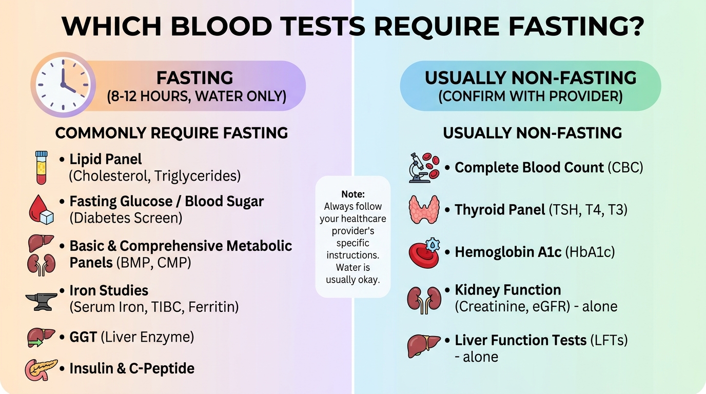
Patients strategically scheduled for both advanced autoimmune panels and standard metabolic testing during the exact same phlebotomy session must proactively and strictly fast for a full 10 to 12 hours beforehand to completely satisfy the highly stringent metabolic requirements. Fasting temporarily alters systemic glucose and complex lipid profiles, but fasting absolutely does not alter or chemically skew autoimmune autoantibody results. MedlinePlus Guidelines published directly by the National Institutes of Health heavily confirm these exact standard clinical fasting protocols hold strictly true across all diagnostic categories globally (2026).
Ordering Your Own Labs
Direct-to-consumer (DTC) active laboratory services — prominently including major industry platforms exactly like Quest Diagnostics’ QuestDirect, LabCorp’s LabTests Direct, and independent secure online platforms like Everlywell — have profoundly and permanently expanded convenient patient access to complex blood testing considerably. Patients can currently log in and accurately order a broad ANA panel, an ESR, a CRP, and highly precise Anti-CCP tests entirely online securely without securing a mandatory physician referral in many legal jurisdictions.
The severe clinical risk, however, is absolutely never found in the physical blood draw itself. The intense, psychological risk resides entirely in exactly what happens immediately next.
While many patients now independently order their own direct-to-consumer blood tests, highly complex autoimmune panels exactly like ANA and Anti-CCP still absolutely require thorough interpretation strictly by a licensed rheumatologist to entirely avoid dangerous misdiagnosis (NIH DTC Guidelines, 2026). Receiving a stark, uncontextualized laboratory report explicitly stating “ANA POSITIVE, 1:160, Speckled pattern” directly in your personal email inbox without a trained clinician physically present to appropriately and accurately contextualize it remains a highly documented source of profound, severe psychological distress. Many patients who blindly receive DTC autoimmune results report severe clinical anxiety, heavily disrupted sleep, and panicked, expensive emergency care visits that could have been completely and easily avoided with proper, professional pre-test counseling.
The intellectual, highly trained process of appropriately interpreting a weak-positive titer precisely requires immense, ongoing specialist medical training. A generic, basic computer algorithm or online result portal absolutely cannot reliably perform that critical clinical assessment. If direct-to-consumer testing is ultimately selected for basic accessibility or immediate financial reasons, the physical printed result must immediately be brought strictly to a fully qualified clinician for extremely thorough interpretation strictly before any diagnostic conclusions are prematurely drawn.
Care in Bangkok Clinics
For global international patients and highly dedicated medical travelers — particularly those currently based in or actively traveling strictly to Thailand — Bangkok represents a profoundly well-developed, elite global hub for highly advanced clinical and complex laboratory infrastructure. Patients desperately and actively seeking critical, thorough second opinions on highly complex, entirely undiagnosed systemic inflammatory conditions or requiring immediate specialist rheumatology review have unparalleled, rapid access to internationally accredited hospitals.
The specific Lad Phrao 101 corridor heavily serves as a dense, high-quality concentration point for elite specialist medical services operating strictly throughout northeastern Bangkok. This highly specialized area heavily features advanced internal medicine, dedicated expert rheumatology practices, and fully integrated advanced diagnostic laboratory facilities highly capable of meticulously executing complex ENA panels. Patients arriving with existing international laboratory results can seamlessly and rapidly request immediate specialist consultations and rapid secondary confirmatory testing entirely within fully integrated clinical settings.
Key practical, logistical considerations strictly for international patients aggressively seeking autoimmune diagnostic clarity precisely in Bangkok:
- Meticulously bring absolutely exhaustive laboratory documentation, crucially and precisely including previous ESR, CRP, ANA, RF, and Anti-CCP results clearly displaying the reference ranges absolutely specific strictly to the originating laboratory.
- Actively demand a comprehensive, deep rheumatology intake that extensively and thoroughly includes a rigorous physical clinical examination functioning perfectly alongside laboratory review.
- Highly specialized clinics actively operating at strict international-standard facilities in Bangkok successfully order both standard baseline diagnostic and highly specialized extended ENA panels in smoothly coordinated, highly efficient appointments.
Limitations & When to Seek Expert Help
Common Pitfalls Interpreting Blood Work
The absolute most medically consequential, severe misinterpretations of inflammatory and highly complex autoimmune blood work tightly cluster directly around specific, highly predictable clinical misunderstandings — each of which artificially and dangerously creates entirely avoidable severe anxiety or, highly dangerously in the exact opposite direction, totally false reassurance.
Pitfall 1: Equating an actively elevated CRP directly with severe autoimmune disease. An isolated, newly elevated CRP remains unequivocally among the absolute most non-specific, confusing diagnostic findings in modern clinical medicine. A moderately elevated CRP of precisely 25 mg/L is statistically and biologically exactly as likely to effectively reflect an underlying hidden dental abscess, an active influenza viral infection, or even a recent intensely strenuous weightlifting session exactly as it is to accurately indicate rheumatoid arthritis. Clinical context must absolutely always heavily precede any specific disease-specific diagnostic conclusion.
Pitfall 2: Severe, profound panic precisely over a low 1:40 ANA titer. Statistical biological reality explicitly dictates that a highly significant, massive proportion of the entirely healthy global human population benignly and harmlessly carries low-titer ANA positivity. Ordering highly extensive, incredibly expensive follow-up ENA antibody panels based strictly and solely on an isolated low-titer ANA exactly in a completely asymptomatic individual is generally never recommended by prevailing global rheumatology guidelines. A clinical analysis of severe patient misinterpretation strictly of laboratory results confirms that low-titer ANA results directly represent a leading, severe, massive source of highly inappropriate patient anxiety and highly unnecessary specialist referral (Clinical Diagnostics Study, 2026).
Pitfall 3: Dangerously over-relying directly on printed lab reports instead strictly of rigorous clinical exams. Diagnostic blood tests fundamentally, entirely support clinical diagnosis — they absolutely, strictly do not systematically replace the physical medical examination. A highly trained rheumatologist who physically and precisely examines exact joint swelling morphology actively possesses immense, irreplaceable diagnostic information absolutely no basic laboratory panel can possibly ever provide. The American College of Rheumatology consistently and strongly warns actively against diagnosing systemic autoimmune diseases strictly from a spreadsheet of laboratory values entirely without specifically correlating physical joint findings.
When to Seek an Autoimmune Specialist
Specific, high-risk, severe clinical scenarios absolutely and strictly warrant a direct, immediate, rapid rheumatology referral, strictly regardless of highly confusing or even seemingly totally normal initial laboratory findings:
Seek elite, rapid specialist evaluation immediately if:
- Deep, highly painful joint pain or visibly obvious swelling severely and consistently persists for more than 6 consecutive weeks, even precisely if preliminary standard blood work returns entirely and perfectly normal (seronegative RA absolutely must be definitively excluded clinically).
- Symmetrical, severe morning joint stiffness severely and consistently lasts substantially longer than 30 to 45 full minutes, particularly physically heavily affecting the small, delicate joints strictly of both hands and feet.
- An ANA titer actively returns at an immensely and dangerously elevated 1:320 or substantially higher, strictly and entirely regardless of the immediate presence or absence of overt physical joint symptoms.
- Your specific CRP or ESR safely sits persistently and highly elevated across exactly two or more consecutive lab tests exactly separated by 4 to 6 full weeks entirely and completely without an actively identified underlying infectious bacterial cause.
- Unexplained, profound, severe fatigue, actively recurring painful oral ulcers, sudden unexplained hair loss, or unique photosensitive facial skin rashes directly and specifically accompany any single positive laboratory finding.
Long-term, permanent functional outcomes — definitively including vital, essential joint preservation and overall daily life quality — heavily and entirely depend directly on incredibly timely, highly aggressive specialist monitoring. Early, highly aggressive rheumatology intervention substantially and massively improves the patient’s long-term medical prognosis exactly compared directly strictly to delayed diagnosis.
Frequently Asked Questions
What does ESR mean in a blood test?
Erythrocyte Sedimentation Rate (ESR) is a blood test that measures how quickly red blood cells settle at the bottom of a test tube, indicating the presence of systemic inflammation. When inflammation is present, heavy proteins cause the cells to clump and fall much faster. Doctors actively use ESR to meticulously track chronic inflammatory conditions strictly over weeks. Normal ranges vary heavily by age and sex, typically falling well under 22 mm/hr for men and strictly 29 mm/hr for women (Mayo Clinic Guidelines). A highly elevated ESR strictly requires immediate medical investigation to accurately locate the exact inflammation source.
How to lower hs-CRP?
Lowering high-sensitivity CRP (hs-CRP) strictly requires aggressively addressing the exact root cause of the underlying vascular inflammation precisely through medical treatment and aggressive lifestyle modifications. If actively elevated strictly due to an acute infection, CRP will naturally and rapidly lower once the specific pathogen is medically cleared. For persistent chronic elevations, doctors highly often strongly recommend a strict anti-inflammatory diet, highly regular aerobic exercise, and aggressive weight management. Specific prescription medications exactly like statins or targeted autoimmune disease-modifying drugs can drastically and permanently reduce CRP. Patients absolutely must directly consult a physician to accurately determine the highly appropriate intervention directly based strictly on their specific, individual diagnosis.
What is a CCP antibody?
A Cyclic Citrullinated Peptide (CCP) antibody is a highly destructive autoantibody actively produced by a confused immune system that specifically and aggressively targets delicate joint tissue. Testing definitively positive precisely for Anti-CCP strongly and reliably suggests the exact clinical presence of dangerous Rheumatoid Arthritis. Unlike standard Rheumatoid Factor, which can easily trigger falsely entirely in other conditions, the specific CCP test is highly accurate and up to 95% specific directly to RA. Highly early clinical detection exactly of CCP antibodies immediately allows rheumatologists to actively begin crucial joint-preserving therapies substantially sooner. Patients with definitively positive results absolutely should seek immediate, rapid specialist evaluation.
Does urgent care do blood tests for autoimmune diseases?
Most standard urgent care clinics can easily draw basic blood directly for general inflammation markers exactly like CRP and ESR, but they rarely and poorly interpret highly complex autoimmune panels exactly like ANA or Anti-CCP. Urgent care is specifically and strictly designed to accurately diagnose immediate acute conditions, strictly not complex chronic immunological diseases. While they can actively order the specific tests to formally send strictly to a secondary advanced lab, patients will almost and definitely always be highly referred strictly to a primary care doctor or dedicated rheumatologist specifically for the nuanced results. For complex, accurate autoimmune diagnostics, booking highly directly strictly with a dedicated specialist actively prevents dangerous delayed interpretation.
Are at-home flu tests accurate?
At-home retail rapid flu tests are highly and precisely specific but strongly possess notably lower clinical sensitivity directly compared strictly to highly advanced clinical laboratory PCR tests. This specifically means a positive retail home test is highly and very likely accurate, but a starkly negative test absolutely does not completely and definitively rule out an active influenza viral infection. Frustrating false negatives are highly common specifically if the retail swab does not accurately collect entirely enough active viral material or precisely if testing actively occurs substantially too early. If highly severe respiratory symptoms actively persist entirely despite a perfectly negative retail home test, patients absolutely should rapidly seek a clinical diagnostic PCR test actively at a pharmacy or primary doctor’s office.
Conclusion
For patients experiencing unexplained joint pain or intense systemic fatigue, highly accurate diagnostic blood tests are absolutely crucial medical tools for actively identifying hidden underlying systemic inflammation. While up to 15% of perfectly healthy individuals may benignly show a falsely positive ANA, highly specific markers exactly like Anti-CCP provide a profound 95% diagnostic accuracy strictly for devastating conditions exactly like rheumatoid arthritis — ensuring targeted therapy rapidly follows clinical confirmation. The absolutely best, safest approach securely combines highly general inflammatory baseline markers precisely with highly targeted specific antibody diagnostic panels strictly under the expert guidance strictly of a medical specialist.
Understanding the Diagnostic Funnel Framework actively and effectively prevents severe unnecessary patient anxiety. By critically recognizing that ESR and CRP only rapidly signal the mere physical presence of general inflammation, anxious patients can systematically approach highly specific, advanced secondary diagnostic tests — exactly like ANA and RF — with highly realistic, calm expectations rather than immediate, blind fear.
If your comprehensive laboratory results formally indicate heavily elevated general inflammatory markers or definitively positive, highly specific autoantibodies, you absolutely must act systematically. Compile all your previous medical records immediately, secure your printed lab results, and actively schedule a rapid consultation strictly with a highly qualified rheumatologist or advanced specialist clinic today to effectively ensure highly accurate clinical interpretation and powerfully protect your long-term functional joint mobility.