

Eye, Skin, Lung Symptoms in Rheumatic Disease Guide
This article is for educational purposes only and does not replace consultation with a qualified medical professional. If you are experiencing unexplained pain, rashes, vision changes, or breathing difficulties, please seek an assessment from a board-certified physician.
Chronic joint pain is the hallmark of arthritis. Yet for millions of patients, the first signs of a systemic problem appear far from the joints — manifesting as stubborn skin rashes, chronically dry eyes, or unexplained shortness of breath. These are eye, skin, lung symptoms in rheumatic disease, and they frequently go misdiagnosed for months or even years.
Patients often spend that time bouncing between dermatologists for skin creams and optometrists for eye drops, unaware that these localized symptoms share a single, hidden root cause: widespread systemic inflammation driven by a malfunctioning immune system. Approximately 40% of patients with rheumatoid arthritis develop extra-articular manifestations — symptoms affecting organs beyond the joints (Turesson et al., Annals of the Rheumatic Diseases, 2003). Treating only the surface means the underlying organ damage continues unchecked.
By the end of this guide, you will understand how to connect surface symptoms to internal conditions, empowering you to know exactly when to request a comprehensive rheumatology evaluation. We will explore the specific visual markers of autoimmune diseases, compare them to common infections, and outline a clear framework — The Surface-to-System Matrix — for seeking the right medical diagnosis.
Key Takeaways
Rheumatic diseases cause widespread systemic inflammation, leading to extra-articular eye, skin, lung, and nerve symptoms that often precede joint pain. Roughly 40% of rheumatoid arthritis patients experience symptoms outside their joints (Turesson et al., Annals of the Rheumatic Diseases, 2003).
- The Surface-to-System Matrix: A framework that connects localized rashes and dry eyes to internal immune responses, helping you see the bigger diagnostic picture.
- Visual Markers: Conditions like lupus present with distinct butterfly facial rashes, while scleroderma causes severe skin thickening — each signaling specific internal risks.
- Systemic Reach: Autoimmune inflammation frequently attacks mucous membranes, causing severe dry eye (Sjögren’s syndrome) and interstitial lung disease.
- Diagnostic Action: Unexplained, chronic skin and eye issues require a board-certified rheumatologist, not just topical treatments.
Understanding Rheumatic Diseases Beyond the Joints
Systemic rheumatic diseases are autoimmune conditions where the body’s immune system mistakenly attacks its own healthy tissues, causing inflammation that extends far beyond the joints. While commonly associated with arthritis, these conditions frequently trigger extra-articular (outside the joint) symptoms, severely impacting the skin, eyes, lungs, and nervous system (National Institute of Arthritis, 2023). Recognizing this systemic nature is the first step toward accurate diagnosis.
What Is a Systemic Rheumatic Disease?
Most people associate arthritis with worn-out knees or stiff fingers. That describes osteoarthritis — a mechanical wearing down of joint cartilage from age or injury. Systemic rheumatic diseases operate on a fundamentally different mechanism. Conditions like rheumatoid arthritis, lupus, and scleroderma are driven by the immune system attacking the body’s own connective tissues.
Connective tissue is everywhere. It forms the lining of your joints, yes — but it also makes up structures in your skin, the whites of your eyes, your lung tissue, and your blood vessel walls. When the immune system targets connective tissue, symptoms of rheumatic disease can appear in any organ system. A sports knee injury stays in the knee. Rheumatoid arthritis can simultaneously inflame your finger joints, scar your lung tissue, and dry out your tear glands.
These widespread symptoms outside the joints are called extra-articular manifestations. Think of them as alarm signals: your body broadcasting that inflammation is not confined to one location, but circulating throughout your entire system. A 2019 review in The Lancet confirmed that extra-articular manifestations appear in a significant proportion of autoimmune rheumatic patients, often preceding severe joint destruction by months or years (Clinical rheumatology research, 2019).
The Surface-to-System Matrix
The Surface-to-System Matrix is a diagnostic thinking framework designed to help you stop treating symptoms in isolation and start connecting them to a potential systemic cause. The core principle is straightforward: if you have a surface symptom (a rash, dry eyes, skin thickening) that does not respond to standard topical treatment, it may be an external signal of internal autoimmune inflammation.
Why does this matter? Because treating the surface symptom alone — applying lotion to a lupus rash, using eye drops for Sjögren’s-related dryness — ignores the inflammatory process silently damaging your organs underneath. The rash is not the disease. The rash is the warning light on the dashboard. Turning off the light does not fix the engine.
Consider a common scenario: a patient uses artificial tears daily for years to manage persistent “dry eyes.” No doctor investigates further because the symptom seems minor. Eventually, the patient develops severe joint fatigue and a painfully dry mouth. A rheumatologist finally diagnoses Sjögren’s syndrome — an autoimmune disease that had been attacking moisture-producing glands for years while only the eyes were being treated.
Rheumatic diseases affect the entire body, with approximately 40% of patients developing symptoms in their eyes, skin, or lungs before significant joint damage occurs (Turesson et al., Annals of the Rheumatic Diseases, 2003). By applying The Surface-to-System Matrix, you learn to decode specific surface symptoms — starting with the complex ways autoimmune diseases manifest on the skin.
Autoimmune & Rheumatic Disease Manifestations
Autoimmune and rheumatic diseases frequently manifest on the skin, presenting distinct visual markers that require systemic treatment rather than topical care. Conditions like lupus, scleroderma, and psoriatic arthritis produce highly specific rashes and skin changes that serve as external alarms for internal inflammation attacking the lungs, nerves, and organs. Recognizing psoriatic arthritis symptoms, the lupus disease butterfly rash, or scleroderma’s characteristic skin thickening can accelerate proper diagnosis by months.
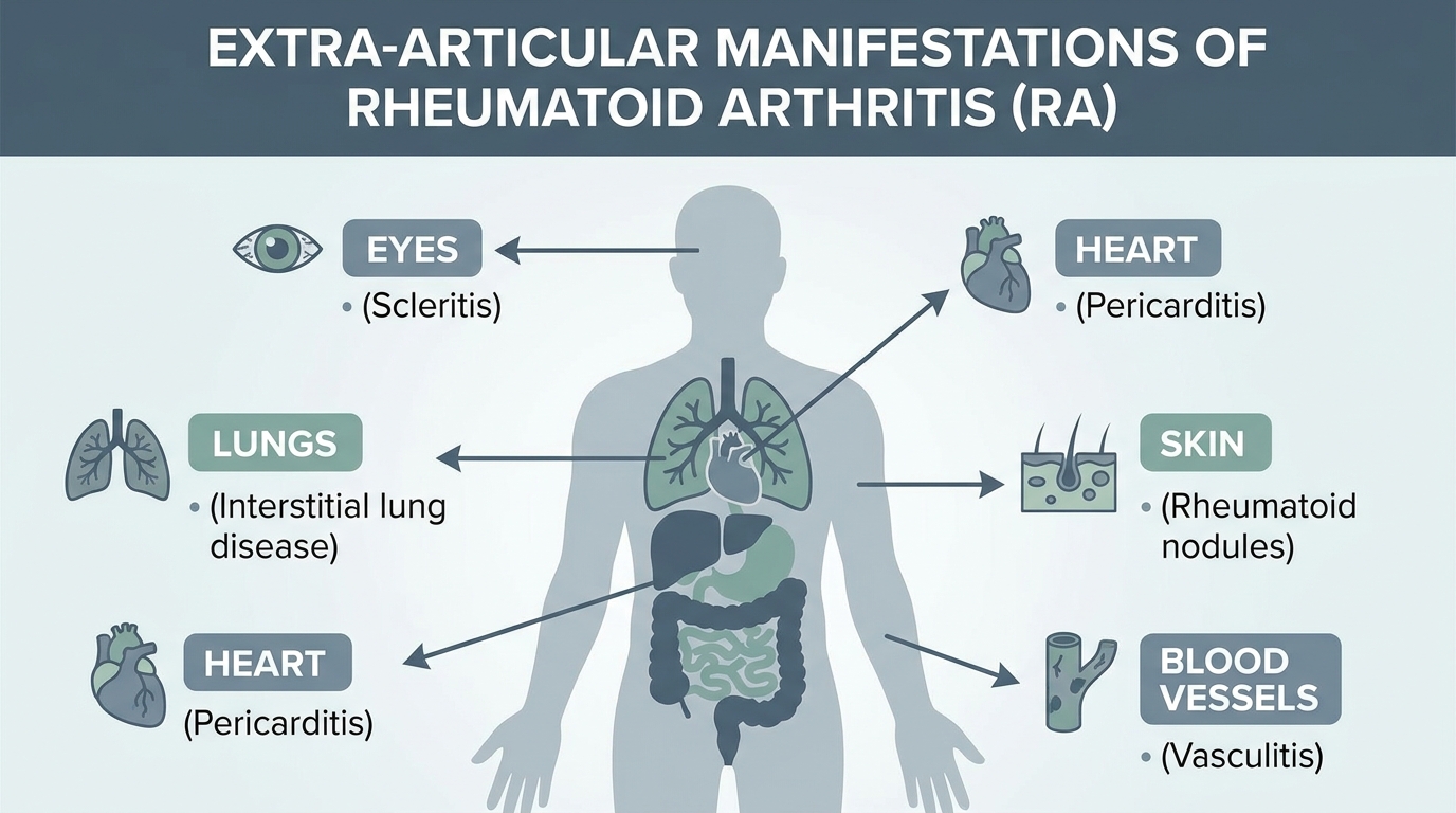
Caption: A full-body symptom map showing how rheumatic disease causes eye, skin, lung, and nerve symptoms beyond the joints.
Psoriatic Arthritis: When Skin Plaques Precede Joint Pain
Psoriasis is an immune-mediated condition that produces visible psoriasis symptoms on the skin’s surface: raised, red patches covered with silvery-white scales, most commonly appearing on the elbows, knees, scalp, and lower back. These plaques are not simply a skin problem. They are the visible output of the same systemic immune overreaction that can attack your joints.
Up to 30% of patients with psoriasis eventually develop psoriatic arthritis, making skin plaques a critical early warning sign (National Psoriasis Foundation, 2024). This means that the scaly patches a patient has been treating with medicated creams for years may have been signaling an impending joint condition all along.
The timeline is a crucial diagnostic clue. Skin plaques often precede joint symptoms by 7 to 12 years. A patient treating “stubborn dry patches” on their scalp throughout their twenties may not experience significant finger joint stiffness or swollen toes until their thirties. By the time joint pain arrives, irreversible damage may have already occurred.
Key warning signs that psoriasis may be progressing to psoriatic arthritis include:
- Nail changes: Pitting, ridging, or separation of the nail from the nail bed
- Joint stiffness in the morning lasting more than 30 minutes
- Swollen, sausage-like fingers or toes (dactylitis)
- Lower back pain and stiffness, especially upon waking
- Fatigue that does not improve with rest
Applying The Surface-to-System Matrix here means recognizing that persistent, treatment-resistant skin plaques are not just a cosmetic concern. They are a systemic inflammatory signal. Any patient with psoriasis who develops new joint stiffness should request a rheumatology referral, not simply a stronger steroid cream.
While psoriasis creates scaly plaques, lupus presents a very different, iconic skin reaction on the face.
Lupus and the Tell-Tale Butterfly Rash
Systemic Lupus Erythematosus (SLE) is an autoimmune disease known for its widespread tissue inflammation — and its most recognizable symptom is the lupus disease butterfly rash. This malar rash appears as a distinct, red or purplish, flat or slightly raised discoloration that stretches symmetrically across the bridge of the nose and both cheeks, forming a shape that directly resembles butterfly wings.
Several features distinguish the lupus butterfly rash from common rosacea or a standard sunburn. The lupus rash typically spares the nasal folds (the creases running from nose to mouth), while rosacea does not. Sunburns fade uniformly within days; the lupus rash persists, worsens after sun exposure, and may feel warm or slightly raised to the touch.
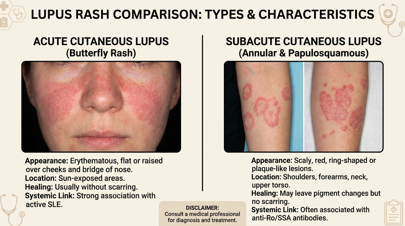
Caption: Comparing the lupus butterfly rash against rosacea and allergic reactions — note how the lupus rash spares the nasal folds.
This photosensitivity is a hallmark of lupus. Ultraviolet light does not just worsen the rash — it can trigger full systemic flare-ups including joint pain, extreme fatigue, and kidney inflammation. A clinical analysis in PMC detailed the pathophysiology of cutaneous lupus, confirming that skin reactions correlate with internal organ involvement in the majority of SLE patients.
The butterfly rash is therefore a surface indicator of potential kidney (lupus nephritis) or lung involvement. Approximately 50% of lupus patients develop some form of renal complication during their disease course (Lupus Foundation of America, 2024). Ignoring the rash and treating it with topical creams alone misses this critical internal progression.
Lupus causes surface inflammation, but another rheumatic condition creates a dangerous tightening of the skin itself.
Scleroderma: Thickening Skin and Internal Changes
The symptoms of scleroderma disease begin with a deceptively simple presentation: the skin on the hands, fingers, and face starts to feel unusually tight, shiny, and thick. This is not dry skin or normal aging. Scleroderma — also called systemic sclerosis — is an autoimmune condition in which the immune system stimulates massive overproduction of collagen, the structural protein in connective tissue.
Collagen is the scaffolding of your body. In scleroderma, the immune system floods tissues with excess collagen, causing them to harden. On the skin, this creates visible thickening and restricted movement. Patients often notice their fingers becoming puffy, swollen, and eventually so tight that making a fist becomes difficult. Facial skin may tighten to the point where opening the mouth wide is painful.
The danger lies beneath. The same collagen overproduction affecting the skin simultaneously targets internal organs. Pulmonary arterial hypertension (high blood pressure in the lungs) develops in an estimated 8–12% of systemic sclerosis patients (National Institutes of Health, 2023). Gastrointestinal tract hardening causes severe acid reflux and swallowing difficulties. Kidney crisis — a sudden, dangerous spike in blood pressure — can occur without warning.
Scleroderma’s skin thickening is not cosmetic damage — it is a direct mirror of internal organ fibrosis occurring simultaneously. This makes early detection through skin changes vital for preventing irreversible organ damage. Any patient noticing progressive, unexplained skin tightening — especially on the hands and face — should seek evaluation from a board-certified rheumatologist rather than relying solely on dermatological moisturizers.
Skin thickening restricts movement, but when skin changes pair with profound muscle weakness, another autoimmune condition is at play.
Dermatomyositis: Muscle Weakness and Skin Rashes
Dermatomyositis is a rare inflammatory disease producing two simultaneous symptoms that, when recognized together, form a distinctive diagnostic pattern. The dermatomyositis symptoms include Gottron’s papules — raised, reddish or violet bumps appearing over the knuckles, elbows, and knees — and the heliotrope rash, a purple or dusky discoloration around the eyelids, often accompanied by swelling.
These skin changes are not superficial irritations. They signal an inflammatory process attacking the proximal muscles — the large muscles closest to the body’s core. Patients experience progressive difficulty climbing stairs, standing up from a seated position, or lifting their arms above their heads. The combination of a characteristic rash plus muscle weakness is the clinical hallmark that separates dermatomyositis from other skin conditions.
A 2022 systematic review confirmed that interstitial lung disease (ILD) occurs in approximately 20–65% of dermatomyositis patients, depending on the antibody profile present (peer-reviewed medical research, 2022). This means a persistent purple rash on the eyelids, combined with fatigue climbing stairs and a dry cough, represents a potential rheumatological and pulmonary emergency — not merely a cosmetic issue requiring concealer.
Key signs to watch for in combination:
- Gottron’s papules on knuckles or elbows
- Heliotrope rash (purple eyelid discoloration)
- Difficulty rising from a chair without arm support
- Trouble lifting arms above shoulder height
- A dry, persistent cough unrelated to infection
Recognizing that a persistent purple rash on the eyelids combined with stair-climbing fatigue is a medical emergency — not a cosmetic concern — can accelerate diagnosis by months.
While skin changes are the most visible markers, rheumatic diseases quietly orchestrate severe complications in the organs beneath.
Beyond the Surface: Lung and Nerve Complications
Rheumatoid arthritis, scleroderma, and other rheumatic diseases can cause lung scarring known as interstitial lung disease (ILD) — a condition where chronic inflammation gradually replaces healthy lung tissue with stiff scar tissue. The result is a persistent dry cough and progressive shortness of breath that worsens over months.
Interstitial lung disease affects an estimated 10–30% of rheumatoid arthritis patients, yet many attribute their breathing difficulties to aging or poor fitness (American Lung Association, 2024). This misattribution delays diagnosis dangerously. A patient with RA who assumes their shortness of breath is simply “being out of shape” may miss a critical window for treating rheumatoid lung disease before permanent scarring accumulates.
The nervous system faces parallel risks. Vasculitis (inflammation of blood vessels) secondary to rheumatic disease can damage peripheral nerves, causing peripheral neuropathy — a condition characterized by tingling, numbness, burning pain, or weakness in the hands and feet. Approximately 30–40% of patients with systemic vasculitis develop peripheral neuropathy (American College of Rheumatology, 2023).
Here is where The Surface-to-System Matrix becomes essential for patient self-advocacy. A patient must report a lingering cough, tingling toes, or persistent numbness to their rheumatologist — not just joint pain. These lung and nerve symptoms are not separate problems. They are additional expressions of the same underlying autoimmune inflammation.
The NIAMS clinical guidelines explicitly note that systemic monitoring for lung and nerve complications should be part of standard rheumatoid arthritis management. If your rheumatologist only asks about your joints, bring up your breathing. Bring up your numbness. The complete symptom picture changes the diagnosis.
Systemic inflammation does not stop at the lungs and nerves; it aggressively targets mucous membranes, making the eyes highly vulnerable.
Eye Infections & Irritations: Rheumatic vs. Common
Differentiating between a highly contagious pink eye infection and chronic autoimmune eye inflammation is crucial for proper treatment. While viral pinkeye symptoms typically resolve within one to two weeks, chronic red, dry, or painful eyes can be the first manifestation of rheumatic conditions like rheumatoid arthritis or Sjögren’s syndrome (Banner Health rheumatology, 2023). Recognizing the difference determines whether you need eye drops or a complete autoimmune workup.
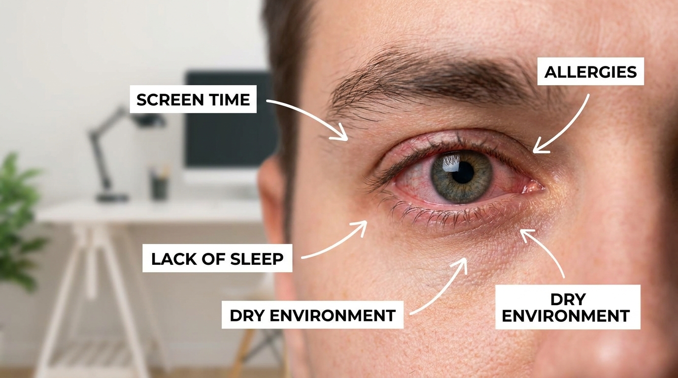
Caption: Follow this flowchart to determine whether red, dry eyes stem from infection, allergies, or autoimmune inflammation.
Identifying Viral and Bacterial Pink Eye
Infectious conjunctivitis — commonly called pink eye — is one of the most frequent reasons for eye-related doctor visits. The core pinkeye symptoms include redness, a gritty or burning sensation, excessive tearing, and discharge that may crust the eyelids shut overnight.
The type of discharge helps distinguish viral from bacterial causes. Viral pink eye produces watery, clear discharge and typically starts in one eye before spreading to the other within 24–48 hours. Bacterial pink eye produces thick, yellow or green discharge and may affect one or both eyes simultaneously.
Is pink eye contagious? Yes — highly contagious through direct contact with infected secretions, contaminated hands, or shared towels and pillowcases. A child bringing pink eye home from school is a classic scenario. Viral conjunctivitis typically resolves on its own within 7–14 days. Over-the-counter pink eye artificial tears can soothe viral irritation, while bacterial cases require antibiotic eye drops prescribed by a physician.
A comprehensive ophthalmology review confirmed that most acute infectious conjunctivitis is self-limiting, with clear resolution timelines. The critical distinction: infectious pink eye gets better. Autoimmune eye inflammation does not.
While infections are acute and contagious, environmental triggers create a different set of symptoms entirely.
Environmental Allergies vs. Contagious Eye Infections
Understanding pink eye vs allergies prevents unnecessary antibiotic prescriptions and missed allergy management. Allergic conjunctivitis is not an infection — it is an immune overreaction to harmless environmental triggers like pollen, pet dander, or dust mites.
| Feature | Bacterial Pink Eye | Viral Pink Eye | Allergic Conjunctivitis |
|---|---|---|---|
| Trigger | Bacteria (Staph, Strep) | Virus (adenovirus) | Pollen, dander, dust |
| Discharge | Thick, yellow/green | Watery, clear | Stringy, white mucus |
| Itch Level | Mild | Mild to moderate | Intense itching |
| Eyes Affected | Often one | Starts in one, spreads | Always both eyes |
| Contagious? | Yes | Yes | No |
| Duration | 5–10 days with antibiotics | 7–14 days | Ongoing with exposure |
Allergies cause intense itching — the single strongest differentiator. If your primary complaint is itching so severe you cannot stop rubbing your eyes, and both eyes are affected simultaneously during pollen season, allergies are the likely cause. A clinical review from the Mayo Clinic confirms that bilateral itching with stringy mucus discharge strongly suggests allergic rather than infectious conjunctivitis (Mayo Clinic, 2024).
If your eye irritation is not contagious and is not caused by pollen, you may be facing an autoimmune attack on your tear ducts.
Autoimmune Dry Eye and Scleritis
Sjögren’s syndrome is an autoimmune disease that specifically attacks the body’s moisture-producing glands — particularly the lacrimal glands (which produce tears) and salivary glands (which produce saliva). The result is severe, unrelenting dryness of the eyes and mouth that standard artificial tears cannot adequately treat.
Approximately 10–30% of patients with rheumatoid arthritis develop secondary Sjögren’s syndrome, leading to severe, chronic dry eye that damages the corneal surface over time (American College of Rheumatology, 2023). This is not the mild, occasional dryness caused by screen time. Sjögren’s dryness feels gritty, burning, and persistent — as if sand is permanently trapped under the eyelids.
More dangerous is scleritis — a painful, deep inflammation of the sclera (the white part of the eye). Scleritis causes a dull, boring pain that worsens at night and may radiate to the jaw or forehead. Unlike pink eye, scleritis is strongly correlated with systemic autoimmune diseases, particularly rheumatoid arthritis. Left untreated, it can lead to vision loss.
While pink eye artificial tears provide temporary comfort, autoimmune eye conditions require systemic immunosuppressive therapy prescribed by a rheumatologist. A patient whose “gritty, burning eyes” persist for months, accompanied by severe dry mouth and joint fatigue, is exhibiting textbook Sjögren’s syndrome. Applying The Surface-to-System Matrix means recognizing that the dry eyes and dry mouth are not two separate problems — they are one autoimmune disease expressing itself through two organ systems.
The Banner Health rheumatology resource details how rheumatoid arthritis specifically damages ocular structures, reinforcing the necessity of ophthalmological screening for all RA patients.
Just as systemic inflammation alters eye moisture, failing internal organs produce distinct, visible crisis signals on the skin.
Systemic Diseases Reflecting on Skin
Internal organ dysfunction and systemic infections frequently produce secondary symptoms on the surface of the skin. Recognizing kidney disease skin symptoms, the distinctive rashes of late-stage systemic STDs like syphilis, or cutaneous signs of lung cancer is vital, as these skin changes indicate severe internal disease progression requiring immediate specialist intervention (National Institutes of Health, 2023). Visible skin changes can serve as an early diagnostic window into conditions that might otherwise go undetected until organ damage is advanced.
Syphilis and Systemic STD Rashes
Secondary syphilis produces one of the most distinctive systemic rashes in medicine: rough, reddish-brown spots that characteristically appear on the palms of the hands and soles of the feet. This syphilis skin rash location is unusual — most common rashes avoid these areas — making it a strong diagnostic indicator.
The biological mechanism explains the rash’s systemic nature. Primary syphilis begins as a single, painless sore (chancre) at the infection site. If untreated, the Treponema pallidum bacteria enters the bloodstream and disseminates throughout the body. The secondary syphilis rash indicates the infection has become systemic — it is no longer a localized problem.
The CDC’s clinical guidelines confirm that the secondary stage rash typically appears 2–8 weeks after the primary chancre and may be accompanied by fever, swollen lymph nodes, and patchy hair loss (Centers for Disease Control and Prevention, 2024). A patient ignoring a painless sore, only to develop a full-body rash weeks later, is witnessing bacterial systemic spread in real time.
By contrast, trichomoniasis symptoms — another common STD — are primarily mucosal, affecting the genital tract with discharge, itching, and irritation. Trichomoniasis does not produce systemic skin rashes, illustrating the critical difference between localized infections and those that enter the bloodstream.
While bacteria use the bloodstream to spread rashes, failing kidneys use the bloodstream to accumulate toxins, leading to devastating skin reactions.
Kidney Disease and Uremic Pruritus
Among the most distressing kidney disease skin symptoms is uremic pruritus (intense, relentless itching without an obvious visible rash). This itching is not caused by a skin condition. It is caused by internal chemical failure.
When kidneys lose their ability to filter waste products from the blood, toxins like phosphorus, urea, and uremic toxins accumulate in the bloodstream and deposit in the skin. The result is itching so severe that patients scratch until they bleed, yet no lotion, antihistamine, or moisturizer provides lasting relief.
Up to 40–70% of patients with advanced chronic kidney disease experience uremic pruritus, an intense itching driven by internal toxin buildup rather than any external skin irritant (Simonsen et al., BMC Nephrology, 2017). This makes pruritus one of the most common yet underrecognized symptoms of renal decline.
In extreme late-stage kidney failure, a rare phenomenon called uremic frost can occur — urea crystals actually precipitate on the skin’s surface, creating a white, powdery residue. While rare in modern medicine due to dialysis availability, uremic frost remains a stark visual reminder that internal organ failure produces external signals.
A diabetic patient who develops full-body itching that no topical treatment can resolve should have their kidney function evaluated immediately. The itching is not a skin problem. It is a kidney problem expressing itself through the skin.
Toxin buildup is a mechanical failure, but some internal diseases, like lung cancer, release rogue proteins that hijack the skin’s biology.
Lung Cancer and Paraneoplastic Skin Syndromes
Lung cancer symptoms on skin are among the least recognized warning signs in oncology. Paraneoplastic syndromes are rare disorders triggered by an altered immune system response to a neoplasm (tumor). The tumor produces proteins, hormones, or antibodies that provoke distant skin reactions — sometimes appearing before the cancer itself is detected through imaging.
One notable manifestation is acanthosis nigricans — dark, velvety patches of thickened skin, most commonly appearing on the neck, armpits, or groin. While mild acanthosis nigricans can be associated with insulin resistance, sudden onset of severe, rapidly spreading patches in a patient without metabolic risk factors warrants oncological screening. Research published in the Journal of Clinical Oncology confirms an association between sudden-onset acanthosis nigricans and internal malignancies, particularly gastric and lung cancers (Curth, JAMA Dermatology, 1976; updated reviews through 2024).
Cutaneous metastasis represents a more direct skin involvement: lung cancer cells spread through the bloodstream and lodge in the skin, forming firm, painless nodules. These subcutaneous nodules can appear anywhere on the body but often cluster on the chest wall or scalp.
Recognizing that a sudden outbreak of dark, velvety patches on the neck in a lifelong smoker warrants an immediate oncology screening — not a dermatological cream — can be the difference between early-stage detection and a delayed, advanced diagnosis. Paraneoplastic skin signs are the body’s alarm system for internal malignancy.
Having explored how systemic rheumatic and organ diseases cause skin symptoms, we must also rule out common external factors — starting with general benign rashes.
General Skin Symptoms, Bumps, & Rashes
Not every rash signals a systemic autoimmune condition. Many skin changes are benign, localized reactions to friction, heat, minor infections, or clogged pores. A small boil on the skin — a painful, red lump caused by a bacterial infection of a hair follicle — typically resolves with warm compresses and does not indicate any deeper immune dysfunction.
However, certain patterns demand attention. Mottled skin — a blotchy, net-like pattern of red and purple discoloration — can indicate vascular problems, circulatory disturbances, or systemic inflammation. While mottling can be harmless in cold temperatures (especially in infants), persistent mottling in warm environments warrants cardiovascular evaluation.
The key distinction lies in petechiae (pronounced peh-TEE-kee-ee) — tiny, pinpoint red or purple dots caused by bleeding under the skin from broken capillaries. Unlike standard rashes, petechiae do not blanch (turn white) when you press on them. They can signal low platelet counts, blood clotting disorders, infections like meningitis, or autoimmune vasculitis. Petechiae always require urgent medical evaluation because they may indicate a serious hematological or systemic condition.
The ability to differentiate between a harmless heat bump and a concerning petechial pattern is foundational to patient self-advocacy. A comprehensive guide to identifying skin rashes covers these distinctions in greater depth.
Tick-Borne Illnesses & Bacterial Infections
Some of the most dangerous skin rashes are caused not by the immune system attacking itself, but by external bacterial invaders. Lyme disease, caused by the Borrelia bacterium transmitted through tick bites, produces the hallmark Erythema migrans rash — a distinctive “bullseye” pattern of concentric red rings expanding outward from the bite site. This rash appears in approximately 70–80% of Lyme disease cases within 3–30 days of infection (CDC, 2024).
Lyme disease requires rapid antibiotic intervention. Without treatment, the Borrelia bacterium can spread to the joints, heart, and nervous system — creating symptoms that mimic rheumatic disease and complicate autoimmune diagnoses.
Dangerous bacterial skin infections like Staphylococcus aureus (Staph) or MRSA (methicillin-resistant Staph) present differently. These cause rapidly spreading, painful boils, abscesses, or cellulitis — warm, swollen areas of skin that can progress to systemic sepsis without antibiotic treatment. The key contrast: tick-borne and bacterial infections are acute, treatable with antibiotics, and resolve with proper care. Autoimmune rashes are chronic, require immune-modulating therapy, and recur.
A guide to identifying tick-borne illnesses and bacterial infections provides detailed visual references for distinguishing these acute infections from chronic autoimmune skin manifestations.
Viral Rashes & Skin Conditions
Viral infections produce distinct rash patterns that differ markedly from both bacterial infections and autoimmune plaques. The Herpes Zoster virus — the same virus that causes chickenpox — can reactivate decades later to cause shingles symptoms: a painful, blistering rash that follows a single nerve pathway on one side of the body. The pain often begins before the rash appears, creating a confusing diagnostic window.
Shingles produces pain described as burning, stabbing, or electric-shock-like, persisting along a dermatomal (nerve-based) distribution. This nerve-specific pattern distinguishes shingles from autoimmune rashes, which tend to appear bilaterally (on both sides of the body) and without the intense nerve pain.
Common childhood viral rashes like Hand, Foot, and Mouth disease (caused by Coxsackievirus) produce small blisters on the palms, soles, and mouth — areas that overlap with secondary syphilis rash locations. This overlap underscores why accurate medical evaluation matters: surface appearance alone cannot determine whether a rash is viral, bacterial, or autoimmune.
The critical distinction is duration. Viral rashes are acute and self-limiting, typically resolving within one to three weeks. Chronic autoimmune plaques persist for months or years. A complete guide to viral rashes and skin conditions explores these viral patterns in detail.
Skin Cancer Recognition & Early Signs
Identifying potentially malignant spots on the skin requires an entirely different visual framework than recognizing autoimmune rashes. Skin cancer — including melanoma, basal cell carcinoma, and squamous cell carcinoma — presents as isolated, evolving spots rather than widespread rashes or plaques.
The ABCDEs of melanoma provide a screening checklist:
- A — Asymmetry: One half of the mole does not match the other
- B — Border: Irregular, ragged, or blurred edges
- C — Color: Uneven color with shades of brown, black, tan, red, or blue
- D — Diameter: Larger than 6mm (the size of a pencil eraser)
- E — Evolving: The mole is changing in size, shape, or color over time
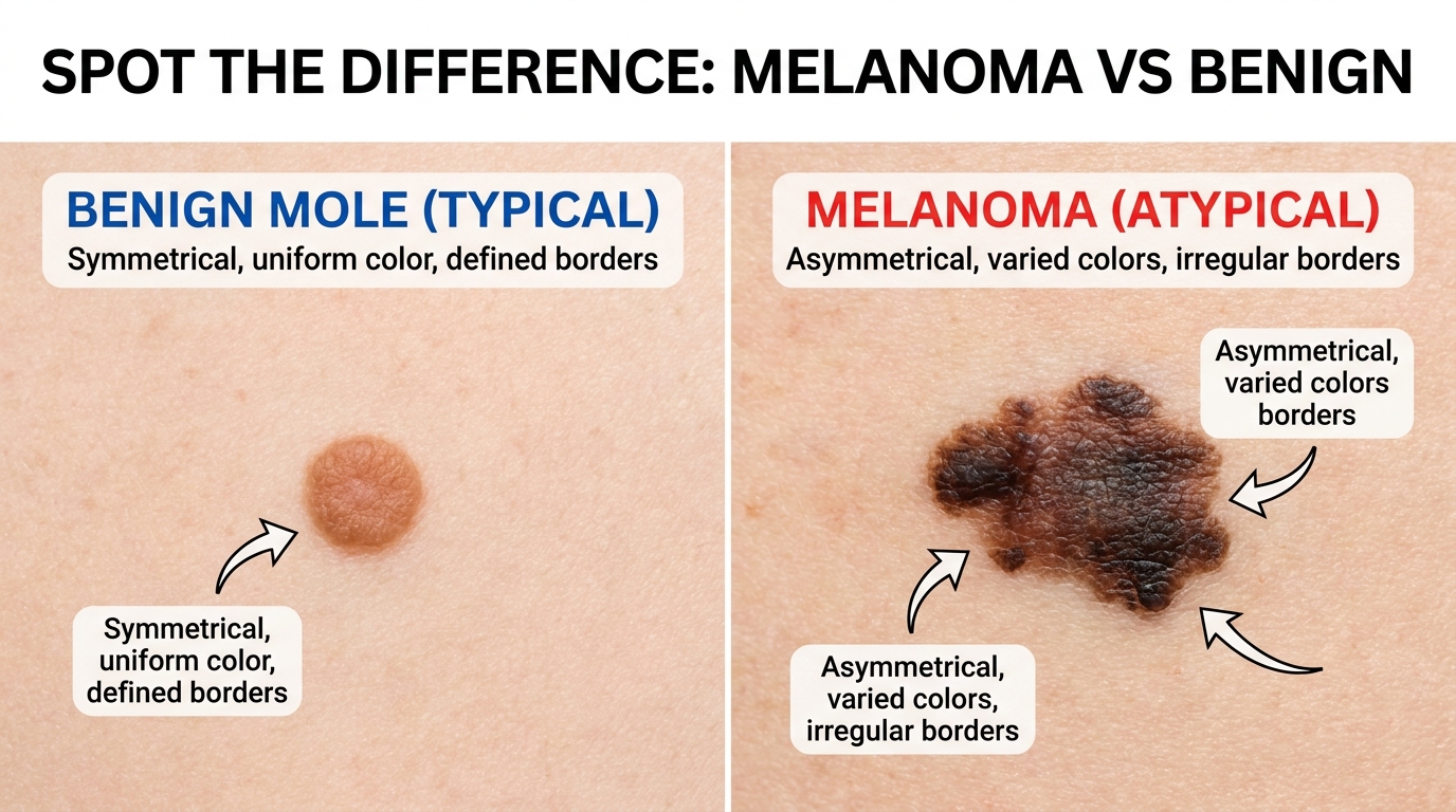
Caption: Comparing melanoma, benign moles, and autoimmune rheumatoid nodules — early detection through visual assessment saves lives.
Any mole meeting two or more of these criteria warrants an immediate dermatological biopsy. Autoimmune rheumatoid nodules — firm bumps appearing near joints — can be visually confused with subcutaneous skin cancer. A professional biopsy provides definitive differentiation. A guide to skin cancer recognition and early signs covers these distinctions comprehensively.
Fungal Skin Infections
Fungal infections represent another category of skin conditions that must be differentiated from autoimmune rashes. Candida (yeast infections) typically appear in warm, moist skin folds — under breasts, in the groin, or between fingers — as bright red patches with satellite lesions (smaller spots around the main rash). Ringworm (Tinea) creates characteristic circular, ring-shaped red patches with a clear center, commonly on the trunk, scalp, or feet.
Both conditions respond well to antifungal medications, distinguishing them from autoimmune rashes that require immunosuppressive therapy. However, one fungal condition bridges the gap between skin and lungs. Histoplasmosis — caused by inhaling Histoplasma fungus spores from contaminated soil — can infect the lungs and produce respiratory symptoms that mimic autoimmune interstitial lung disease. In immunocompromised patients, histoplasmosis can disseminate systemically and cause skin lesions.
This overlap between systemic fungal infections and autoimmune lung symptoms highlights why accurate laboratory testing — not visual assessment alone — is essential for correct diagnosis. A guide to identifying fungal skin infections provides deeper coverage of these conditions.
Allergies, Hives, & Environmental Rashes
Allergic skin reactions operate on a fundamentally different immune mechanism than autoimmune diseases, even though both involve the immune system. An allergy skin rash results from the immune system overreacting to a harmless external trigger — poison ivy, a food allergen, a medication — rather than attacking the body’s own tissues.
Contact dermatitis (from substances like poison ivy, nickel, or latex) produces localized redness, blistering, and intense itching at the contact site. Hives (urticaria) appear as raised, itchy welts that can migrate across the body within hours. Both conditions resolve once the allergen is removed, unlike autoimmune rashes that persist regardless of environmental changes.
The critical emergency distinction is anaphylaxis — a severe, whole-body allergic reaction causing throat swelling, difficulty breathing, rapid pulse, and a dangerous drop in blood pressure. Anaphylaxis is a life-threatening emergency requiring immediate epinephrine injection and emergency medical services. Unlike autoimmune conditions that develop gradually, anaphylaxis escalates within minutes.
A guide to allergies, hives, and environmental rashes covers allergen identification, emergency protocols, and the distinction between allergic and autoimmune immune pathways.
Bites & Parasites
Skin reactions from bites and parasites are acute, externally caused events — the opposite of internally driven autoimmune conditions. Spider bite symptoms range from minor local redness and swelling (most spiders) to severe necrotic tissue damage (brown recluse) or systemic neurotoxic effects (black widow), including muscle cramps, nausea, and difficulty breathing.
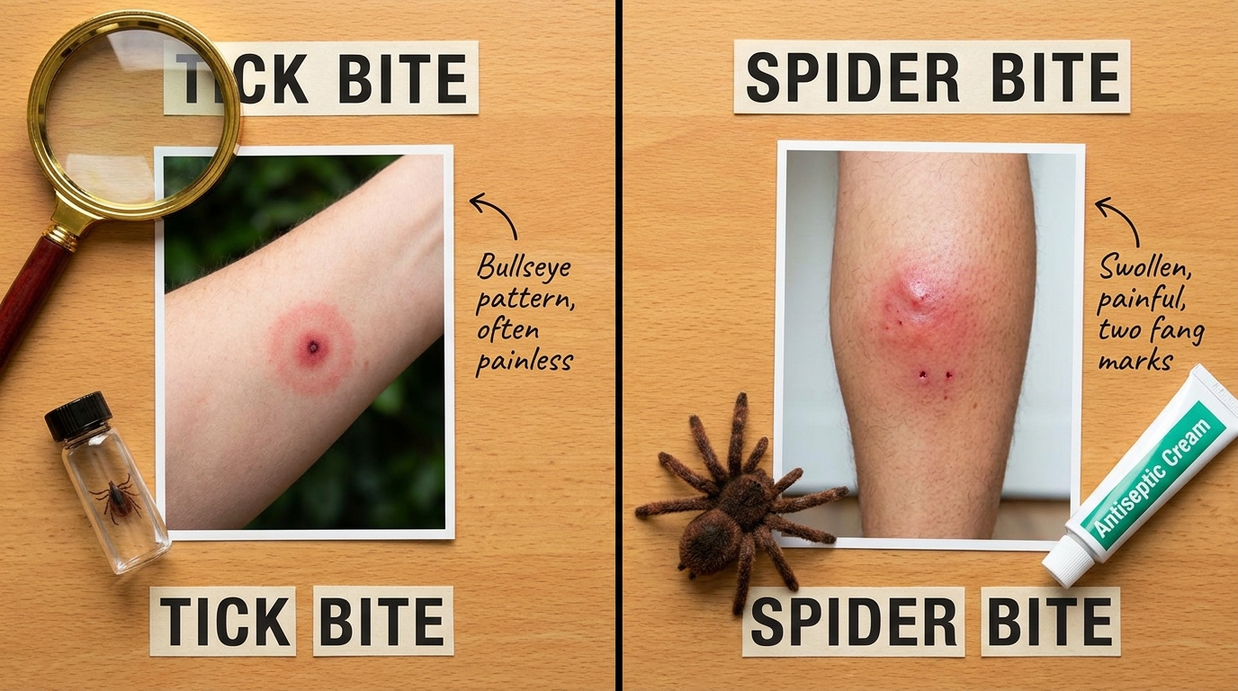
Caption: Visual comparison of tick bite bullseye rash versus venomous spider bite reactions — each requiring different emergency responses.
Skin mites (Sarcoptes scabiei) cause scabies — an intensely itchy condition caused by microscopic mites burrowing into the upper layer of skin, creating visible, winding trails. Scabies itching worsens dramatically at night and requires prescription antiparasitic treatment.
The key contrast with autoimmune conditions: bites and parasites produce acute, localized reactions with a clear external cause. Autoimmune diseases produce chronic, systemic symptoms driven by internal immune dysfunction. However, severe spider envenomation can trigger systemic inflammatory responses that overlap with rheumatic symptoms, reinforcing the importance of accurate clinical assessment. A guide to identifying spider bites and skin parasites provides first-aid protocols and emergency care guidelines.
Celebrity Skin Conditions & Pigmentation Disorders
Public curiosity about celebrity skin conditions has brought valuable awareness to autoimmune pigmentation disorders. The most notable example is vitiligo — the skin condition Michael Jackson publicly disclosed in 1993. Vitiligo causes the immune system to destroy melanocytes (the cells that produce skin pigment), resulting in white spots on skin that gradually expand over time.
Vitiligo is an autoimmune depigmentation disorder, directly connecting it to the broader theme of immune system dysfunction explored throughout this guide. Approximately 1–2% of the global population has vitiligo (American Academy of Dermatology, 2024). Patients with vitiligo have higher rates of other autoimmune conditions, including thyroid disease and rheumatoid arthritis — further evidence that autoimmune conditions frequently cluster together.
It is important to differentiate vitiligo from tinea versicolor (a fungal infection causing lighter-colored patches) or post-inflammatory hypopigmentation (lighter areas following skin healing). While tinea versicolor responds to antifungal treatment and post-inflammatory changes resolve over time, vitiligo is a chronic autoimmune condition requiring ongoing management. An explainer on celebrity skin conditions and pigmentation disorders covers these distinctions and treatment advances in depth.
Diagnostic Pitfalls and When to Seek Expert Help
Common Diagnostic Pitfalls to Avoid
Misdiagnosis is one of the greatest risks for patients whose rheumatic disease presents primarily through surface symptoms. Three diagnostic pitfalls are particularly dangerous:
Pitfall 1: Mistaking a lupus butterfly rash for sunburn or rosacea. Because the malar rash appears across sun-exposed facial skin, primary care physicians and patients alike sometimes dismiss it as routine sun sensitivity. This delay means systemic lupus continues damaging kidneys, lungs, and joints while only the skin is being treated with sunscreen and moisturizer. The distinguishing feature — the rash sparing the nasal folds — is a clinical clue that should prompt an antinuclear antibody (ANA) blood test.
Pitfall 2: Treating autoimmune dry eye with over-the-counter artificial tears alone. Sjögren’s syndrome destroys moisture-producing glands progressively. While artificial tears provide temporary symptom relief, they do nothing to stop the underlying glandular destruction. Without systemic treatment, patients risk corneal damage, dental decay from chronic dry mouth, and lymphoma — a known complication of long-standing Sjögren’s.
Pitfall 3: Dismissing shortness of breath as deconditioning. Patients with rheumatoid arthritis who develop a persistent dry cough or gradually worsening exercise intolerance frequently assume they are simply “out of shape.” In reality, interstitial lung disease related to RA can cause progressive, irreversible lung scarring. Pulmonary function testing should be part of routine RA monitoring, yet many patients never receive it because they never mention their breathing to their rheumatologist.
When to Seek a Rheumatologist
Three specific scenarios should prompt you to escalate beyond your primary care physician or dermatologist to a board-certified rheumatologist:
-
A persistent rash accompanied by joint pain, chronic fatigue, or unexplained fever. When skin symptoms and systemic symptoms co-occur, you need a specialist who evaluates the entire immune system — not just the visible patch of skin.
-
Dry eyes and dry mouth that persist for months despite standard treatments. If artificial tears and lip balm are not resolving your dryness, and you are also experiencing joint stiffness or fatigue, request Sjögren’s syndrome antibody testing.
-
Any combination of skin thickening, muscle weakness, and breathing difficulty. This triad suggests serious connective tissue disease — scleroderma, dermatomyositis, or overlap syndromes — requiring specialized rheumatological and pulmonary workup.
Early intervention prevents permanent joint deformity and irreversible organ damage. A rheumatologist can order comprehensive bloodwork (ANA, anti-CCP, RF, specific antibody panels), imaging, and biopsies to accurately diagnose the underlying autoimmune condition — rather than treating each surface symptom in isolation.
Frequently Asked Questions
What autoimmune disease affects the eyes and lungs?
Rheumatoid arthritis and Sjögren’s syndrome are the primary autoimmune diseases that severely affect both the eyes and lungs. These systemic conditions cause the immune system to attack moisture-producing glands, leading to severe dry eye or scleritis. Simultaneously, the inflammation targets lung tissue, resulting in interstitial lung disease and progressive scarring. Patients experiencing chronic dry eyes alongside shortness of breath should request a rheumatological evaluation, as early treatment significantly improves outcomes compared to delayed diagnosis.
What does a lupus butterfly rash look like?
A lupus butterfly rash (malar rash) appears as a distinct, red or purplish, flat or slightly raised rash that stretches across the bridge of the nose and both cheeks. Its shape directly resembles the wings of a butterfly. This rash frequently worsens after direct sun exposure — a hallmark called photosensitivity — and generally spares the nasal folds (the creases beside the nose). Because the malar rash indicates systemic lupus erythematosus, a medical biopsy and blood test are required for formal diagnosis rather than topical treatment alone.
What are the skin symptoms of scleroderma?
The primary skin symptom of scleroderma is severe hardening, thickening, and tightening of the skin, most commonly affecting the fingers, hands, and face. This tightening occurs due to an autoimmune overproduction of collagen, the structural protein in connective tissue. Patients often notice their skin appears unusually shiny, and they may experience restricted movement in their fingers or difficulty opening their mouth wide. This surface hardening frequently mirrors corresponding internal tissue fibrosis, making early rheumatological evaluation essential for monitoring organ involvement.
How to tell the difference between pink eye and allergies?
Allergies typically cause intense itching, stringy mucus, and affect both eyes simultaneously, whereas infectious pink eye causes a gritty feeling with heavy yellow or green crusting, often starting in one eye. Allergic conjunctivitis worsens in specific environments like high pollen counts, while viral and bacterial pink eye spread through direct contact regardless of environment. The absence of itching strongly suggests infection rather than allergy. If symptoms persist beyond two weeks or are accompanied by joint pain or dry mouth, an autoimmune cause such as Sjögren’s syndrome should be evaluated.
What do small red dots on the skin mean?
Small, pinpoint red dots on the skin, known as petechiae, indicate bleeding under the skin caused by broken capillaries. Unlike standard rashes, these dots do not turn white (blanch) when pressed. Petechiae can result from minor physical trauma, certain medications, or severe systemic issues including low platelet counts, blood clotting disorders, or autoimmune vasculitis. Because petechiae can signal a hematological disorder, they always require immediate medical evaluation — particularly when accompanied by fatigue, bruising, or joint pain suggesting an underlying rheumatic or blood condition.
Conclusion
For patients struggling with unexplained symptoms, understanding that eye, skin, lung symptoms in rheumatic disease are interconnected — not isolated — is vital for securing a proper diagnosis. With approximately 40% of rheumatoid arthritis patients experiencing extra-articular manifestations (Turesson et al., Annals of the Rheumatic Diseases, 2003), ignoring a stubborn rash or persistent dry eye can delay crucial treatment while internal organ damage progresses silently.
The Surface-to-System Matrix exists because the traditional approach — treating each surface symptom with a separate specialist and separate topical medication — fails patients with systemic autoimmune disease. Your rash, your dry eyes, and your shortness of breath may share a single root cause. Recognizing that connection transforms how you communicate with your medical team and accelerates accurate diagnosis.
Prepare for your next medical appointment by documenting all of your symptoms — including ones that seem unrelated to your primary concern. Bring a written timeline of when each symptom started, note which treatments have failed, and specifically request evaluation for systemic autoimmune conditions if multiple surface symptoms persist. A board-certified rheumatologist can order the comprehensive bloodwork and imaging necessary to see the full picture that individual specialists may miss.