

Imaging Review: X-Ray, MRI, and Ultrasound Guide
This article is for educational purposes only and does not replace consultation with a qualified medical professional.
Every year, millions of patients are prescribed a diagnostic imaging scan without fully understanding why a specific machine was chosen or what the results will mean for their health. Staring down a referral for a neurology or rheumatology assessment can cause severe anxiety—especially when terms like “radiation,” “contrast,” and “claustrophobia” enter the conversation.
The fear of the unknown often makes medical visits feel overwhelming. Whether you are dealing with chronic joint pain, sudden back issues, or inexplicable headaches, knowing exactly what your prescribed test involves is the first step toward effective treatment. By the end of this Imaging Review guide, you will understand the exact differences between X-rays, MRIs, CT scans, and ultrasounds so you can approach your appointment with confidence. We will objectively synthesize the science behind these modalities (types of medical machinery), explain their specific uses for musculoskeletal and neurological conditions, and outline essential safety protocols.
Key Takeaways
Diagnostic imaging uses targeted physics—from sound waves to magnetic fields—to visualize internal structures and guide evidence-informed treatment plans.
- The Structural-Functional Imaging Spectrum: Classifies scans by purpose; X-rays capture hard anatomy, while MRIs reveal soft tissue and fluid.
- Radiation differences: Ultrasound and MRI use zero ionizing radiation, making them preferred for specific continuous monitoring.
- Preparation matters: Bringing medical records and understanding “open MRI” options improves patient safety and scan quality.
What Is Diagnostic Imaging?
Diagnostic imaging is the medical practice of using various technologies to look inside the body, allowing doctors to perform suitability assessments for treatments. According to radiological diagnostic guidelines from the National Institutes of Health, these technologies are divided by their physics—using either sound waves, magnetic fields, or ionizing radiation. This allows clinicians to target specific tissues, from fractured bones to neuroinflammatory diseases.
Structural-Functional Imaging Spectrum
Understanding what these machines actually do requires a clear framework. The Structural-Functional Imaging Spectrum is a critical concept that classifies scans based on their specific diagnostic purpose. Imaging is never a one-size-fits-all solution; it exists along a spectrum ranging from mapping dense architecture to visualizing dynamic physiological activity.
Structural imaging tools, such as X-rays and CT scans, effectively map the hard, dense architecture of the body, functioning much like architectural blueprints. Functional or soft-tissue imaging devices, like MRIs and ultrasounds, map muscles, ligaments, and fluid dynamics, providing insight into how the body’s systems interact.
For example, if you visit a rheumatologist (a doctor treating autoimmune and joint conditions), they might order an MRI for joint inflammation rather than an X-ray. This is because standard X-rays simply cannot show the soft-tissue swelling that characterizes early rheumatoid arthritis.
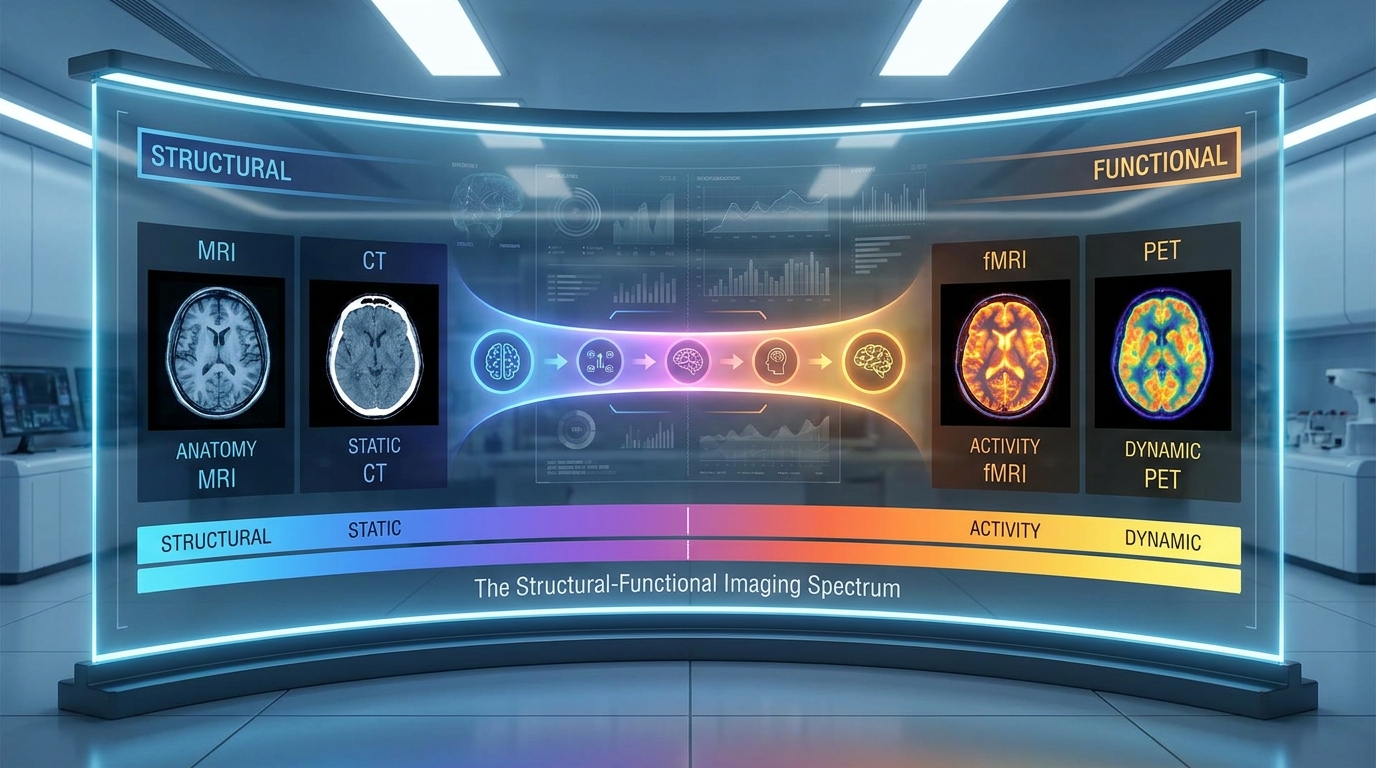
Caption: The spectrum illustrates how X-rays target dense bone while MRIs visualize soft tissue and fluid.
To understand this spectrum, we must first look at the most common tools for structural imaging.
X-Ray & CT: The Structural Baseline
X-rays provide basic two-dimensional shadows of dense materials inside your body, primarily your bones. When a standard X-ray is not detailed enough, your doctor might order ct imaging (Computed Tomography). A CT scan takes hundreds of individual X-rays from different angles to create a highly detailed, three-dimensional structural model of your bones and organs, essentially creating a 360-degree topographical map of your anatomy.
Both methods rely on ionizing radiation, which understandably causes patient anxiety. However, evidence-informed approaches show that the radiation from a standard chest X-ray is equivalent to just a few days of natural background radiation from the environment, according to the Journal of Radiological Protection. These structural tools remain the best choice for identifying acute bone fractures, structural spine issues, or severe joint degeneration.
While these tools are excellent for mapping bone, they cannot effectively show the softer tissues surrounding them.
MRI vs. X-Ray: Soft Tissue Analysis
This brings us to the crucial mri vs x ray difference. Magnetic Resonance Imaging (MRI) uses powerful magnets and radio waves to create highly detailed images of water and fat inside your body. It acts as an incredibly precise soft-tissue scanner. Alternatively, ultrasound uses high-frequency sound waves as another radiation-free option. Unlike standard photographs, ultrasound provides real-time, moving images of your body’s internal functions.
X-rays utilize ionizing radiation for dense bone architecture, whereas MRIs rely on powerful magnets to map soft tissues—eliminating radiation exposure entirely.
Because of their unique physics, MRIs are the gold standard for neurological assessments, such as evaluating brain tissue and the spinal cord for complex conditions. They are also the definitive choice for identifying soft tissue injuries like torn ligaments or compromised cartilage, as supported by the American College of Radiology.
Because MRIs are the definitive choice for soft tissue, understanding how to navigate the specific procedure is crucial for patient comfort.
MRI Procedures & Safety (Summary)
The MRI experience can be intimidating, especially when you are enclosed inside a large tube surrounded by loud knocking noises. These precise scans typically take between thirty minutes and a full hour depending on the complexity. During this extended time, the powerful magnetic fields are systematically aligning protons in your body to generate incredibly detailed cellular maps.
Because the magnet is constantly active and incredibly powerful, there are strict contraindications (reasons to withhold a medical treatment) regarding metallic implants. Patients with pacemakers, certain cochlear implants, or embedded shrapnel must follow rigorous safety rules. Before the scan begins, technologists will perform a thorough suitability assessment to verify that you have no hidden metals, including those in zippers or underwire. They may also administer a contrast agent (a specialized dye injected into your vein) to highlight specific tissues for better clarity.
If you experience severe claustrophobia (fear of confined spaces), proactively ask your doctor about “Open MRI” machines. These evidence-informed alternatives use specialized magnets that do not fully enclose your body. Open MRIs are highly beneficial for specific body types and highly anxious patients, ensuring that clinical comfort does not compromise the ultimate diagnostic accuracy.
Ultrasound Imaging Capabilities
While most people immediately associate ultrasound services with obstetrics, this technology is a crucial pillar of the structural-functional imaging spectrum. Ultrasound uses high-frequency sound waves—completely free of ionizing radiation—to create real-time, live-action images of internal organs, blood flow, and soft tissues.
Musculoskeletal (MSK) Ultrasound Uses
One of the most rapidly expanding applications is msk ultrasound (musculoskeletal ultrasound). Its primary superpower is dynamic assessment. Unlike an MRI where the patient must stay perfectly still for a long duration, an ultrasound allows a doctor to evaluate a joint while it actually moves.
During the examination, the physician might ask you to move your shoulder or bend your knee. This real-time movement evaluation lets them see exactly when and where a tendon pinches or rubs against a bone. Musculoskeletal ultrasound provides real-time, dynamic visualization of tendons and joints, allowing immediate motion assessment without exposing patients to ionizing radiation.
This technology plays a massive role in rheumatology for detecting early joint inflammation. It is also used to guide needle injections safely into tight joint spaces. Evidence-informed clinical guidelines suggest using ultrasound for evaluating rotator cuff tears, Achilles tendon issues, and rheumatoid arthritis flare-ups, per research in the Journal of Clinical Ultrasound.
The ability to compare the symptomatic side of your body with the asymptomatic side in real-time gives your doctor immediate comparative data. Once you and your physician determine which of these modalities fits your condition, the next step is locating an appropriate facility.
Finding an Imaging Center (Summary)
Getting a prescription from your doctor is only half the battle; finding a high-quality diagnostic imaging center is the crucial next step in your healthcare journey. Many patients simply do not realize that they have distinct options regarding exactly where their prescribed scan takes place.
You generally have two primary choices: a hospital imaging department or an independent outpatient imaging center. Having a scan at a large hospital is often significantly more expensive due to facility overhead and may involve considerably longer wait times. Alternatively, outpatient clinics typically offer substantially lower costs, easier parking, and a much more streamlined daily patient experience.
Before booking any appointment, you must proactively verify your insurance networks. Always look for fully accredited facilities that maintain strict technical quality control. Major national networks, such as SimonMed or American Health Imaging, often house the absolute latest machinery, including advanced 3T MRIs and premium high-resolution ultrasound units. Taking the essential time to research your local facility ensures you receive clear, highly accurate diagnostic results without facing unexpected medical bills. Your doctor’s office can usually send your necessary referral directly to the facility of your ultimate choice.
Careers in Medical Imaging (Summary)
The accuracy of your diagnostic imaging relies heavily on the skilled technologists and sonographers operating the machinery. These professionals are the backbone of the imaging process, ensuring that patients remain safe while capturing the precise anatomical views doctors need for accurate diagnoses.
Becoming an ultrasound tech, MRI technologist, or radiologic technician requires specialized, rigorous schooling. Enrolling in an accredited ultrasound tech school or specialized radiologic technology program provides the critical foundational knowledge required for these roles. The educational pathways differ significantly depending on the technology. X-ray technicians focus heavily on radiation physics and skeletal anatomy, while ultrasound sonographers study the acoustic properties of sound waves and complex fluid dynamics.
Regardless of their specialty, these healthcare professionals must pass strict registry board exams. Organizations like the American Registry of Radiologic Technologists (ARRT) demand extensive clinical hours and theoretical knowledge before granting certification. This high educational standard exists to guarantee patient safety and exceptional image quality across all modalities. These demanding careers require both technical precision and deep compassion for patients experiencing pain or anxiety.
Medical Limitations & Safety
While imaging technologies are remarkable, they possess distinct limitations that patients must understand to set realistic expectations for their care.
Common Pitfalls in Imaging
One of the most frequent pitfalls in modern medicine is treating the scan instead of the patient. MRI and CT scans frequently reveal incidental findings—such as mild disc degeneration or slight cartilage wear—that cause absolutely zero physical pain. Fixating on these asymptomatic abnormalities can lead to unnecessary anxiety and invasive procedures that offer no functional benefit.
Another common pitfall involves misunderstanding radiation risks. Anxious patients might completely avoid necessary X-rays, risking a dangerous misdiagnosis. Conversely, some patients demand unnecessary CT scans for minor issues, exposing themselves to radiation without clinical justification. Evidence-informed care relies on balancing these risks with the potential diagnostic rewards, aligned with guidance from the American Medical Association.
When to Seek Expert Help
Diagnostic imaging should never be interpreted in a vacuum. A scan alone does not provide a definitive diagnosis; it merely provides a map of your anatomy at one specific moment in time.
Always review your results in conjunction with a physical clinical examination by a qualified physician. A specialist will correlate the images with your actual symptoms, mobility, and medical history. Remember that functional outcomes and an improved quality of life are the ultimate goals. You should seek expert help if your imaging results seem to contradict the physical pain you are experiencing, as a secondary medical review or different imaging modality may be necessary.
Frequently Asked Questions
MRI examination expectations
During your scan, you will lie flat on a motorized table that slowly slides into a cylindrical magnetic resonance imaging magnet. Because the machine utilizes powerful magnetic fields and radio waves, it generates loud tapping or thumping noises. To manage this discomfort, technologists typically provide noise-canceling headphones or earplugs. Most standard examinations last between thirty to sixty minutes depending on the specific targeted anatomical area. If your physician ordered a contrasted study, a technologist will also administer a specialized dye into your vein to properly highlight specific internal tissues.
Imaging results: helpful or harmful?
Diagnostic imaging is incredibly helpful for guiding targeted, evidence-informed treatment plans when the results are interpreted correctly. However, these sophisticated scans can sometimes cause unnecessary anxiety if patients fixate on minor, age-related structural changes. For instance, a spinal MRI might show slight disc degeneration that does not actually cause any physical pain. A qualified physician will always carefully correlate your imaging abnormalities directly with your current physical symptoms. This comprehensive approach helps clinicians prevent harmful overtreatment and keeps the focus strictly on functional outcomes.
Finding an outpatient clinic
Start by calling your insurance provider directly to request a comprehensive list of approved in-network outpatient imaging facilities in your area. You can then deliberately ask your prescribing doctor for specific recommendations regarding accredited local clinics. It is important to prioritize facilities that feature modern, well-maintained equipment and specialized certified technical staff. Independent outpatient centers usually offer significantly easier scheduling and noticeably lower out-of-pocket costs than large, busy hospital departments. Finally, if you suffer from severe claustrophobia, be absolutely sure to verify whether the recommended clinic currently houses an open MRI machine.
fMRI vs. standard MRI differences
The main distinction between these two powerful imaging modalities is exactly what kind of biological activity they are designed to measure. A standard MRI essentially maps the static physical structure of soft tissue, ligaments, and organs at a frozen moment in time. In distinct contrast, a functional MRI (fMRI) measures dynamic, real-time brain activity by detecting metabolic changes associated with localized blood flow. Neurologists rely heavily on fMRI scans to observe which specific areas of the brain actively illuminate during complex mental tasks. Ultimately, your specialist will choose between them based on whether they need to analyze your physical anatomy or your cognitive function.
Conclusion
For patients requiring diagnosis, diagnostic imaging delivers precise internal visualization. X-rays use small amounts of ionizing radiation to capture dense structures like bone, whereas MRIs use powerful magnets to visualize soft tissues without radiation, according to comprehensive clinical imaging reviews. The best approach combines matching the right modality—X-ray, MRI, or Ultrasound—to the specific tissue type.
Understanding The Structural-Functional Imaging Spectrum allows you to navigate the healthcare system as an informed patient. By recognizing that X-rays show hard anatomy while MRIs and ultrasounds map functional soft tissue, you can successfully manage procedural anxiety and set realistic expectations for your clinical care.
Take an active role in your suitability assessment by preparing thoroughly for your appointment. Bring your previous medical records to your consultation and ask your provider which imaging modality is the most evidence-informed choice for your specific symptoms to ensure optimal functional outcomes.