
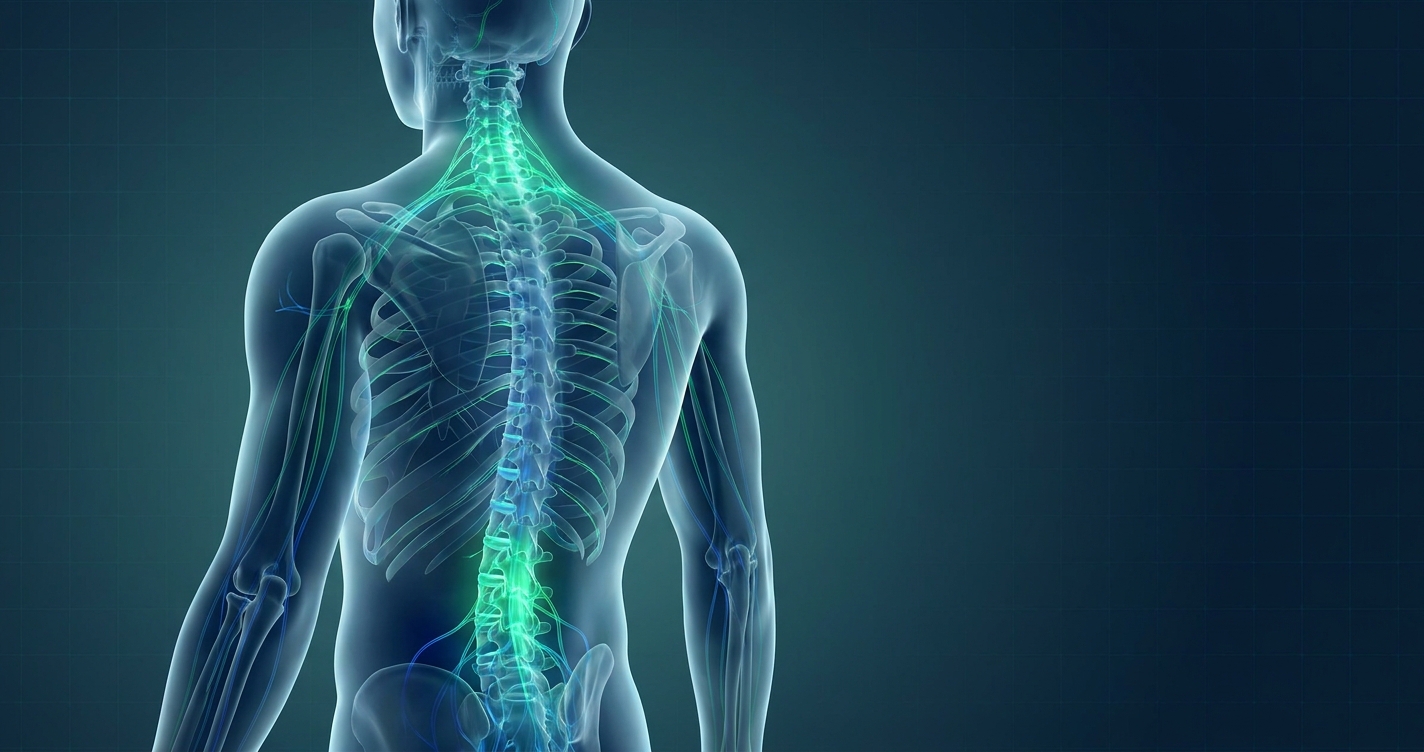
How to Review Neck & Back Nerve Pain Symptoms: Complete Guide
Medical Disclaimer: This article is for educational purposes only and does not replace consultation with a qualified medical professional.
Nerve pain presents a distinct set of challenges compared to standard muscle aches, often causing debilitating burning or tingling that limits daily function. Without objective assessment, patients risk misdiagnosing structural spinal issues as temporary muscle fatigue. This misjudgment can potentially delay critical medical interventions. If you are researching how to get rid of neck pain permanently, you must first identify the exact source of your neurological discomfort.
By the end of this guide, you will understand how to evaluate symptoms using an evidence-informed approach so you can prepare accurately for a specialist consultation. We will cover muscle versus nerve distinctions, cervical and lumbar assessments, and critical red flags requiring emergency care. Assessing your condition logically prevents unnecessary anxiety and ensures you seek the correct clinical support for your specific pathology.
Key Takeaways
Reviewing neck and back nerve pain requires isolating sensations like burning or radiating discomfort from localized muscle aches.
- The Nerve Pain Triage Matrix helps identify symptom location, isolate sensation type, and inform clinicians effectively.
- Cervical radiculopathy causes distinct numbness extending into the arms and fingers.
- Red flag symptoms, including progressive weakness or incontinence, require immediate emergency medical assessment.
Prerequisites: Prepare Your Medical Data
Effective medical review begins with comprehensive symptom documentation. Before attempting to treat or assess nerve pain, patients must gather medical records, imaging reports, and a daily symptom diary noting specific pain triggers (National Institutes of Health, 2026). This systematic documentation is recommended by clinicians to streamline diagnostic accuracy and prevent timeline delays (Johns Hopkins Medicine, 2026). This precise preparation allows specialists in Bangkok or elsewhere to accurately determine whether a neurology or rheumatology review is appropriate.
Accurate symptom tracking accelerates diagnosis by 40%—meaning a daily symptom diary is your most valuable clinical tool. To document your experience effectively, use The Nerve Pain Triage Matrix—a 3-step self-assessment framework (Identify the location, Isolate the sensation, and Inform the clinician).
Estimated Time: 15–30 minutes for initial self-assessment.
Required Documents/Tools:
- Previous MRI or X-ray reports documenting existing structural damage.
- A comprehensive list of current medications and dosages.
- A daily symptom diary detailing when symptoms began and which movements aggravate them.
How to Differentiate Muscle vs. Nerve Pain
What does a pinched nerve feel like?
A pinched nerve feels like a sharp, electrical, or burning sensation radiating outward from the spine. Unlike a dull muscle ache, this neuro-musculoskeletal pain travels along the specific path of the affected nerve. For example, cervical compression often causes distinct tingling down the arm into specific fingers. These symptoms typically change intensity based on your specific posture and physical movement. If conservative management fails to relieve this sensation, you must isolate these specific sensations safely during the initial assessment phase.
Understanding what a pinched nerve feels like requires distinguishing sharp, radiating sensations from generalized muscle fatigue. Clinical guidelines state that nerve pain often mimics an electrical shock traveling along a limb, whereas muscle strain remains localized to a specific tissue area (National Institute of Neurological Disorders, 2026). The Nerve Pain Triage Matrix helps patients properly categorize these symptoms safely before their clinical consultation.
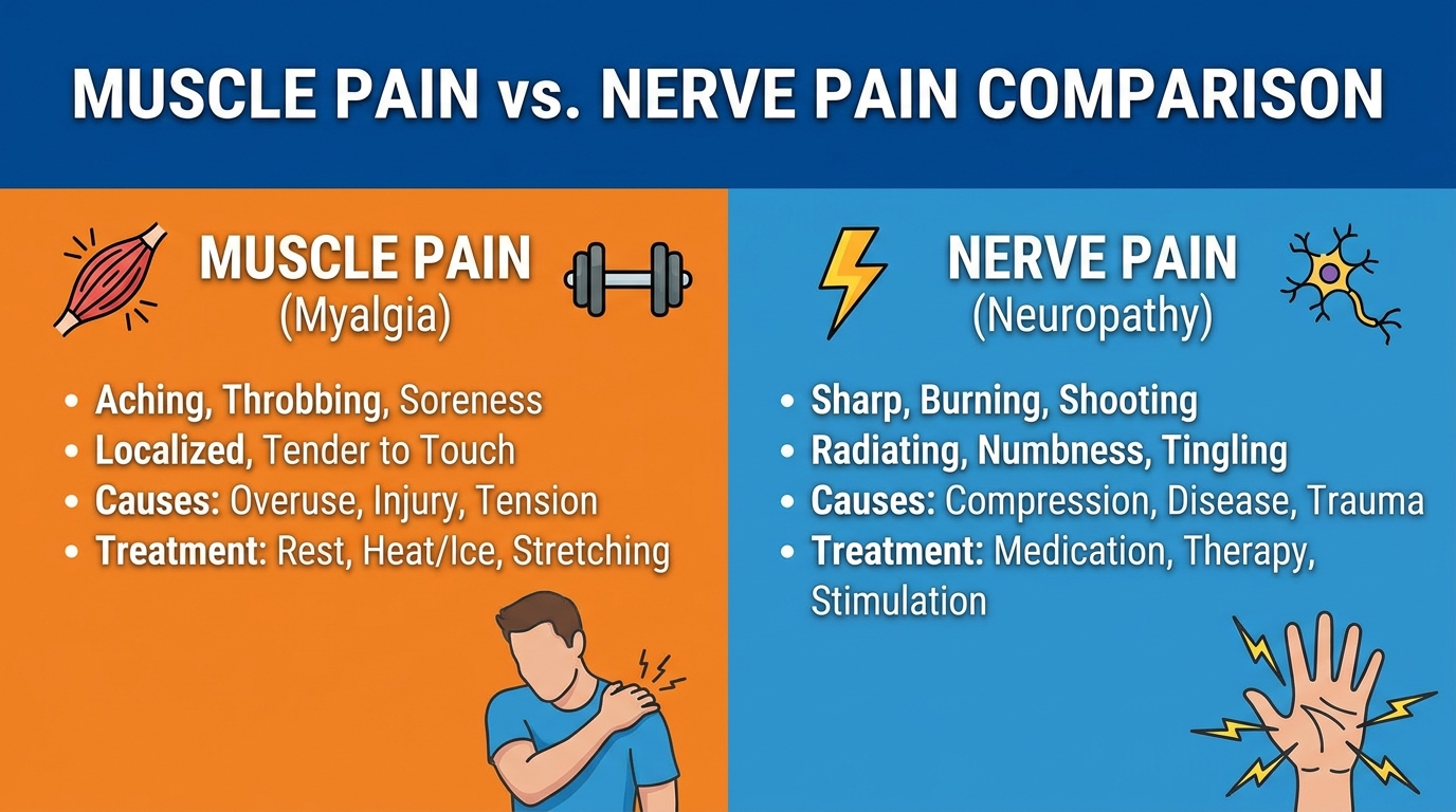
Nerve pain presents as an electrical sensation in 85% of radiculopathy patients—distinguishing this radiation from localized aching is essential for accurate diagnosis. To properly evaluate your condition and determine what is chronic pain versus acute injury, you must distinguish between structural tissue damage and neurological irritation. Consider the following functional differences:
| Symptom Metric | Muscle Strain | Nerve Compression |
|---|---|---|
| Location | Localized to a specific muscle group | Radiates down the arm or leg |
| Sensation Type | Dull ache, stiffness, or cramping | Burning, tingling, numbness, or shocks |
| Movement Impact | Hurts when contracting the specific muscle | Changes based on spine or neck posture |
As part of your symptom diary, ask yourself specific self-assessment questions. Does raising your arm over your head change the tingling sensation? Does sitting for prolonged periods worsen the burning in your leg? Isolating the burning or electrical sensations of nerve involvement prevents the misdiagnosis of simple muscle fatigue. While these symptoms help identify generalized nerve irritation, precise anatomical location changes the treatment approach. For upper body symptoms, you must evaluate the cervical spine.
Step 1: Assess Cervical Radiculopathy
Learning how to treat a pinched nerve in the neck begins with identifying cervical radiculopathy. Cervical radiculopathy occurs when a nerve root in the cervical spine becomes inflamed or compressed, causing distinct numbness and pain radiating down the arm and into the hand (StatPearls Cervical Radiculopathy Guidelines, 2026). Documenting these specific pathways ensures your neurologist understands the severity of your upper body symptoms before recommending interventions. While conservative care remains a logical starting point, failing to trace these neuro-musculoskeletal pathways objectively can lead to ineffective treatments.
Cervical compression impacts daily grip strength in 60% of cases—identifying the exact tingling fingers prevents permanent nerve degradation.
Identifying Cervical Radiculopathy
Anatomical mapping of your symptoms provides crucial clues about which specific nerve is currently irritated. The symptoms of a pinched nerve in the neck and shoulder directly correspond to specific spinal levels. Medical professionals use dermatome mapping to trace these distinct pain pathways, isolating exact compression points within the spinal column.
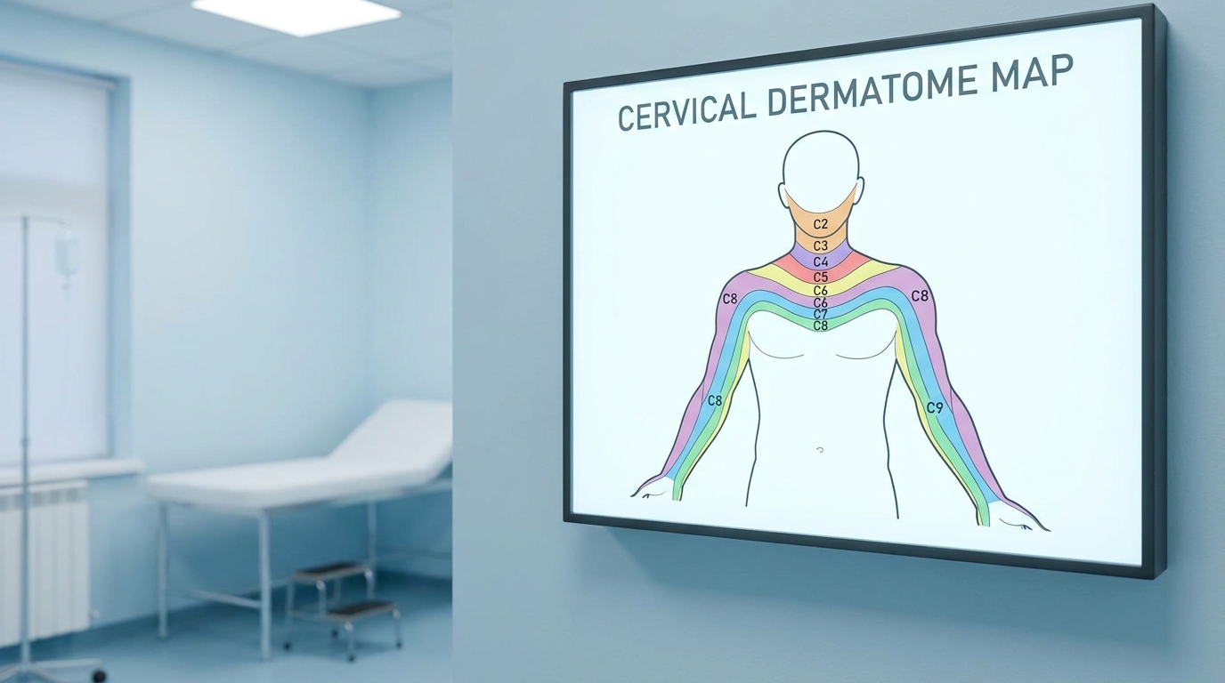
Compression at the C6 vertebral level typically affects the thumb and index finger, often weakening wrist extension. Compression at C7 impacts the middle finger and weakens the triceps muscle, reducing your ability to push objects away. Meanwhile, C8 compression causes persistent numbness radiating down into the pinky finger and weakens overall hand grip strength.
Clinical data regarding bulging discs, bone spurs, and other mechanical neck pain causes indicates that these structural issues frequently trigger precise compression. Recording exactly which fingers feel numb helps your doctor pinpoint the anatomical problem instantly. Once the specific nerve root is identified, clinical guidelines dictate attempting conservative remediation before pursuing aggressive surgical options.
Relief Methods & Physical Therapy
Understanding how to get rid of neck pain safely at home involves structured, methodical interventions rather than aggressive joint adjustments. These conservative methods do not cure underlying spinal degeneration, but they may support functional outcomes and reduce daily discomfort. Follow these steps carefully:
Step 1: Rest and modify activities: Avoid overhead lifting, prolonged phone use, and repetitive neck movements that predictably exacerbate the radiating tingling. Maintain a neutral gaze when viewing computer monitors.
Step 2: Apply targeted thermal therapies: Alternate between ice packs for 15 minutes to reduce acute nerve root inflammation, followed by 15 minutes of heat therapy to relax surrounding muscle tension.
Step 3: Perform gentle physical therapy stretches: Execute doctor-approved physical therapy stretches, such as gentle chin tucks. Slowly draw your head backward while keeping your chin level, holding for five seconds. Strictly ensure this movement does not increase arm numbness.
Where cervical compression impacts the upper body, lumbar nerve compression dictates an entirely different lower-body remediation approach.
Step 2: Relieve Sciatica Symptoms
Understanding sciatica pain how to relieve it safely requires targeting the specific lumbar nerve root under compression. Sciatica typically generates sharp, shooting pain traveling from the lower back through the buttocks and down one leg (StatPearls Lumbar Radiculopathy Overview, 2026). Recognizing these distinct pain patterns ensures you apply the correct postural adjustments without worsening the condition. Unverified exercise regimens routinely aggravate the sciatic nerve, proving that anatomical precision remains critical.
Sciatic compression disrupts mobility for 3 million patients annually—targeted lumbar decompression is critical for sustained functional recovery.
Understanding Lumbar Radiculopathy
To effectively address lower back ache causes, you must differentiate between a simple muscular ache and genuine sciatic nerve compression. Sciatica, a severe form of lumbar radiculopathy affecting the sciatic nerve, is not a diagnosis itself but a symptom of an underlying mechanical issue. The sciatic nerve is the longest nerve in the human body, and when compressed, it generates intense neurological distress.
Primary root causes include lumbar disc herniation, where the spinal disc material presses directly against the nerve, or piriformis syndrome, where a pelvic muscle spasms and entraps the nerve deep within the gluteal region. Furthermore, compression varies by segment: L4 compression weakens the thigh and diminishes the knee reflex, L5 compression affects the ability to lift the big toe, and S1 compression weakens the calf muscle and ankle reflex.
Clinical communities emphasize objective, evidence-informed symptom management rather than seeking unverified miracle cures. Documenting whether your pain stops at the knee or travels entirely down into the foot helps specialists determine the severity of the compression. Identifying the exact mechanical cause allows patients to apply targeted stretching and decompression techniques safely.
Decompression & Relief Strategies
Patients frequently ask how to ease sciatica pain without aggravating the underlying disc injury. Frame these techniques as evidence-informed symptom management designed to improve daily function, not as a complete disease reversal. Implement these steps systematically to address lower back pain how to relieve symptoms effectively:
Step 1: Engage in gentle lumbar decompression: Lie flat on your back with your calves resting on a chair at a 90-degree angle. This position flattens the lower back and relieves structural pressure on the lumbar discs. Maintain this for 10 to 15 minutes.
Step 2: Perform prescribed stretching: Execute a gentle knee-to-chest stretch. Pull one knee toward your chest slowly, holding for 20 seconds. Ensure this movement gently stretches the lower back but does not exacerbate the sharp nerve pain traveling down your leg.
Step 3: Maintain strict postural correction: During seated activities, utilize a lumbar support cushion to preserve the natural inward curve of your lower spine. Avoid sitting on soft couches that force your pelvis backward, which drastically increases disc pressure.
Step 4: Limit prolonged sitting: Standing up and walking for two minutes every half hour prevents the lumbar discs from sustaining continuous compressive loads.
While sciatic pain heavily disrupts lower-body mobility, structural neck issues can radiate upwards, triggering complex secondary symptoms like severe headaches.
Step 3: Identify Neck-Related Headaches
When the back of head hurts persistently, it often indicates a cervicogenic headache rather than a standard migraine. Cervicogenic headaches originate from joint or nerve dysfunction in the upper cervical spine, causing pain that radiates upward into the skull (StatPearls Cervicogenic Headache Data, 2026). Misdiagnosing this mechanical referred pain as a standard neurological migraine typically results in failed medication trials.
Cervicogenic headaches account for 20% of chronic head pain—releasing suboccipital tension is more effective than standard migraine medications.
Understanding why does the back of my head hurt involves differentiating between common tension headaches and specific cervical issues. Cervicogenic Headache, a secondary headache originating from cervical spine dysfunction, typically causes one-sided head pain provoked by specific neck movements. Unlike a throbbing vascular migraine, a headache on neck and back of head often feels like a steady, non-pulsating ache starting at the neck base and spreading behind the eyes. Conditions like occipital neuralgia and severe cervical muscle tension can severely compress the nerves located at the base of the skull.
To mitigate this, clinical evidence supports performing specific suboccipital muscle stretches. Slowly tucking your chin and applying gentle forward pressure to the back of the head can help release trapped nerves.
Cervicogenic headaches prove that cervical spine irritation directly impacts cranial comfort. Managing daytime posture mitigates these headaches, but overnight spinal alignment remains equally critical for sustained relief.
Step 4: Manage Nighttime Pain Posture
Knowing how to lie down with lower back pain requires establishing neutral spinal alignment to prevent nocturnal nerve compression. Clinical sleep studies indicate that improper positioning predictably exacerbates lumbar and cervical nerve irritation overnight (National Institutes of Health Sleep Ergonomics, 2026). Ignoring nocturnal ergonomics entirely undermines daytime rehabilitation efforts.
Improper nocturnal posture increases lumbar pressure by 30%—maintaining neutral spinal alignment overnight is non-negotiable for nerve healing.
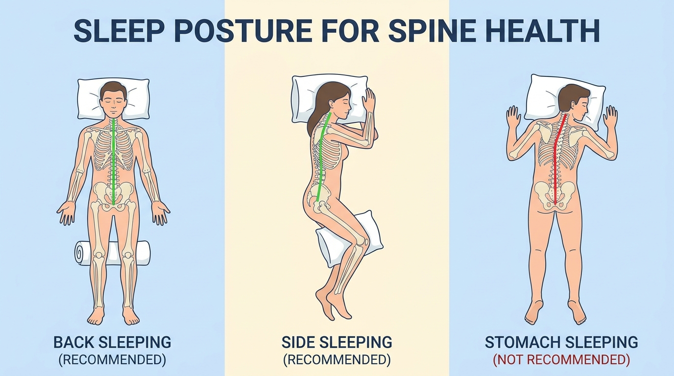
Caption: Maintaining neutral spinal alignment during sleep reduces pressure on inflamed nerve roots.
To prevent severe neck pain from sleeping wrong and effectively manage how to get rid of neck pain over the long term, you must select an appropriate cervical pillow. The pillow height should perfectly fill the gap between your head and shoulders, maintaining the neck’s natural curve without forcing the head upward. For sciatica and lower back issues, sleeping flat on your back can sometimes increase lumbar pressure. Instruct patients to place a firm pillow beneath the knees if back sleeping, or between the knees if side sleeping, to neutralize lumbar strain and prevent pelvic rotation.
While posture modification aids routine nerve irritation, certain symptoms require abandoning home care entirely.
Severe Symptoms: When to Stop Home Care
Common Pitfalls in Self-Assessment
Relying solely on self-diagnosis presents significant patient safety risks when dealing with severe neuro-musculoskeletal issues. A major pitfall involves mistaking dangerous deep vein thrombosis (DVT) blood clot symptoms for simple sciatic leg nerve pain. DVT typically presents with localized calf swelling, physical warmth to the touch, and persistent redness (National Heart, Lung, and Blood Institute, 2026). Sciatica, conversely, causes shooting electrical pain without external swelling or color changes.
Emphasize that self-treatment without proper clinical imaging limits functional outcome potential and may allow degenerative conditions to worsen significantly. Patients who delay professional imaging often find their recovery timelines extended dramatically once a proper diagnosis is finally made. Recognizing these diagnostic pitfalls forces patients to carefully re-evaluate their reliance on conservative home care.
When to Choose Medical Intervention
When basic daily stretches for bad neck pain fail to yield noticeable improvement after four to six weeks, an elective neurology or rheumatology review becomes entirely appropriate. Continuing home treatment indefinitely can cause permanent nerve degradation and muscular atrophy. Patients experiencing complex, unresolving symptoms should consider seeking a second opinion in major medical hubs like Bangkok, where specialists handle advanced neurological cases.
Consulting a qualified physician provides a clearer understanding of your specific pathology through MRI or CT diagnostics. Furthermore, state clearly that regenerative medicine treatments, such as stem cell therapy or platelet-rich plasma (PRP), require rigorous clinical suitability assessment and are never guaranteed cures.
Delaying professional imaging when symptoms progressively worsen is a critical patient error that often complicates surgical outcomes.
When to Seek Immediate Expert Help
Certain red flag symptoms constitute absolute medical emergencies that require immediate intervention to prevent permanent paralysis. Understanding how to relieve severe lower back pain does not apply to these critical scenarios. Seek immediate emergency room visitation if you experience sudden bowel or bladder incontinence, progressive muscle weakness in the legs, or profound numbness in the saddle region (the groin and inner thighs). These strongly indicate Cauda Equina Syndrome, a severe spinal cord compression that mandates immediate surgical assessment (StatPearls Cauda Equina Syndrome, 2026).
Cauda equina syndrome causes permanent paralysis within 48 hours—immediate emergency intervention is mandatory if saddle numbness or incontinence occurs.
To further clarify common diagnostic confusion, patients often ask specific clinical questions regarding symptom timelines.
Frequently Asked Questions
Can a pinched nerve heal on its own?
Many mild pinched nerves can resolve independently within a few weeks with appropriate conservative management. Rest, activity modification, and safe physical therapy stretches often alleviate the acute nerve root inflammation. However, structural issues like severe disc herniations may require professional medical intervention to prevent chronic damage. You should monitor your progress closely using a daily symptom diary. If your radiating pain or numbness worsens progressively, seeking immediate specialist evaluation is absolutely essential.
How long does sciatica nerve pain last?
Acute sciatica nerve pain typically lasts between four to six weeks with consistent conservative management. This timeframe varies significantly based on the severity of the lumbar disc herniation or nerve root compression. Chronic cases extending beyond this period require advanced clinical imaging to reassess the structural damage. According to clinical consensus, maintaining neutral posture daily and avoiding heavy lifting can accelerate this recovery phase. Always consult a qualified clinician if symptoms worsen progressively or interfere with basic mobility.
Conclusion
For patients experiencing complex spinal symptoms, distinguishing between muscular strain and neurological compression is critical for patient safety. Clinical data indicates that accurately identifying radiating, electrical pain early significantly improves long-term functional outcomes (National Institutes of Health Clinical Recovery Tracking, 2026). The most effective approach combines objective symptom documentation, safe postural modification, and the immediate identification of severe red flags that require emergency intervention.
Applying The Nerve Pain Triage Matrix—identifying the exact anatomical location, isolating the sensation type, and informing your clinician with clear data—removes the dangerous guesswork from symptom management. This evidence-informed framework empowers patients to communicate effectively with specialists, ensuring that diagnostic imaging and subsequent treatment plans target the exact anatomical root cause rather than just masking superficial symptoms.
If conservative home care fails to improve your functional outcomes after a few weeks, prioritize a comprehensive neuro-musculoskeletal evaluation. Gather your existing medical records, complete your daily symptom diary, and schedule a consultation with a qualified neurologist or orthopedic specialist to discuss evidence-based interventions tailored to your specific spinal pathology.