
How to Manage Nerve Pain After Injury: Long-Term Guide
This article is for educational purposes only and does not replace consultation with a qualified medical professional.
Nerve pain after a physical injury can be highly alarming, especially when the discomfort feels like unpredictable electric shocks or severe burning months after the initial trauma. Many patients endure this confusing pain without realizing that understanding the biological timeline of nerve regeneration is the only way to differentiate normal healing sensations from permanent tissue damage. Without a clear recovery timeline, critical windows for effective surgical intervention are often missed, leading to long-term complications.
By the end of this comprehensive guide, you will understand how to accurately track your sensory symptoms and know precisely when a specialist review is medically necessary for your safety. We will evaluate the stages of peripheral nerve healing, identify the clear red flags for permanent damage, and outline long-term monitoring strategies to keep your recovery evidence-informed.
Key Takeaways
Managing nerve pain after an injury requires distinguishing between natural nerve regeneration and permanent neuropathic damage. Peripheral nerves regenerate at roughly 1 inch per month (NIH, 2023).
- Tracking progress: Use The Functional Nerve Tracking Framework to log pain types and motor function.
- Healing signs: Electric shock sensations often indicate active nerve regeneration, not a worsening injury.
- Critical window: A complete lack of clinical improvement within 6 months typically requires an immediate surgical evaluation.
Understanding Nerve Healing: Why Recovery Takes Time
Nerve pain after a traumatic event signals that the body’s electrical wiring has been disrupted. Peripheral nerves have the remarkable ability to regenerate themselves following an injury, growing back at a rate of approximately 1 inch per month (NIH, 2023). This slow biological pace means recovery requires extended patience and precise long-term monitoring.
How Peripheral Nerves Regenerate
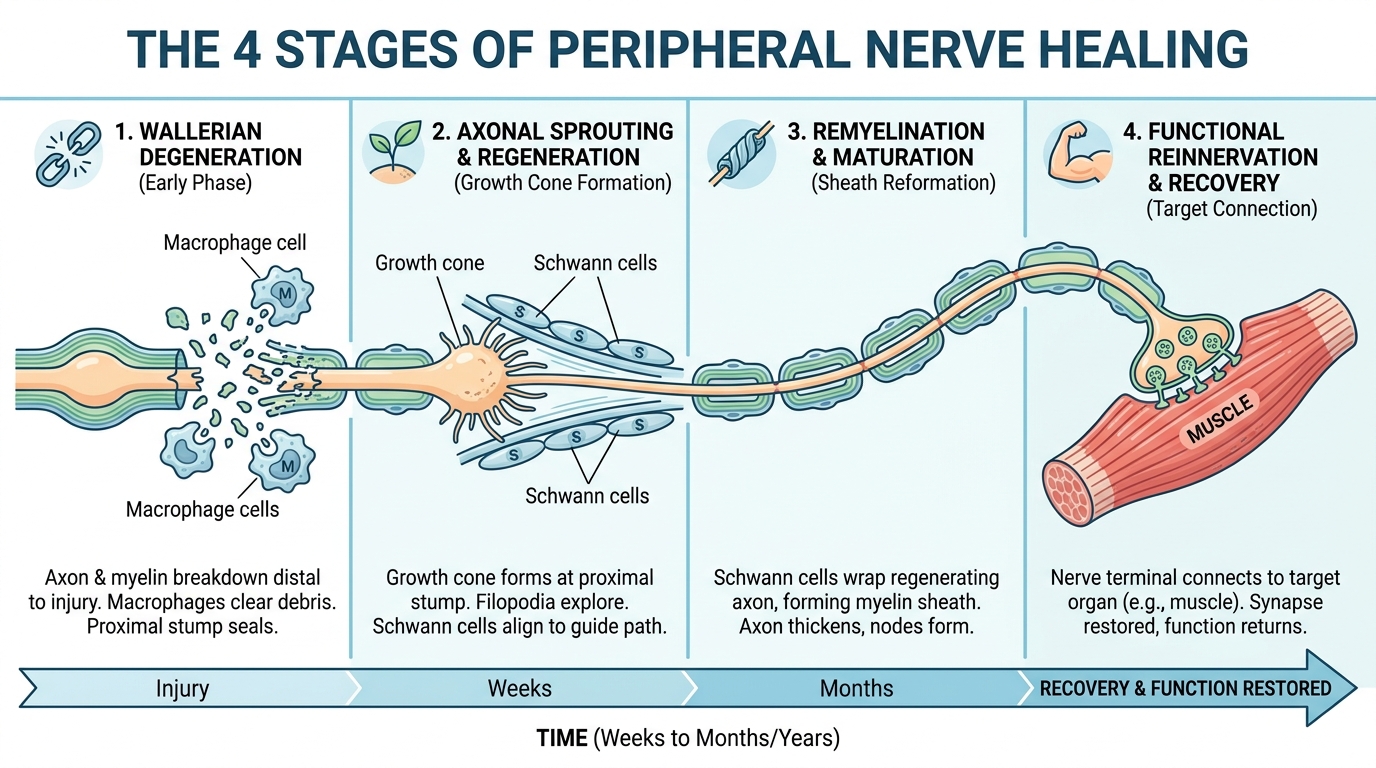
Peripheral nerves (the pathways connecting your brain to your extremities) undergo a specific repair cycle. When severed or crushed, they experience Wallerian degeneration (the biological process where damaged nerve fibers clear away before rebuilding). Following this phase, recovery is painstakingly slow. Published daily axonal regeneration metrics show that axons typically repair at a cellular speed of 1 to 3 millimeters per day (PubMed, 2017).
This biological speed translates directly to NIH data on nerve regeneration rates, measuring about 1 inch per month. Think of this process like a damaged fiber-optic cable slowly repairing itself from the injury site down to your fingertips.
`
`
Because this process is so slow, understanding the difference between healing pain and worsening damage is vital for patient safety.
Progressive vs. Degenerative Patterns
Monitoring your symptoms requires identifying the specific direction and nature of the pain. Progressive nerve patterns indicate healing, where advancing sensations—like a tingling feeling—physically move further down the limb as the nerve root reconnects. In contrast, degenerative nerve patterns represent dying tissue. These are marked by worsening numbness, ascending pain moving toward the spine, or a steady loss of muscle control.
To prevent further trauma during the early healing stages, clinicians often recommend a nerve cast (a specialized splint for immobilization) to stabilize the surrounding structures while fragile tissue repairs. General clinical guidelines strongly emphasize early immobilization to protect regenerating fibers (Mayo Clinic). Recognizing these distinct biological patterns allows patients to successfully identify whether their specific sensations are normal milestones or medical red flags requiring intervention.
Signs of Nerve Regeneration vs. Permanent Damage
Effective Nerve Pain After Injury: Long-Term Review And Monitoring relies on distinguishing between the uncomfortable sensations of healing and the warning signs of permanent tissue death. A lack of clinical improvement after 3 to 6 months may necessitate surgical options such as nerve grafting (Johns Hopkins Medicine, 2023). Patients must track sensory changes closely.
What Normal Nerve Healing Feels Like
Counterintuitively, nerve regeneration is an inherently painful biological process. Johns Hopkins guidelines on normal regeneration pain indicate patients experiencing active nerve recovery often feel severe tingling, “pins and needles,” or sharp electric shocks. For example, an unpredictable electric shock traveling down the leg after a crush injury often indicates axons are successfully firing and reconnecting to the muscle.
Clinicians regularly check for Tinel’s sign (localized tingling triggered by lightly tapping over the injured nerve), which serves as an objective indicator of active growth. While shocks can be encouraging, other specific symptoms serve as urgent warnings that the delicate nerve is failing to heal properly.
Red Flags for Permanent Nerve Damage
Not all discomfort indicates positive regeneration. If the nerve is compressed or dying, the body presents specific warning signs demanding immediate medical escalation. Evidence-informed clinical guidelines for long-term neuropathic pain monitoring stress carefully tracking localized hyperalgesia (extreme sensitivity to pain) to evaluate the exact extent of permanent peripheral nerve damage (The Lancet).
Watch carefully for these distinct red flags:
- Profound, unchanging numbness that does not fluctuate.
- Visible muscle atrophy (shrinking) in the affected limb.
- A complete loss of motor function (e.g., being unable to lift your foot).
- Neuroma formation (a painful, disorganized tangle of nerve fibers attempting to heal improperly).
If these severe red flags present themselves, patients must acknowledge the biological clock ticking on potential surgical interventions.
The Critical 6-Month Intervention Window
The first 3 to 6 months represent the absolute most critical window for peripheral nerve injury management. A strict clinical timeline for identifying neuroma formation shows that it is crucial to accurately distinguish between normal regeneration and symptomatic neuroma development within the first 6 months to determine if surgical intervention is necessary (PMC).
If a neuroma is left untreated beyond this specific window, the disconnected muscles begin experiencing permanent atrophy. A surgeon evaluating a severe crush injury at month five can often still perform a successful nerve graft. However, waiting 12 to 18 months may render the limb permanently paralyzed, as muscles without a nerve supply undergo irreversible biological degeneration. This rigid medical timeline applies across a wide variety of localized nerve injuries, from the lower back to the extremities.
Specific Nerve Conditions to Monitor (Summary)
Sciatica and Spinal Nerve Compression
Sciatica exemplifies how a localized nerve root compression creates radiating symptoms down an entire limb. Rather than a traumatic laceration, this condition occurs when surrounding spinal structures pinch the sciatic nerve. Between 80% and 90% of patients experiencing sciatic nerve pain achieve successful relief without surgery within six weeks through conservative management (non-surgical sciatica recovery statistics, 2023).
This rapid recovery timeline contrasts sharply with severe traumatic nerve severances, which take months to repair. If conservative physical therapy does not alleviate the compression within this timeframe, a targeted specialist review is warranted. While sciatica originates in the lower spine, identical entrapment mechanisms frequently occur in the upper extremities.
Peripheral Entrapment Syndromes
Beyond the spine, isolated nerves in the arms and legs are highly vulnerable to post-injury compression. Ulnar nerve entrapment (compression at the elbow or arm) and peroneal nerve injuries (often causing a debilitating foot drop) are among the most common. According to medical consensus on entrapment, these localized injuries follow the exact same 1-inch-per-month healing rule if they are successfully decompressed, whether surgically or conservatively (Mayo Clinic).
Recognizing these localized syndromes ensures that a pinched nerve in the elbow is not misdiagnosed as a generalized muscle strain. Regardless of whether the injury originates in the spine or the elbow, objectively tracking the recovery requires clinical diagnostic data.
Long-Term Monitoring & Rehabilitation Strategies
Long-term nerve recovery cannot be measured by pain levels alone, as pain often increases during the active healing phase. Accurate long-term monitoring utilizing electromyography and quantitative sensory testing is essential to successfully halt progressive nerve degeneration (diagnostic testing for progressive nerve damage, 2023). Effective rehabilitation combines clinical diagnostics with diligent patient symptom tracking.
Why Electromyography (EMG) Matters
Electromyography (EMG) and nerve conduction studies are the gold standard diagnostic tools for evaluating ongoing nerve health. These clinical tests precisely measure the electrical activity of your muscles and the speed of your nerve signals. Establishing a baseline EMG at 3 to 4 weeks post-injury, followed by a reassessment at the 3-to-6-month mark, is standard clinical practice.
An EMG can definitively tell a neurologist if an electrical signal is successfully making it past a severe crush injury site in the leg, even if the patient cannot yet physically feel the sensation. This objective data prevents doctors from relying solely on subjective patient pain reports. While neurologists rely heavily on EMGs, patients must take an active, daily role in documenting their functional changes between medical appointments.
The Functional Nerve Tracking Framework
To monitor daily changes objectively, patients should utilize The Functional Nerve Tracking Framework—a 3-step evidence-gathering methodology designed specifically for patient use prior to specialist reviews.
- Log Pain Qualities: Record your specific sensations daily. Is it burning, tingling, or entirely numb? Note if the sensation physically moves further down the limb over time.
- Map Motor Function: Test specific mechanical movements. Can you actively lift your toe or successfully pinch your fingers together? Record short video evidence weekly to track minute improvements.
- Record Clinical Milestones: Compile your baseline EMG results, imaging reports, and medication responses into a single organized file.
Arriving at a neurology consultation with this structured framework ensures the clinician has objective data rather than vague emotional reports of pain. Diligent tracking prevents patients from falling victim to unsafe or unproven medical treatments.
Safety Precautions & When to Seek a Specialist
Common Pitfalls in Neuropathy Management
Patient safety depends entirely on avoiding critical neuropathy management errors. The most common pitfall is ignoring progressive numbness, falsely assuming the tissue will naturally “take time” to heal without clinical intervention. Another dangerous trap is spending thousands on unverified “miracle” regenerative stem cell treatments that lack FDA approval or peer-reviewed medical evidence (standard medical safety guidelines). You must remember that no over-the-counter treatment can instantly reverse dead nerve tissue; consistent, evidence-informed monitoring is absolutely essential.
When to Seek Urgent Neurological Review
Certain biological warning signs require immediate escalation beyond standard physical therapy. If you observe a rapid onset of visible muscle atrophy in the injured limb, seek professional help immediately. Furthermore, a sudden loss of bowel or bladder control combined with a severe lower back injury strongly indicates Cauda Equina Syndrome. You must discuss these specific symptoms immediately with a qualified clinician or visit emergency care to prevent permanent paralysis.
Frequently Asked Questions
What does nerve regeneration pain feel like?
Nerve regeneration pain typically feels like sharp electric shocks, intense tingling, or a “pins and needles” sensation. This occurs as recovering axons begin firing signals again. While highly uncomfortable, these sensations often indicate positive biological healing (NIH, 2023). Individual experiences may vary based on injury severity.
What are the signs that a nerve is permanently damaged?
Permanent nerve damage is characterized by profound, unchanging numbness, complete loss of motor function, and visible muscle atrophy. Unlike the tingling of a healing nerve, permanent damage often results in a complete absence of sensation or severe, unyielding neuropathic hyperalgesia that does not improve over months.
Can an injury cause peripheral neuropathy?
Yes, a physical injury is a primary cause of peripheral neuropathy. Traumatic impacts, lacerations, or crush injuries can sever or compress peripheral nerves, disrupting the electrical signals between the brain and the body. Prompt medical evaluation is required to accurately assess the severity of the initial physical trauma.
How do you encourage nerve regeneration?
You can encourage nerve regeneration through early medical decompression, targeted physical therapy, and strict long-term symptom monitoring. Maintaining a healthy neural microenvironment by controlling localized inflammation and managing blood sugar levels is also critical (PubMed). Always consult a neurologist for a personalized, evidence-informed rehabilitation plan.
Conclusion
For patients recovering from physical trauma, effectively managing nerve pain requires distinguishing between active biological regeneration and permanent tissue damage. Peripheral nerves heal painstakingly slowly at approximately 1 inch per month (NIH, 2023). The safest approach combines early clinical diagnostics, conservative physical therapy, and rapid surgical intervention if no clinical improvement occurs within 6 months.
Utilizing The Functional Nerve Tracking Framework allows patients to objectively map their motor function and distinct pain qualities over time. This rigorous, evidence-based tracking removes emotional guesswork and provides neurologists with the exact, verifiable data needed to make critical surgical or medical management decisions.
If you are approaching the critical 3-to-6-month post-injury window without any measurable sensory improvement, compile your daily symptom logs immediately. Schedule a comprehensive diagnostic review with a qualified neurologist or neuro-musculoskeletal specialist to ensure your long-term functional safety.