
How to Spot Neuroinflammation: Complete Beginner’s Guide
This article is for educational purposes only and does not replace consultation with a qualified medical professional. Always discuss symptoms and treatment options with a licensed clinician.
If your standard blood tests keep coming back “normal” but you are still battling persistent brain fog and fatigue, the missing piece of the puzzle might be hiding in your central nervous system. Most routine lab work measures inflammation in the bloodstream — not inside the brain — leaving thousands of patients without answers, without a diagnosis, and without a clear path forward.
This is one of the most frustrating blind spots in modern medicine. You feel unwell. Your cognition is slower. Your energy is gone. Yet every report on the doctor’s desk says you are fine. Neuroinflammation — an inflammatory process occurring within the brain itself — may be the explanation your current tests simply cannot detect.
By the end of this guide, you will understand exactly what neuroinflammation is, how specialists attempt to diagnose it, and how to have a focused, informed discussion with your neurologist. We will define the underlying biology, outline the specific diagnostic biomarkers that matter, review key management strategies, and explain when professional referral is essential.
Key Takeaways
Neuroinflammation is an inflammatory response within the brain or spinal cord, acting as a critical bridge between acute injury and chronic disease. When the brain’s immune system fails to switch off, progressive cellular damage follows.
- The Neuroinflammation Cascade Framework separates acute, protective inflammation from chronic, destructive states — helping patients and clinicians identify which stage is driving symptoms.
- Standard blood tests often fail to detect central nervous system inflammation; specialists rely on specific cytokine markers, cerebrospinal fluid (CSF) analysis, and advanced MRI or PET neuroimaging.
- Emerging evidence shows that addressing gut health, reducing ultra-processed food intake, and targeted anti-inflammatory interventions may support neurological recovery alongside medical treatment.
Neuroinflammation: What Patients Should Know
Neuroinflammation is the activation of the brain’s immune response, typically managed by specialized cells called microglia. Within the fields of neurology, neuroimmunology, and neuroinflammation research, clinicians recognize that this process is initially designed to protect the central nervous system (CNS — the brain and spinal cord together) from infections or injury. However, when this response fails to shut off, it contributes to long-term cognitive and functional decline.
Understanding why this happens — and how to detect it — begins with one critical distinction: the difference between a healthy, short-term immune response and a damaging, long-term one. The medical community relies heavily on this distinction to guide patient treatment plans.
What Does Brain Inflammation Feel Like?
Patients frequently describe the physical sensation of neuroinflammation as attempting to process information through a thick, heavy mental fog. Unlike standard physical exhaustion that improves after a good night’s rest, the fatigue associated with an inflamed central nervous system feels profoundly cellular and relentless.
This localized inflammation rarely causes sharp, traditional headaches. Instead, it manifests as a constellation of invisible symptoms. Individuals often experience a noticeable slowing in their cognitive processing speed, struggling to recall simple words or follow multi-step instructions. Many patients report migrating neuropathic tingling — a sensation of pins and needles without any clear physical injury — alongside a sudden, uncharacteristic inability to regulate their emotions.
Because these symptoms are entirely invisible to outside observers, patients often endure severe psychological frustration. They look physically healthy, yet their central nervous system is operating under massive systemic distress. Understanding that these are physical, biologically driven sensations is vital for patients seeking accurate medical validation.
Defining Brain Inflammation
Not all brain inflammation is harmful. In fact, acute neuroinflammation — short-term, rapid activation of the brain’s immune defenses — is a strictly necessary survival mechanism. When you sustain a concussion, contract a localized infection, or experience minor neural trauma, your brain immediately dispatches immune cells to clear cellular debris, neutralize the damage, and begin structural repairs.
Swelling occurs at the microscopic level. Noticeable symptoms, like a temporary headache or mild fatigue, typically follow. But within days or weeks, the response subsides completely, and normal function returns.
Acute neuroinflammation is entirely protective. It is time-limited, highly targeted, and automatically resolved once the biological threat is neutralized. The clearing process efficiently sweeps away damaged proteins, allowing healthy neurons to reestablish their functional connections. Think of it like a sprained ankle: it swells and hurts, but the swelling serves a distinct physiological purpose — immobilizing the joint so it can eventually heal. Once the ankle is stable, the body signals the “all-clear” and the swelling vanishes.
Chronic neuroinflammation operates as an entirely different, highly destructive process. In a chronic state, the brain’s immune response never fully switches off. Immune cells remain aggressively active long after the initial trigger has passed. Rather than protecting healthy neural tissue, they become confused and begin attacking it.
This mechanism is analogous to a chronic autoimmune condition attacking a healthy joint. Instead of healing the injury, the immune system compounds the damage, week after week. The distinction matters enormously for patients navigating chronic illness. Chronic neuroinflammation is not a temporary inconvenience; it is a persistent, low-grade assault on neurons (brain cells) and their delicate synaptic connections.
Over 60% of patients with chronic brain inflammation report significant mood disruptions — highlighting how cellular damage actively alters neurotransmitter production. This ongoing cellular damage contributes directly to progressive cognitive symptoms. Patients frequently report slowed cognitive processing, profound memory gaps, sudden emotional dysregulation, and severe fatigue that standard medical tests routinely miss. Understanding this shift from a protective response to a destructive condition is best visualized through a specific staging model.
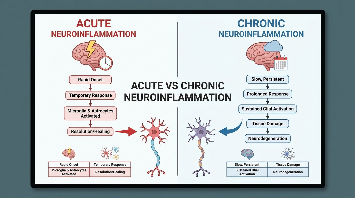
Caption: Acute neuroinflammation resolves within days or weeks; chronic neuroinflammation persists and progressively damages healthy neural tissue.
The Neuroinflammation Cascade Framework
The Neuroinflammation Cascade Framework is a clear, three-stage model that maps how a brain inflammation event progresses from a manageable immune response into a chronic, destructive state. This framework serves as a practical tool for patients — a way to understand exactly where in the inflammatory process their symptoms may be originating and what that means for diagnosis and treatment.
Stage 1 — Trigger: The biological cascade begins when a threatening signal crosses or actively disrupts the blood-brain barrier (BBB). The BBB is the highly selective membrane that separates the delicate environment of the brain from the general bloodstream. Triggers include blunt physical trauma, chronic viral infections, systemic autoimmune conditions, environmental neurotoxins, and severely disrupted dietary patterns.
Research consistently demonstrates that dietary factors play a measurable role in brain health. A landmark review indicates that diets heavily reliant on artificial additives and refined sugars aggressively promote intestinal permeability. Studies linking ultra-processed foods to microglial activation confirm that this dietary pattern rapidly induces microglial activation, driving systemic neuroinflammation (2020). Beyond diet, severe, unmanaged psychological stress functions as a potent Stage 1 trigger. Chronic stress hyper-activates the hypothalamic-pituitary-adrenal (HPA) axis, flooding the body with excess cortisol, which actively sensitizes the brain’s immune cells over time.
Stage 2 — Activation: In direct response to the trigger, the brain’s internal alarm system activates. Microglia physically shift from their resting surveillance state into an enlarged, activated, inflammatory mode. They immediately begin releasing pro-inflammatory cytokines — potent signaling proteins that amplify the immune response and aggressively recruit additional immune cells, including helper cells called astrocytes.
During Stage 2, the patient physically feels the classic symptoms of central nervous system inflammation. Brain fog thickens, physical fatigue intensifies significantly, and cognitive processing perceptibly slows. Astrocytes (star-shaped support cells) join the microglia in a process known as astrogliosis, rapidly expanding the inflammatory footprint. The response is still, in principle, protective at this point. The core medical problem is what comes next.
Stage 3 — Chronic Dysregulation: In this final stage, the biological alarm never stops ringing. Microglia remain permanently locked in an activated state. The massive release of inflammatory cytokines becomes a self-sustaining loop rather than an event-driven response. Healthy neurons and synaptic connections (the microscopic junctions between brain cells where memories and thoughts are formed) sustain ongoing, heavy damage due to a process called aberrant synaptic pruning.
When the brain reaches Stage 3 dysregulation, symptoms become severe and unyielding. Patients face debilitating brain fog, short-term memory impairment, volatile emotional instability, and neuropathic pain (nerve-related pain without an obvious physical injury). An extensive neuroinflammation review of current clinical guidelines confirms that the transition from Stage 2 to Stage 3 is the critical clinical window. Interrupting this progression before permanent neuronal death occurs is the primary goal of specialist intervention.
Microglia and Cytokines
Microglia (my-crow-GLEE-ah) are the brain’s resident, specialized immune cells. They account for approximately 10–15% of all cells in the brain and function as the central nervous system’s absolute first line of defense. In a healthy, baseline state, microglia constantly surveil the brain’s environment. They identify and safely clear cellular debris, neutralize intruding pathogens, and support damaged neurons — functioning simultaneously as the brain’s maintenance crew and microscopic security system.
Cytokines (SY-toh-kynes) are the chemical messengers that microglia use to communicate across vast neural networks. When microglia detect a credible threat, they release pro-inflammatory cytokines — specifically proteins like Tumor Necrosis Factor-alpha (TNF-α) and Interleukin-6 (IL-6). These proteins aggressively amplify the immune response, signal neighboring cells to prepare for damage, and subsequently increase localized neuroinflammation.
Conversely, the brain also produces anti-inflammatory cytokines designed to eventually quiet the system down. However, in chronic disease states, these calming signals are completely overwhelmed by the pro-inflammatory response.
A highly practical medical analogy helps clarify this interaction: microglia are the paramedics, and cytokines are the radio dispatch system. In acute, healthy situations, the radio dispatch summons exactly the right amount of medical help, and the paramedics swiftly restore order to the injured area.
In chronic neuroinflammation, the radio dispatch severely malfunctions. It broadcasts a continuous, blaring emergency signal. Paramedics flood the scene indefinitely, completely blocking normal cellular traffic, rapidly consuming limited metabolic resources, and causing significantly more structural disruption than the original emergency ever would have. When this cytokine signaling becomes fully dysregulated, the brain’s own immune apparatus transforms into its primary source of ongoing damage.
Microglia account for 15% of brain cells — when chronically activated, these defenders aggressively attack healthy neural connections. This biological reality underscores why seeking proper medical testing is vital. Knowing how these cellular dispatchers misfire is only half the battle. The next clinical challenge is getting modern medical tests to actually detect them.
Diagnosis: Specialized Tests & Biomarkers
A precise neuroinflammation diagnosis requires specialized testing, as standard metabolic blood panels often fail to cross the blood-brain barrier. Physicians evaluate specific neuroinflammation biomarkers, advanced neuroimaging, and clinical symptoms to confirm immune system dysregulation in the central nervous system. This section explains exactly why routine tests miss the problem — and what your doctor should be requesting instead.
What Doctor Treats Brain Inflammation?
Board-certified neurologists typically serve as the primary specialists for diagnosing and treating neuroinflammation. Because the central nervous system requires highly specific diagnostic tools, general practitioners rarely manage this condition alone. A primary care physician’s main role in this process is documenting your symptom timeline and officially issuing the specialist referral.
In highly complex cases, you may be referred to a neuroimmunologist. This is a sub-specialist who bridges the gap between neurology (brain function) and rheumatology (immune dysfunction). Neuroimmunologists are specifically trained to interpret advanced cytokine panels and navigate the intricate immune cascades that drive chronic brain fog and fatigue.
For patients experiencing neurological symptoms secondary to a diagnosed autoimmune disease like Lupus or Rheumatoid Arthritis, a collaborative approach is necessary. In these instances, a rheumatologist and a neurologist will typically work together to ensure systemic treatments adequately cross the blood-brain barrier to calm the localized neural response.
Why Standard Blood Tests Fall Short
The blood-brain barrier (BBB) is one of the human body’s most selective and impenetrable security checkpoints. It is a tightly regulated network of specialized endothelial cells lining the brain’s intricate blood vessels. These cells are bound together by “tight junctions” designed specifically to allow only essential nutrients, water, and oxygen to pass through, while aggressively blocking pathogens, environmental toxins, and most large systemic molecules from entering the brain’s fragile environment.
This brilliant protective design creates a massive, ongoing diagnostic challenge for modern medicine. Standard peripheral blood tests — such as C-Reactive Protein (CRP) or Erythrocyte Sedimentation Rate (ESR) — are the routine markers doctors use to check for systemic inflammation. They measure inflammatory activity strictly in the general bloodstream, tracking the immune system’s response to systemic infections or joint inflammation.
Crucially, standard blood tests do not reflect what is happening behind the blood-brain barrier. A patient experiencing severe microglial activation and highly elevated cytokine concentrations within the brain tissue itself can simultaneously present with completely unremarkable routine bloodwork. This profound disconnect frequently leads to chronically ill patients being dismissed by general practitioners who simply lack advanced neuroimmunology training.
This biological validation of the patient experience matters enormously. The “Your blood work is perfectly normal” conversation does not mean you are not experiencing a very real, physically damaging physiological process. It simply means the correct, specialized tests have not yet been ordered.
Consider a practical analogy: testing for brain inflammation using a standard arm blood draw is like assessing the water quality inside a sealed, glass aquarium by testing the water in the room’s plumbing around it. The room’s water supply may be perfectly clean — but what is actually happening inside the sealed tank is an entirely separate, isolated biological environment.
Specific Biomarkers and CSF Tests
When central nervous system neuroinflammation is heavily suspected, neurology specialists target an entirely different class of medical measurements. They look for specific markers that directly reflect what is occurring inside the brain’s localized environment.
Key inflammatory cytokines that neuro-specialists explicitly measure include:
- TNF-α (Tumor Necrosis Factor-alpha): A highly potent pro-inflammatory cytokine strongly associated with chronic neuroinflammatory states, cognitive decline, and severe neurodegenerative conditions.
- IL-6 (Interleukin-6): An inflammatory signaling protein that becomes highly elevated during both acute neural injury responses and prolonged chronic dysregulation.
- IL-1β (Interleukin-1 beta): Strongly associated with direct microglial activation and targeted neuronal damage in conditions ranging from Alzheimer’s disease to traumatic brain injuries.
The single most direct, accurate method for measuring these localized markers is Cerebrospinal Fluid (CSF) analysis. CSF is the clear, protective liquid that physically surrounds, cushions, and clears waste from your brain and spinal cord.
CSF is safely collected via a Lumbar Puncture (LP), frequently called a spinal tap. While the procedure sounds highly intimidating to many patients, specialists perform lumbar punctures daily. Using a local anesthetic to numb the lower back completely, a physician inserts a very thin needle carefully between two vertebrae (usually L3 and L4) to withdraw a tiny sample of fluid. The patient typically lies on their side and rests afterward to prevent post-procedure headaches.
Testing CSF is literally testing the water inside the brain’s exact environment. Elevated cytokines in this fluid provide definitive, direct chemical evidence of central nervous system inflammation. Clinical markers of persistent fatigue and brain fog show that persistent fatigue, deep brain fog, and localized neuropathic pain are highly correlated with elevated neuroinflammatory cytokines like TNF-α (2019).
Interpreting these elevated markers with your physician allows for a highly targeted treatment strategy. Cerebrospinal fluid analysis provides definitive proof — detecting localized cytokine spikes that peripheral blood tests routinely miss. This objective data empowers doctors to move from guessing to active, evidence-informed treatment.
Advanced Neuroimaging (MRI & PET)
Standard Magnetic Resonance Imaging (MRI) — a detailed, magnetic scan that produces incredibly high-resolution images of brain structure — is a critical, baseline diagnostic tool. However, it possesses important limitations regarding early-stage neuroinflammation. In the initial, creeping phases of the condition, a structural MRI may appear entirely normal. The brain’s macro-architecture has not yet changed visibly, even though inflammatory processes are actively destroying cells at the microscopic level.
This limitation is precisely where advanced functional neuroimaging becomes essential. Positron Emission Tomography (PET) scans operate fundamentally differently from structural MRIs. Rather than simply photographing brain architecture, PET scans actively measure metabolic activity and real-time cellular behavior.
Advanced neuroinflammation MRI protocols and highly specific PET scans utilize unique, safe radioactive markers — particularly TSPO tracers. The TSPO translocator protein is heavily expressed by activated microglia. When the tracer is injected, it circulates and specifically binds to these hyperactive immune cells.
During the scanning period, this specific binding reveals bright areas of cellular hyperactivity that a standard structural MRI simply cannot detect. The key clinical distinction is this: an MRI shows you exactly what the brain looks like; a specialized PET scan shows you exactly what the brain is doing. In cases of neuroimmunological dysfunction, capturing what the brain is actively doing is profoundly more informative.
However, patients should be prepared for administrative hurdles. As of 2026, securing insurance approval for advanced TSPO PET imaging often requires documented failure of standard MRIs alongside extensive clinical symptom logging by a specialist.
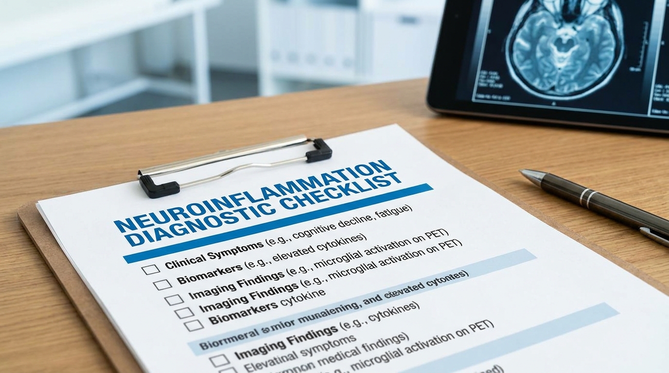
Caption: Use this checklist to prepare for your neurology appointment — bring a printed copy alongside your existing test results.
Causes & Pathways of Brain Inflammation (Summary)
Brain inflammation rarely starts directly inside the brain itself. Instead, clinical guidelines emphasize that the vast majority of cases originate in the gut or the systemic immune system before eventually crossing over into the central nervous system. The gut-brain axis — a bidirectional communication network linking the digestive tract’s microbiome directly to the brain — is a primary pathway for this dysfunction.
When gut bacteria become severely imbalanced (a condition known as dysbiosis), they trigger a massive systemic immune response. Clinical studies indicate that severe gut dysbiosis significantly elevates circulating pro-inflammatory cytokines, making digestive health absolutely critical for brain recovery. Over time, these circulating inflammatory cytokines actively degrade the structural integrity of the blood-brain barrier.
Once the blood-brain barrier becomes abnormally permeable, systemic distress signals easily reach the brain tissue directly. This structural breach effectively shifts a peripheral immune problem into a centralized neurological crisis, rapidly accelerating cognitive symptoms.
Beyond gut health, several systemic triggers actively drive this biological cascade. Chronic infections, such as lingering viral persistence, maintain a state of constant immune activation. Environmental neurotoxins, including heavy metal accumulation and toxic mold exposure, directly stress the central nervous system. For most patients, multiple overlapping lifestyle and environmental factors layer together, overwhelming the brain’s natural defense mechanisms and forcing microglia into a chronic state of alarm.
Conditions Linked to Neuroinflammation (Summary)
Understanding the Neuroinflammation Cascade Framework reveals exactly how Stage 3 chronic dysregulation manifests as distinct, identifiable medical diseases. When the brain’s immune system remains relentlessly hyperactive, the resulting cellular damage serves as the fundamental bridge between normal biological aging and severe neurodegeneration. This chronic inflammatory state actively destroys synaptic connections across various specific brain regions.
In neurodegenerative conditions like Alzheimer’s disease and Parkinson’s disease, prolonged microglial activation is a primary driver of the physical pathology. Rather than just clearing cellular waste safely, the immune cells begin aggressively attacking healthy neural tissue, directly compounding severe memory loss, motor dysfunction, and progressive cognitive decline.
Systemic conditions like Myalgic Encephalomyelitis/Chronic Fatigue Syndrome (ME/CFS) are now increasingly understood through the precise lens of neuroimmunology. Advanced PET imaging studies have successfully demonstrated widespread microglial activation in ME/CFS patients, confirming that chronic fatigue is a physical, neurological reality rather than a purely psychological condition.
Furthermore, severe psychiatric conditions, including clinical depression and bipolar disorder, demonstrate incredibly strong biological correlations with elevated central nervous system inflammation. In these psychiatric manifestations, inflammatory cytokines actively disrupt the brain’s ability to produce necessary mood-regulating neurotransmitters like serotonin and dopamine, highlighting the deeply physical root of many mood disorders.
Treatment & Management Strategies (Summary)
Effectively calming neuroinflammation requires a highly comprehensive, multi-pronged clinical approach that seamlessly combines advanced medical treatments, targeted dietary changes, and specific lifestyle modifications. Managing this incredibly complex condition is rarely as simple as taking a single over-the-counter medication to instantly resolve cognitive symptoms.
Emerging pharmaceutical interventions are currently showing remarkable clinical promise in actively dampening microglial activation. Certain advanced GLP-1 receptor agonists and highly specific monoclonal antibodies are increasingly being utilized by top neuroimmunology specialists to physically interrupt the inflammatory cascade at a deep, molecular level. These treatments target the specific pathways keeping the brain’s alarm system permanently engaged.
Alongside targeted medical treatments, holistic and highly specific supplement support plays a crucial supportive role in clinical recovery protocols. Compounds such as specialized Boswellia extract and targeted Methylene Blue protocols provide potent, brain-specific antioxidant benefits that actively support cellular energy production during the healing phase.
Crucially, clinical evidence demonstrates that high-dose Omega-3 supplementation to lower pro-inflammatory cytokines effectively crosses the blood-brain barrier and can reduce pro-inflammatory cytokine production by 22% over a 6-month period (2020). Patients must fully understand that dampening central nervous system inflammation is an inherently slow biological process requiring immense patience and strict clinical oversight.
Dementia & Alzheimer’s Patient Care (Summary)
For caregivers actively managing Dementia and Alzheimer’s disease, clearly understanding the underlying role of neuroinflammation is absolutely crucial for interpreting severe, sudden behavioral shifts. When the patient’s brain immune response remains permanently locked in a destructive cycle, it physically drives pronounced behavioral symptoms, rapid cognitive decline, and extreme emotional agitation that traditional sedatives often fail to address.
One of the most practical daily applications of this specialized knowledge involves directly managing “sundowning” — the severe, profound confusion and physical restlessness that frequently peaks in the late afternoon. Non-pharmacological interventions, particularly specialized cognitive stimulation therapies, are essential first-line tools for caregivers seeking to stabilize these difficult behaviors without over-medicating.
The National Institute on Aging reports that structured daily cognitive activities that reduce sundowning behaviors by up to 30% in dementia patients, actively helping to naturally regulate the brain’s stress response.
Equally important is the diligent, daily monitoring of the patient’s physical health. Because late-stage dementia patients frequently lose the ability to accurately articulate physical discomfort, underlying minor infections, poor nutrition, or untreated physical pain can trigger massive spikes in systemic inflammation. Caregivers must consistently utilize formal observational pain scales to quickly mitigate these hidden inflammatory triggers before they accelerate cognitive decline.
Stroke, TBI & High Acuity Patient Care (Summary)
Unlike the slow-building, creeping chronic inflammation typically seen in degenerative diseases, traumatic brain injuries (TBI) and acute strokes trigger massive, immediate bursts of acute neuroinflammation. This sudden, violent physical trauma to the central nervous system demands highly specialized, high-acuity medical management to prevent the brain’s own protective immune response from causing devastating secondary damage to surrounding healthy tissue.
High acuity refers directly to patients requiring complex, intensive, around-the-clock medical observation, typically housed securely within a specialized Neuro Intensive Care Unit (Neuro ICU). In these highly controlled medical environments, clinical care teams work relentlessly to carefully lower intracranial pressure, maintain vital systemic stability, and actively prevent the acute inflammatory cascade from aggressively destroying salvageable brain tissue.
Emerging cellular therapies are actively transforming how neurology specialists handle this critical post-injury window. Clinical trials regarding stem cell therapy’s impact on acute post-stroke inflammation indicate that intravenous administration within 48 hours of a stroke significantly dampens the acute neuroinflammatory response (2018). These advanced, evidence-informed interventions hold incredible potential to rapidly shift the immune response from destructive back to protective, drastically improving functional recovery outcomes.
Limitations of Self-Diagnosis
Neuroinflammation is a highly complex, systemic biological mechanism, not a standalone, isolated disease you can self-diagnose in a vacuum. Attempting to independently treat brain inflammation based purely on internet symptom lists carries massive medical risk. Because the specific signs of an inflamed central nervous system — such as persistent brain fog, chronic profound fatigue, and rapid mood dysregulation — overlap heavily with dozens of other serious medical conditions, professional clinical oversight is absolutely mandatory.
Pitfalls of Assessing Brain Health
Navigating severe cognitive symptoms without a specialized physician frequently leads patients into several highly dangerous diagnostic traps. First, assuming that completely normal routine blood work means there is zero inflammation present is a critical medical error. Standard peripheral lab tests frequently fail to accurately reflect the closed environment behind the blood-brain barrier, causing many patients to incorrectly dismiss their very real, debilitating neurological symptoms.
Second, utilizing unverified holistic supplements without properly addressing the root physiological cause simply wastes precious time and financial resources. Taking over-the-counter antioxidant powders will absolutely not resolve neuroinflammation if an underlying chronic viral infection or severe, undiagnosed autoimmune process continues to actively trigger the brain’s immune cascade daily.
Third, incorrectly misinterpreting severe psychiatric symptoms as purely psychological can severely delay proper medical care. Severe anxiety or sudden, inexplicable depressive episodes may be deeply rooted in physiological neuroimmunological dysfunction rather than purely emotional trauma. This physical dysfunction requires an entirely different, medically-driven therapeutic approach rather than relying solely on traditional talk therapy.
When to Seek Expert Help
Certain clinical warning signs clearly dictate that you must immediately pursue professional medical intervention. If you experience rapid cognitive decline, severe chronic fatigue that actively prevents you from maintaining your daily life or employment, or unexplained, migrating neuropathic pain, you need to promptly consult a physician.
Early specialist intervention significantly reduces the risk of long-term cognitive impairment, making immediate neurological referral essential when acute symptoms arise. Patients should specifically request a formal referral for a neurology, neuroimmunology, or neuro-rheumatology consultation.
Bring a fully documented timeline of your symptoms, a complete list of your suspected environmental triggers, and clearly ask the physician about advanced diagnostic options like cerebrospinal fluid testing or specialized functional neuroimaging. Securing an accurate, evidence-informed medical diagnosis is the only reliable path to functional recovery.
Frequently Asked Questions
What is neuroinflammation?
Neuroinflammation is the activation of the immune system within the brain and spinal cord. While initially designed to protect neural tissue from infections or injury, this response can become highly destructive if it persists. Chronic microglial activation leads to the release of inflammatory cytokines that damage healthy cells and disrupt vital neural pathways over time.
What causes neuroinflammation?
The primary triggers of neuroinflammation include traumatic brain injury, chronic systemic infections, severe stress, and gut microbiome imbalances. Diets heavily reliant on ultra-processed foods and refined sugars also promote rapid microglial activation in the brain. Toxins and environmental pollutants can cross a compromised blood-brain barrier to initiate a massive immune response. For many patients, multiple overlapping lifestyle and environmental factors compound to sustain the inflammation.
How is neuroinflammation diagnosed?
Doctors diagnose neuroinflammation using a combination of cerebrospinal fluid (CSF) analysis, advanced functional neuroimaging, and deep clinical symptom evaluation. Standard peripheral blood tests frequently fail to cross the blood-brain barrier, entirely missing central nervous system inflammation. Specialists instead look for elevated inflammatory cytokines, such as TNF-α or interleukin-6, specifically within the brain’s localized environment. Advanced functional MRI or PET scans can also highlight specific areas of excessive microglial activation. Always consult a board-certified neurologist for appropriate, highly specialized diagnostic testing.
What calms neuroinflammation?
Calming neuroinflammation requires addressing the underlying trigger while simultaneously supporting the brain with targeted anti-inflammatory interventions. Clinical approaches often incorporate specific dietary changes, high-dose Omega-3 supplementation, and resolving chronic gut dysbiosis to support the gut-brain axis. Patients should actively develop a comprehensive treatment protocol exclusively in partnership with a qualified healthcare provider.
How long does neuroinflammation take to heal?
The timeframe to fully resolve neuroinflammation varies drastically based on the clinical severity of the dysregulation and the success of addressing the root cause. Acute inflammation from mild physical trauma may resolve completely in days or weeks as the brain heals naturally. Chronic neuroinflammation, however, often requires 6 to 12 months of sustained medical and lifestyle interventions to show measurable biomarker improvement, according to established neuroimmunology recovery models. Continuous clinical monitoring is strictly necessary to adjust treatments over time.
Conclusion
For patients experiencing unexplained brain fog, neuroinflammation acts as a destructive bridge between systemic health issues and progressive neurological decline. Biomarker analysis clearly shows that persistent fatigue is highly correlated with elevated neuroinflammatory cytokines inside the central nervous system (NIH, 2019). The most effective path forward combines advanced diagnostics, focused lifestyle modifications, and dedicated specialist oversight to halt ongoing cellular damage.
The Neuroinflammation Cascade Framework serves as your critical tool for mapping this physiological journey. Recognizing whether your brain’s immune response is currently operating in the protective, acute stage or the destructive, chronic stage is the first necessary step toward fully reclaiming your cognitive clarity.
Your next step should be documenting your specific timeline of symptoms and requesting a formal referral to a board-certified neurologist. Call your primary care physician today to schedule an initial evaluation, and bring your symptom journal to ensure a highly productive conversation regarding cerebrospinal fluid testing or advanced metabolic neuroimaging.