
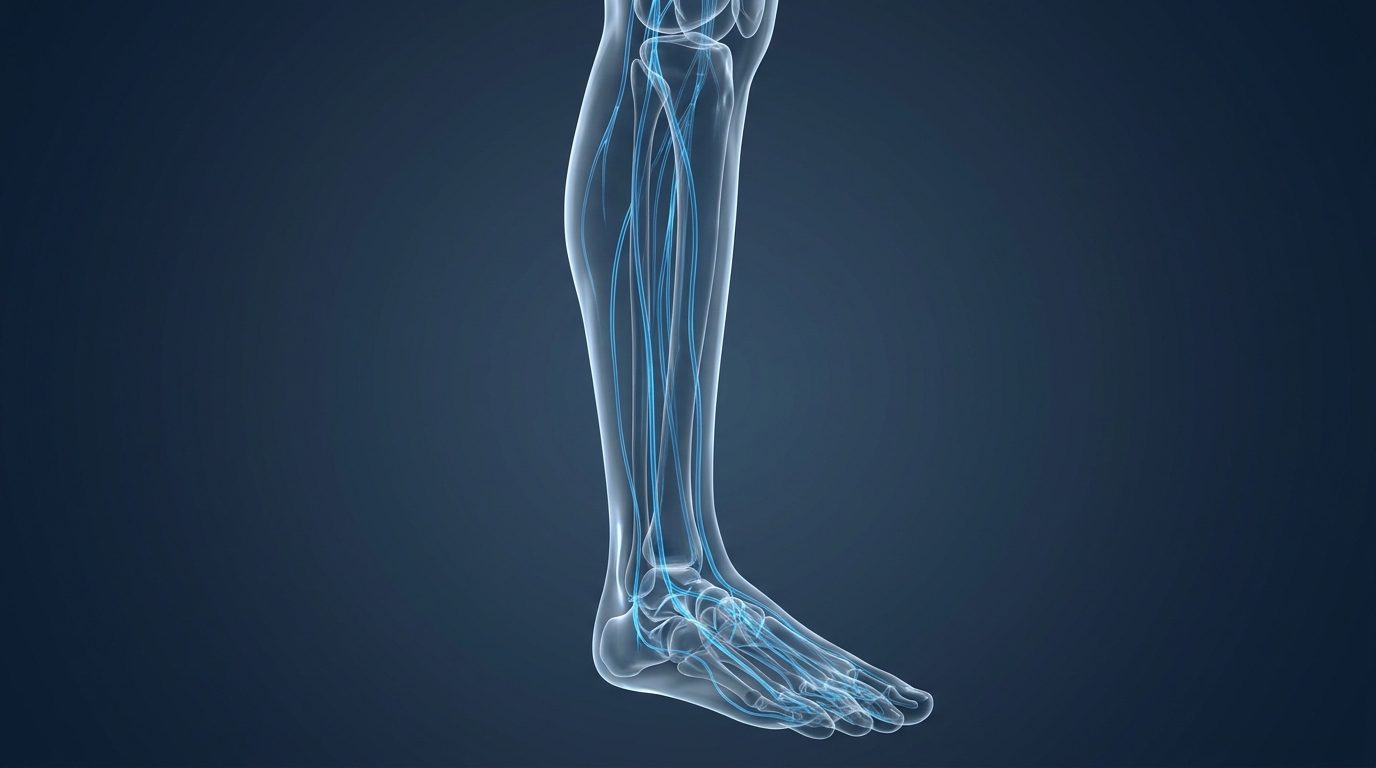
Peripheral Neuropathy: Symptoms, Causes & Assessment
Epidemiological data shows that approximately half of all individuals living with diabetes will eventually develop some form of peripheral nerve damage (CDC, 2026)—but blood sugar is far from the only culprit. Vague symptoms like tingling, burning, or sudden numbness create immense anxiety, as patients struggle to determine whether they are experiencing progressive peripheral damage or a life-threatening acute neurological emergency.
By the end of this guide, you will understand the precise mechanisms of nerve damage, exactly how clinicians assess it, and how to utilize the Nerve-Triage Matrix to navigate your differential diagnosis safely. This evidence-informed analysis synthesizes the core symptoms, primary toxic and metabolic causes, and the critical clinical boundaries between peripheral neuropathy and central nervous system disorders.
This article is for educational purposes only and does not replace consultation with a qualified medical professional. Always discuss symptoms with a qualified clinician.
Key Takeaways
Peripheral neuropathy causes nerve damage manifesting as sensory, motor, or autonomic symptoms, requiring clinical assessment for accurate diagnosis.
- The Nerve-Triage Matrix separates slow-progressing peripheral symptoms from acute, one-sided central emergencies like strokes.
- Primary triggers include uncontrolled diabetes, specific neurotoxic chemotherapies (like vincristine), and vitamin B12 deficiencies.
- Clinical evaluation relies on Electromyography (EMG) paired with nerve conduction velocity tests to objectively measure electrical nerve damage.
- Management focuses on suitability assessment, addressing underlying causes, and evidence-informed interventions like alpha-lipoic acid.
Symptoms & Clinical Assessment
Peripheral neuropathies symptoms occur when the peripheral nervous system—the communication network transmitting information between the central nervous system and the rest of the body—sustains damage. This degradation disrupts neural signaling, leading to progressive sensory loss, pain, and autonomic dysfunction. Understanding these mechanics is the first step in medical triage.
The peripheral nervous system acts as the body’s primary communication highway. It carries complex signals between the brain, spinal cord, and every organ, muscle, and patch of skin. When any part of that highway degrades—whether from a metabolic insult, a toxic compound, or physical trauma—signal transmission becomes highly unreliable. Patients frequently experience pain where there is no physical injury, total numbness where deep sensation should exist, or sudden muscle weakness without any structural lesion visible on imaging.
This inherent diagnostic complexity makes clinical assessment incredibly challenging and underlines exactly why objective electrodiagnostic testing remains indispensable. Accurately assessing nerve degradation requires professional neurological evaluation, utilizing both comprehensive physical exams and advanced electrodiagnostic tools. Clinical consensus indicates that sole reliance on subjective pain reporting fundamentally undermines early clinical intervention for nerve damage. An electromyography (EMG) paired with a nerve conduction velocity (NCV) test is considered the clinical gold standard for measuring the exact electrical activity and damage level of peripheral nerves (Johns Hopkins, 2026).
Clinical guidelines dictate that early detection directly correlates with better functional outcomes. Once the delicate axons begin dying, the recovery window narrows significantly.
Defining the Triad of Nerve Damage
A neuropathy definition that resonates deeply with patients begins here: peripheral nerves fall into three functionally distinct categories, and damage to each produces an entirely different constellation of clinical symptoms. Understanding which specific nerve type is affected helps clinicians pinpoint the precise location, severity, and appropriate medical intervention required to halt progression.
Sensory nerves function as the body’s critical alarm system. They transmit essential information about touch, temperature, vibration, and pain from the skin back to the central brain. When sensory fibers degrade, patients report a predictable cascade of distress. Initial intermittent tingling or “pins and needles” is quickly followed by burning pain that can become severe, and ultimately profound numbness as the nerve fibers die. Loss of temperature sensation—failing to feel hot bathwater, for instance—is a particularly telling sign of severe sensory nerve damage.
Motor nerves act as the human body’s steering wheel. They transmit the brain’s complex movement commands outward to the peripheral muscles. Damage here produces a highly different clinical picture: progressive muscle weakness that begins subtly, involuntary twitching known clinically as fasciculations, painful muscle cramping, and eventually the classic gait abnormality known as “drop foot.”
Autonomic nerves govern the body’s automatic climate control. These fibers manage complex physiological processes that run entirely without conscious thought. Blood pressure regulation, cardiac rhythm, digestion, bladder control, and sweat production are exclusively managed by autonomic pathways. According to the NINDS outline of progressive neuropathy stages, peripheral neuropathy damages the peripheral nervous system, and symptoms often progress through various sequential stages ranging from mild sensory loss to severe autonomic disruption (2026).
How to Assess Nerve Damage
Neuropathy symptoms rarely present clearly in the initial clinical setting. Physicians begin with a highly structured neurological physical examination long before any advanced technology is deployed. This hands-on exam evaluates deep tendon reflexes tapped with a hammer at the knee and ankle, actively tests muscle strength against physical resistance, and meticulously assesses the patient’s ability to feel specific vibration frequencies when a 128 Hz tuning fork is held against the bony prominences of the feet and ankles. A standard 10-gram monofilament test quantifies protective sensation loss.
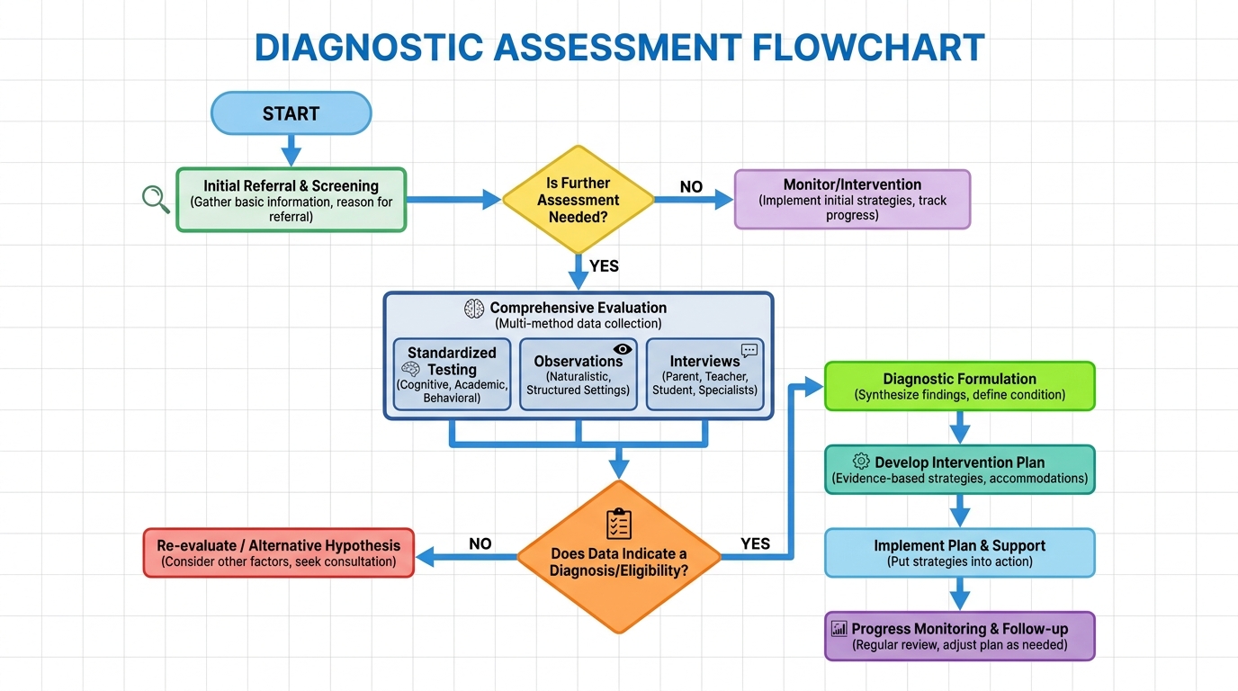
Electromyography (EMG), a diagnostic procedure evaluating the health of muscles and the nerve cells that control them, operates as the diagnostic gold standard for peripheral damage. This procedure inserts microscopic electrodes directly into muscle tissue to record resting and contractile electrical activity. The resulting wave patterns definitively separate neurogenic from myogenic disorders. The Johns Hopkins patient guide to EMG assessments indicates that an electromyography (EMG) paired with a nerve conduction velocity (NCV) test is considered the clinical gold standard for measuring exact electrical activity (2026).
The NCV component specifically measures the exact speed at which electrical signals travel through a specific nerve. Healthy large fibers conduct at 50–70 meters per second; drastically slowed conduction directly indicates severe myelin sheath destruction (demyelination), while reduced signal amplitude points to axonal loss. For patients reviewing their own medical records, the relevant ICD-10 diagnostic codes include G60 (Hereditary and idiopathic neuropathy), G61 (Inflammatory polyneuropathy), and E11.40–E11.49 (Diabetic neuropathy subtypes).
According to clinical expectations for nerve regeneration rates, the ‘rule of 3’ in nerve regeneration estimates that a damaged nerve grows back at a rate of approximately 1 millimeter per day, or 1 inch per month (2026). This sets realistic patient expectations regarding recovery timelines. Immediate symptom resolution is biologically impossible for severe structural nerve injuries.
With diagnostic data confirming the absolute degradation of the peripheral nerves, the clinical focus immediately shifts to identifying the specific metabolic or physiological root cause driving the destruction.
Primary Causes & Subtypes
While polyneuropathy symptoms manifest similarly across diverse patient populations, the underlying medical triggers driving the damage are remarkably vast. Clinical literature formally identifies over 200 distinct causes of peripheral neuropathy, ranging from chronic metabolic disorders to aggressive autoimmune attacks. Identifying the specific disease subtype is critical for determining appropriate clinical suitability assessments and mapping long-term functional outcomes.
The sheer breadth of potential causes is exactly why a systematic, highly categorical approach is absolutely essential. No single blood test or imaging scan identifies every possible trigger. Instead, clinicians must systematically work through the most statistically probable categories first—metabolic, toxic, autoimmune, and hereditary—before officially concluding that a patient’s case is entirely idiopathic. The Nerve-Triage Matrix categorizes whether a neuropathy is slow-onset and bilateral versus rapid and asymmetric, ensuring high-risk systemic conditions are ruled out early.
Approximately 50% of all individuals living with diabetes develop nerve damage—making strict glycemic control the ultimate neuroprotective strategy (CDC, 2026). Dismissing early tingling as a normal part of human aging aggressively denies patients their most critical intervention window.
Metabolic Causes: Diabetes and High Blood Sugar
Persistently elevated blood glucose acts incredibly like a slow-acting corrosive agent. It aggressively damages the microvascular system—the intricate network of tiny blood vessels (capillaries) that supply vital oxygen and nutrients to the peripheral nerves. Think of the peripheral nerve exactly like a living electrical cable. The surrounding capillaries act as the cable’s required power supply. When those capillaries are progressively destroyed by sugar-thickened blood, the nerve slowly starves.
Diabetic peripheral neuropathy symptoms follow a highly characteristic “stocking-glove” clinical distribution. Damage begins in the absolute longest peripheral nerves first—those running directly to the toes and feet—before slowly progressing upward toward the hands over several years. This specific pattern reflects the geometry of human nerve vulnerability; the further a nerve fiber extends from the spinal cord, the more capillary territory it strictly depends upon.
Persistently elevated HbA1c levels exponentially increase the risk of severe microvascular nerve damage. Strict glycemic control remains the primary neuroprotective strategy for all patients. According to CDC statistics on diabetic neuropathy prevalence, epidemiological data shows that approximately half of all individuals living with diabetes will eventually develop some form of peripheral nerve damage (2026).
The Top 10 Causes of Peripheral Neuropathy:
- Uncontrolled diabetes mellitus (Most prevalent global trigger)
- Chronic alcohol abuse (Direct toxic attack and nutritional depletion)
- Vitamin B12 deficiency (Severe demyelination of neural pathways)
- Chemotherapy drugs (Vincristine, cisplatin toxicity)
- Autoimmune diseases (Lupus, rheumatoid arthritis)
- Kidney failure (Uremic neuropathy from blood toxins)
- Thyroid disorders (Untreated severe hypothyroidism)
- Physical nerve trauma (Herniated discs, compression)
- Hereditary conditions (Charcot-Marie-Tooth disease)
- Idiopathic (Unknown clinical origins requiring constant monitoring)
Toxic Exposure: Alcoholism and Idiopathic Cases
Alcoholism neuropathy symptoms represent one of the most incredibly complex clinical presentations in peripheral neurology simply because the specific damage mechanism is devastatingly twofold. Chronic, heavy alcohol consumption causes severe, direct neurotoxicity. Ethanol and its primary metabolite acetaldehyde are highly chemically harmful to delicate nerve tissue, aggressively impairing mitochondrial energy function within the fragile axons. Furthermore, chronic alcoholism routinely depletes the body of essential thiamine (Vitamin B1), compounding the physical nerve destruction with severe nutritional starvation.
Idiopathic Neuropathy, nerve damage occurring without a clinically identifiable cause, requires the systematic exclusion of over 200 known metabolic, genetic, and toxic triggers. Modern medicine’s inability to definitively identify a specific root cause does not imply that absolutely no cause exists; it simply reflects the current clinical limits of our advanced diagnostic technology.
The clinical approach for idiopathic cases strongly prioritizes strict symptom management, highly structured monitoring, and reassessment at regular intervals. Ongoing neurological monitoring is essential to catch delayed systemic manifestations. Patients receiving an idiopathic diagnosis must practice extreme vigilance, as hidden autoimmune or hematological issues (like monoclonal gammopathies) may surface years after the initial nerve pain begins.
Nerve Fiber Varieties: Small Fiber vs. Polyneuropathy
Small fiber neuropathy causes a uniquely challenging clinical diagnostic puzzle for treating neurologists. This specific condition selectively damages the tiny unmyelinated C-fibers and thinly myelinated A-delta fibers responsible for transmitting pain and temperature signals back to the central brain. The clinical result is often severe, relentless burning pain—frequently described by patients as walking on hot coals—without any accompanying motor weakness or balance disruption.
Because small fiber damage leaves the larger, heavily myelinated nerve fibers entirely intact, a standard EMG and NCV test will very often return completely normal results. This tragically leaves the patient’s severe burning pain clinically dismissed or misattributed to psychological factors. The key diagnostic distinction requires Quantitative Sensory Testing (QST) and a specialized skin punch biopsy, a minimally invasive clinical procedure that measures the exact density of small nerve fibers residing in the epidermis.
Medication & Nutrition Drivers
Neuropathy vitamin deficiency and pharmaceutical toxicity represent highly specific, external drivers of nerve damage. Unlike slow-progressing metabolic conditions, medication-induced neuropathy can onset rapidly during treatments. Understanding these iatrogenic (medically induced) and nutritional triggers allows patients and clinicians to quickly adjust regimens before permanent degradation occurs.
Both rapid toxic inundation from systemic chemotherapy drugs and the prolonged chronic absence of critical neuro-vitamins severely impair essential nerve signal transmission. When properly addressing these complex pharmacological and nutritional deficits, patients must work exclusively with qualified clinicians to formulate highly evidence-informed medical treatment interventions that protect long-term functional mobility.
Specific chemotherapy medications aggressively attack neural microtubules—inducing sudden, severe peripheral neuropathy in oncology patients (NCI, 2026). Separating unavoidable pharmaceutical side effects from easily correctable nutritional blind spots requires rigorous blood chemistry analysis.
Chemotherapy-Induced Peripheral Neuropathy (CIPN)
Chemotherapy-Induced Peripheral Neuropathy (CIPN), a severe side effect of specific cancer-fighting drugs, fundamentally alters oncology treatment plans. Specific oncological drugs, particularly vincristine peripheral neuropathy, forcefully attack the nerve’s delicate intracellular microtubules. This aggressively halts the essential intracellular transport systems required to keep the long axons functionally alive. Unlike the slow suffocation observed in diabetic microvascular damage, CIPN constitutes a direct, aggressive chemical assault on the structural integrity of the peripheral nerve fibers themselves.
Platinum-based chemotherapy compounds, such as cisplatin and oxaliplatin, are notorious for heavily damaging the dorsal root ganglia. This frequently results in profound sensory ataxia and severe balance disruptions. Immediate dose reassessment is often clinically necessary to prevent permanent paralysis when severe sensory deficits arise during active cancer treatment.
Medical experts at the NCI data on neurotoxic chemotherapy drugs report that specific chemotherapy medications, notably vincristine, paclitaxel, and cisplatin, are primary neurotoxic triggers that can induce sudden peripheral neuropathy (2026). Establishing realistic patient expectations regarding CIPN is critical—some sensory symptoms gradually resolve after the oncological treatment ceases entirely, while severe motor deficits frequently become a permanent chronic condition.
Critical Vitamin Deficiencies (B12, Copper)
Vitamin B12, a crucial nutrient for maintaining nerve myelin sheaths, operates as the foundational biological building block for neural insulation. The complete absence of this nutrient starves the peripheral nerves, triggering widespread, aggressive demyelination. Neuropathy vitamin deficiency frequently stems from complex gastrointestinal malabsorption issues, prolonged adherence to strict unsupplemented vegan diets, or the chronic daily use of aggressive antacid medications that heavily suppress the necessary stomach acid required for proper cobalamin absorption.
Secondary nutritional deficiencies, particularly involving elemental copper and Vitamin E, significantly cripple the peripheral nervous system’s essential antioxidant defense mechanisms. Patients who have undergone aggressive gastric bypass surgery are at an exceptionally high clinical risk for rapid copper deficiency. This frequently mimics the exact progressive demyelination patterns classically seen in severe B12 starvation due to the disruption of zinc-copper absorption ratios in the altered gut.
Untreated B12 deficiency can lead to permanent, irreversible nerve demyelination if not addressed promptly. Identifying a specific, correctable nutritional deficiency dictates an incredibly obvious clinical treatment path, but effectively managing idiopathic or advanced neuropathic pain requires a multifaceted, highly evidence-led therapeutic approach.
Treatment & Diet Interventions
Managing intractable neuropathy neuropathic pain requires transitioning from passive medical diagnosis to highly proactive, evidence-informed intervention. Because severe peripheral nerve damage cannot currently be “cured” or instantly reversed with modern medicine, clinicians utilize a complex combination of pharmacological pain blockers, targeted nutritional supplements, and systemic dietary modifications to meaningfully improve functional outcomes and overall patient quality of life.
Optimal, evidence-based neuropathy management relies entirely on safe, carefully measured medical interventions. These strictly prioritize long-term functional mobility and aggressive pain reduction over the wildly unrealistic commercial promises of immediate nerve cures.
The SYDNEY 2 trial demonstrated 600 mg of daily alpha-lipoic acid reduces neuropathic symptom severity by 24%—providing a critical evidence-based intervention (PubMed, 2006). Acknowledging the strict limitations of standard pain medications empowers patients to actively pursue lifestyle changes that fundamentally alter the cellular environment.
Evidence-Based Supplements: Alpha-Lipoic Acid and Benfotiamine
Alpha-Lipoic Acid (ALA), a potent antioxidant studied for nerve repair, heavily anchors clinical supplementation protocols. Clinical data strongly supports the use of specific targeted antioxidants in repairing early-stage metabolic nerve damage. Alpha lipoic acid neuropathy treatments focus heavily on its potent antioxidant properties. ALA aggressively reduces the chronic oxidative stress currently destroying the delicate microvascular capillaries surrounding the starved peripheral nerves. By acting as a powerful free radical scavenger in both water- and fat-soluble cellular environments, ALA actively helps restore the damaged intracellular ecosystem.
Benfotiamine, a highly fat-soluble synthetic form of Vitamin B1, actively blocks the destructive biochemical pathways that cause severe vascular damage in poorly controlled diabetics. Standard water-soluble thiamine struggles to penetrate the nerve cell membrane, but benfotiamine achieves significantly higher intracellular concentrations. This directly inhibits advanced glycation end-products (AGEs), which are the toxic compounds responsible for hardening the delicate blood vessels feeding the peripheral nerves.
Research evaluating clinical trial data on alpha-lipoic acid efficacy confirms that the SYDNEY 2 clinical trial demonstrated that daily supplementation of 600 mg of alpha-lipoic acid significantly reduced neuropathic pain and sensory deficits over a 5-week timeframe (2006). While specific antioxidant supplements successfully attempt to alter the damaged nerve’s toxic microenvironment, achieving immediate, profound symptom relief usually requires highly targeted pharmaceutical intervention.
Pharmacological Pain Management
Gabapentin, an anticonvulsant medication frequently prescribed off-label for neuropathic pain, forms the backbone of standard medical treatment. Standard pharmacological medical treatments for severe neuropathy medication primarily involve complex anticonvulsants and powerful antidepressants heavily repurposed for specific neuropathic pain. Prescription medications like gabapentin, pregabalin, and SNRIs (such as duloxetine) do not physically repair the broken nerve fibers. Instead, they aggressively alter exactly how the central brain interprets the incoming erratic pain signals generated by the damaged peripheral nerves.
Pregabalin directly binds to the specific voltage-gated calcium channels located within the central nervous system. This effectively dampens the excessive release of excitatory neurotransmitters (like glutamate) that cause severe burning pain. Prescribing these agents without concurrently addressing the underlying metabolic root cause merely masks progressive nerve suffocation. Note the immense clinical importance of constant monitoring and rapid reassessment to safely manage common pharmacological side effects, which frequently include severe somnolence, dangerous dizziness, and profound cognitive fog.
Diet and Lifestyle Modifications
Dietary intervention protocols prioritize strict glycemic control to prevent progressive microvascular capillary damage. Implementing a highly anti-inflammatory, low-glycemic neuropathy diet directly stabilizes erratic blood glucose levels, effectively halting the relentless progression of metabolic nerve suffocation. Emphasize highly practical, patient-first lifestyle changes, particularly the daily wearing of specialized protective footwear and mandatory routine visual foot inspections for all diagnosed diabetics.
Strict adherence to a low-glycemic index diet, such as the Mediterranean dietary protocol rich in omega-3 fatty acids, can halt microvascular degradation in highly susceptible prediabetic populations. Dietary intervention is a primary medical treatment rather than an optional lifestyle choice. Incorporating daily, low-impact cardiovascular exercise significantly boosts essential microvascular blood flow directly to the oxygen-starved peripheral extremities. This actively promotes optimal conditions for incredibly slow natural nerve regeneration.
Central vs. Peripheral Triage
Applying the Nerve-Triage Matrix is absolutely vital for ensuring immediate patient safety, as accurately recognizing acute brain bleed symptoms versus chronic peripheral nerve pain can be definitively life-saving. While peripheral neuropathy develops incredibly slowly over months and affects the lower extremities symmetrically, acute cerebrovascular events violently strike the central nervous system instantly, requiring immediate, aggressive emergency medical intervention to prevent permanent brain death.
Chronic tingling in both feet is a peripheral nerve issue requiring a scheduled neurological consult; sudden profound weakness in one arm is a central nervous system emergency demanding an immediate 911 call.
Unlike the gradual, bilateral tingling of peripheral neuropathy, a stroke typically presents with sudden numbness localized heavily to one side of the body (CDC, 2026). Failing to utilize the Nerve-Triage Matrix during acute symptom onset fundamentally endangers patient survival.
Recognizing Acute Cerebrovascular Events (Strokes and Brain Bleeds)
Cerebrovascular Event, a medical emergency involving disrupted blood flow in the brain, destroys central tissue through acute oxygen deprivation. This produces instant, devastating neurological deficits. The fundamental physiological difference is clear: peripheral neuropathy involves localized, extremely slow nerve degradation. Stroke causes involve sudden, massive central oxygen deprivation directly to delicate brain tissue.
Ischemic Stroke, a blockage of a brain artery, creates distinct clinical patterns based on which specific vascular territory is choked. Transient Ischemic Attacks (TIAs), often called minor strokes, can frequently cause sudden, transient numbness that is tragically easily confused with simple pinched peripheral nerves. Ischemic strokes demand intervention within a 4.5-hour critical window, so recognizing unilateral (one-sided) numbness over bilateral (two-sided) tingling saves brain tissue.
The CDC differential diagnosis guidelines for sudden numbness state that unlike the gradual, bilateral tingling of peripheral neuropathy, a stroke typically presents with sudden numbness or weakness localized heavily to one side of the face or body (2026). If severe symptoms occur suddenly, predominantly on one side of the human body, patients must assume central brain pathology and seek immediate emergency care using the F.A.S.T. (Face drooping, Arm weakness, Speech difficulty, Time to call 911) protocol.
| Triage Feature | Stroke / Cerebrovascular Event | Peripheral Neuropathy |
|---|---|---|
| Onset Speed | Instantaneous (Seconds/Minutes) | Gradual (Months/Years) |
| Location | Unilateral (One side of body) | Bilateral (Both sides) |
| Associated Signs | Speech slurring, facial droop | None (Isolated to limbs) |
| Pain Type | Usually painless numbness | Burning, tingling, stinging |
Trauma and Pressure-Related Neurological Symptoms
A severe subdural hematoma symptoms (dangerous brain bleeding often resulting directly from head trauma) aggressively compresses delicate brain tissue. This forceful pressure buildup forcefully projects severe numbness and profound motor weakness straight down the peripheral limbs. Intracranial hypertension (dangerously high fluid pressure confined within the skull) severely disrupts global neurological function. This creates a highly complex clinical presentation that is entirely unlike isolated peripheral nerve damage.
Any formal clinical history of recent, significant head trauma immediately and permanently shifts the diagnostic medical focus entirely away from the peripheral nerves and directly toward the central brain. Severe herniated spinal discs can also cause severe radiculopathy. This shoots immense pain down a single leg, perfectly mimicking peripheral neuropathy, but it actually originates from physical spinal cord compression at the L5-S1 nerve roots. Differentiating spinal impingement from true peripheral polyneuropathy requires dedicated MRI imaging of the lumbar or cervical spine.
Neurodegenerative Disorders
Applying rigorous differential diagnosis frameworks is absolutely crucial when clinically evaluating sudden seizure symptoms or progressive movement disorders, as these complex conditions originate exclusively within the central nervous system. While peripheral neuropathy actively degrades the human body’s external electrical wiring, severe neurodegenerative diseases like Multiple Sclerosis or Parkinson’s systematically destroy the central processing unit, requiring entirely different clinical suitability assessments and specialized therapies.
Central neurodegenerative conditions produce severe spasticity, profound cognitive decline, and central tremors—symptoms entirely and fundamentally distinct from the flaccid weakness and distal symmetrical numbness of standard peripheral neuropathy.
Multiple sclerosis destroys central spinal myelin—creating spastic rigidity and hyperreflexia that fundamentally differs from the flaccid weakness of peripheral damage (NIH, 2026). Conflating central brain demyelination with peripheral axon damage fundamentally misdirects the required therapeutic interventions.
Multiple Sclerosis vs. Peripheral Demyelination
Multiple Sclerosis (MS), an autoimmune disease targeting the central nervous system’s myelin, attacks the central nervous system by deploying autoimmune macrophages against brain and spinal tissue. Both MS and some severe inflammatory neuropathies involve rapid demyelination, but ms symptoms in women and men involve the central nervous system (the brain and spinal cord), absolutely not the peripheral nerves. Multiple sclerosis primarily manifests in patients aged 20 to 40. Young adults presenting with sudden numbness require aggressive central neurological MRI scans.
Central brain damage causes profound spasticity (stiff, highly rigid muscles and hyperactive reflexes). Conversely, peripheral nerve damage definitively causes flaccid weakness and entirely lost reflexes. The specific cellular target differs immensely: MS destroys central oligodendrocytes, while peripheral neuropathy damages external Schwann cells. Severe visual issues, particularly optic neuritis (inflammation of the optic nerve causing sudden vision loss), are incredibly common early indicators in MS but remain exceedingly rare in standard peripheral neuropathy.
Cognitive Decline and Seizure Disorders
Parkinson’s Disease, a progressive neurodegenerative brain disorder affecting movement, degrades the substantia nigra’s ability to produce dopamine. Standard peripheral neuropathy never causes cognitive decline. If severe memory loss directly accompanies peripheral numbness, incredibly complex conditions like Lewy body dementia must be immediately and thoroughly evaluated by a qualified neurologist. Resting central tremors and sudden gait freezing are absolute hallmark Parkinson’s disease symptoms. These remain entirely distinct from the clumsy, slapping gait caused by numb, neuropathic peripheral feet.
Because Parkinson’s is strictly central, the resulting movement disorder demands dopaminergic therapy, not peripheral nerve pain blockers. Furthermore, severe seizure symptoms firmly represent highly dangerous electrical misfires exclusively within the brain cortex. This renders them entirely unrelated to any form of peripheral nerve damage. A damaged nerve in the foot cannot travel backward up the spinal cord to spontaneously trigger a cortical brain seizure. Any manifestation of seizure activity immediately confirms a central nervous system pathology.
Systemic & Infectious Mimics
Systemic bacterial infections and complex myalgic encephalomyelitis symptoms frequently mimic peripheral neuropathy, but their onset timelines and accompanying systemic symptoms are distinctly, clinically different. While metabolic nerve damage progresses incredibly slowly over years without causing systemic illness, infectious neurological attacks like meningitis or Guillain-Barré syndrome present acutely, incredibly often accompanied by high fevers, profound systemic fatigue, and rapidly ascending muscular paralysis.
Integrating the Nerve-Triage Matrix clearly reveals that absolutely any neurological symptom heavily accompanied by a sudden high fever, stiff neck, or rapidly ascending paralysis is a catastrophic systemic emergency, not standard chronic neuropathy.
Guillain-Barré syndrome is an acute form of nerve damage where the immune system attacks peripheral nerves, predominantly triggered by recent infections (WHO, 2026). Delaying emergency medical care during acute systemic infections guarantees severe, often irreversible neurological deficits.
Acute Brain Inflammation (Meningitis and Encephalitis)
Meningitis, a severe inflammation of the fluid and membranes surrounding the brain and spinal cord, dramatically inflames the central nervous system. Meningitis and symptoms specifically manifest as a deadly clinical triad: sudden high fever, incredibly severe headache, and a profoundly stiff neck. While this incredibly dangerous infection causes massive global neurological distress, it strictly remains an infection of the central meninges, absolutely not the peripheral nerves.
Bacterial meningitis can trigger fatal brain herniation within 24 hours. A stiff neck paired with numbness necessitates an immediate emergency room visit. Encephalitis (direct brain inflammation) and severe transverse myelitis (acute spinal cord inflammation) frequently present as acute central emergencies that cause total numbness below a highly specific spinal segment. Standard peripheral neuropathy absolutely does not cause high fevers or aggressively stiff necks.
Post-viral autoimmune syndromes require specialized immunological oversight, far exceeding the scope of standard peripheral neuropathy management.
Post-Viral Syndromes and Autoimmune Presentations
Guillain-Barré Syndrome (GBS), a rare disorder where the immune system attacks the peripheral nerves, operates as a highly unique, terrifyingly acute form of aggressive neuropathy. The body’s own immune system violently attacks the peripheral nerves immediately following a viral or bacterial infection (such as Campylobacter jejuni). This causes profound weakness that starts directly in the feet and rapidly moves upward toward the respiratory muscles.
According to the WHO summary on post-infectious neurological triggers, Guillain-Barré syndrome is an acute form of nerve damage where the immune system attacks peripheral nerves, predominantly triggered by a recent bacterial or viral infection (2026).
Severe Chronic Fatigue Syndrome (ME/CFS) features profound systemic exhaustion and severe brain fog, clearly separating it from isolated, painless peripheral nerve damage. Generalized systemic autoimmune diseases, exactly like Lupus or Rheumatoid Arthritis, can aggressively cause severe peripheral neuropathy. These systemic autoimmune conditions cause vasculitic neuropathy—severe inflammation of the blood vessels feeding the nerves—requiring immediate, specialized rheumatological medical evaluation to halt the immune attack.
Common Pitfalls & Limitations
Patient safety depends entirely on acknowledging when a symptom profile escalates beyond clinical outpatient management into an acute medical emergency. While establishing an accurate neuropathy diagnosis empowers patients, successfully navigating the complex medical landscape requires immense vigilance to avoid common pitfalls, dangerous marketing traps, and potentially fatal triage errors.
Many commercial stem cell clinics operate without FDA approval for neurological treatments, exposing patients to severe financial and physical risks (FDA Consumer Guidance, 2026). Trusting subjective marketing over objective electrodiagnostic data remains a critical threat to long-term functional mobility.
Critical Pitfalls in Neuropathy Management
Unproven regenerative medicine products often flood the market with dangerous, unsubstantiated claims of miraculous nerve cures. Falling for aggressive “cure” marketing remains the most dangerous financial and physical pitfall for neuropathy patients. Commercial regenerative medicine products—such as unverified stem cell injections or aggressive heavy metal chelation—are incredibly often not FDA approved for broad neuropathy claims.
Ignoring the root metabolic causes, such as actively taking gabapentin pain medication while completely failing to strictly control blood sugar, ensures the underlying nerve damage will silently, permanently worsen. Delayed initial clinical assessment invariably leads to completely irreversible axon death; early medical evaluation remains the only reliable method for preserving long-term functional mobility.
When to Seek Immediate Emergency Expert Help
Applying the Nerve-Triage Matrix prevents fatal clinical hesitation. If profound sensory loss occurs incredibly suddenly, presents entirely on one single side of the human body, or is heavily accompanied by severe facial drooping, dangerously slurred speech, or a sudden high fever, patients must bypass the neurologist and immediately go to the Emergency Room. Chronic, bilateral tingling can safely wait for a properly scheduled neurological consultation; acute unilateral motor deficits absolutely cannot.
Frequently Asked Questions
How do you assess for peripheral neuropathy?
Clinicians assess peripheral neuropathy using Electromyography (EMG) and Nerve Conduction Velocity (NCV) tests. These objective diagnostic tools measure the speed and strength of electrical signals traveling through the peripheral nerves. Before electrodiagnostic testing, physicians conduct a comprehensive physical exam to evaluate reflexes, muscle tone, and sensitivity to vibration or temperature. This two-step process accurately confirms nerve degradation. Patients should bring thorough medical records to ensure a precise suitability assessment.
What are the 4 stages of neuropathy?
Peripheral neuropathy typically progresses through four clinical stages of degradation. Stage one involves intermittent numbness or tingling, often ignored by patients. Stage two brings chronic, regular pain and noticeable sensory loss that frequently disrupts sleep. Stage three results in severe pain accompanied by motor weakness and dangerous balance issues. In stage four, patients experience total loss of sensation and significant autonomic dysfunction. Early medical intervention is crucial to halt progression before stage four.
What is the rule of 3 in a nerve injury?
The rule of 3 in clinical neurology estimates that a severed or damaged peripheral nerve regrows at 1 millimeter per day, or approximately 1 inch per month (NIH, 2026). This biological regeneration rate applies only if the nerve pathway remains structurally intact and the underlying cause of damage is fully resolved. Because nerves regenerate incredibly slowly, patients must maintain highly realistic expectations regarding recovery timelines. Immediate symptom resolution is biologically impossible for severe structural nerve injuries.
What are the top causes of neuropathy?
The top global cause of peripheral neuropathy is uncontrolled diabetes mellitus, which chronically damages the microvascular blood vessels supplying the nerves. Secondary primary causes include chronic alcohol abuse, which induces direct neurotoxicity, and specific chemotherapeutic drugs like vincristine. Vitamin B12 deficiency and systemic autoimmune diseases also rank highly as primary metabolic and inflammatory triggers. In many cases, the condition remains idiopathic, meaning modern medicine cannot identify a definitive root cause despite extensive testing.
Conclusion
For patients experiencing vague, confusing nerve pain, peripheral neuropathy requires highly objective, evidence-based medical assessment. Epidemiological data confirms that half of diabetics develop this severe nerve damage (CDC, 2026), making metabolic screening non-negotiable. The safest clinical approach seamlessly combines aggressively identifying the root metabolic or toxic cause with implementing highly evidence-informed lifestyle and dietary interventions.
Applying the Nerve-Triage Matrix remains absolutely critical for ensuring long-term patient safety. Distinguishing incredibly slow, bilateral peripheral symptoms from acute, unilateral central emergencies—like devastating ischemic strokes or sudden brain bleeds—prevents catastrophic triage errors and ensures patients immediately receive the exact level of critical care required.
Patients should thoroughly compile their specific symptom timelines, existing serological bloodwork, and comprehensive family medical history to schedule a thorough, objective diagnostic evaluation with a qualified clinical neurologist. Stop relying strictly on subjective pain management, and demand the objective electrodiagnostic data required to protect your long-term functional mobility.